Features and performance of edible
films, obtained from whey protein isolate
formulated with antimicrobial compounds
Óscar L. Ramos
a,b, Sara I. Silva
a, José C. Soares
a, João C. Fernandes
a, M. Fátima Poças
a,
Manuela E. Pintado
a, F. Xavier Malcata
b,c,⁎
a
CBQF/Escola Superior de Biotecnologia, Rua Dr. António Bernardino de Almeida, P-4200-072 Porto, Portugal
b
Instituto de Tecnologia Química e Biológica, Universidade Nova de Lisboa, Avenida da República, P-2780-157 Oeiras, Portugal
c
ISMAI— Instituto Superior da Maia, Avenida Carlos Oliveira Campos, P-4475-690 Avioso S. Pedro, Portugal
a b s t r a c t
a r t i c l e i n f o
Article history: Received 4 June 2011 Accepted 20 September 2011 Keywords:Whey protein isolate Antimicrobial agents Active packaging Physical properties Food safety
Antimicrobial packaging
The goal of this research effort was to assess the efficacy of edible films produced from whey protein isolate (WPI) and glycerol, including incorporation of lactic acid (LA) and propionic acid (PRO), chitooligosaccharides with nom-inal MW of 3 kDa (COS) and natamycin (NA) as antimicrobial agents. Their features were evaluated in vitro via agar diffusion and viable cell counting, against spoilage microflora often found contaminating cheese surfaces. The effect of incorporating the aforementioned compounds upon thickness, moisture content (MC), solubility (S), density (ρs), water activity (a
w) and water vapor permeability (WVP), as well as upon tensile and optical
prop-erties of thosefilms were also evaluated. Films formulated with LA, PRO or COS exhibited antimicrobial activity against all microorganisms tested, yet the viable cell count assay was more sensitive and reproducible. COS was the most active against Gram-negative bacteria, whereas LA was the most active against Gram-positive ones. NA was not active against bacteria, but displayed the strongest effect against yeasts. Incorporation of said antimicrobial compounds did not significantly (pN0.05) affect film thickness, yet it significantly (pb0.05) reduced tensile strength (TS). Incorporation of LA and NA in particular did not significantly (pb0.05) affect MC, S, ρs, WVP,
elon-gation at break (EB) and Young's modulus (YM) values; however, a statistically significant increase (pb0.05) of MC, S and WVP, together with a statistically significant decrease (pb0.05) of ρswere attained upon incorporation
of PRO or COS. Moreover, PRO produced the highest variation (pb0.05) in EB, TS and YM, whereas COS produced the highest change (pb0.05) in optical properties.
© 2011 Elsevier Ltd. All rights reserved.
1. Introduction
Foods are normally susceptible to physical, chemical and microbi-ological deterioration throughout storage and distribution, both as a function of their composition and the environmental conditions they are exposed to (Cha & Chinnan, 2004). An adequate selection of packaging materials can prevent food quality loss by providing bar-rier, or otherwise protective features thereto (Campos et al., 2011). If packagingfilms are in addition edible — e.g. those manufactured from polysaccharides, proteins or lipids, they will convey an extra set of ad-vantages, viz. biodegradability, non-toxicity and biocompatibility, be-sides esthetic appearance (Bourtoom, 2009; Khwaldia, Perez, Banon, Desobry, & Hardy, 2004; Tharanathan, 2003).
Whey proteins have been successfully employed as raw material for biodegradable packaging because they come from a renewable source and are a by-product of the cheesemaking industry; hence,
they are widely available, relatively easy to handle and essentially in-expensive. Whey protein isolates (WPI) represent the purer form of such whey proteins (Mulvihill & Ennis, 2003), and have shown prom-ising mechanical features, as well as moderate moisture permeability (McHugh, Aujard, & Krochta, 1994) and good oxygen barrier properties— comparable to those exhibited by the best synthetic polymer-basedfilms available e.g. low-density polyethylene (LDPE), high density polyethyl-ene, ethylene vinyl alcohol, vinyl alcohol, polyvinylidene chloride (PVDC), cellophane and polyester (Khwaldia et al., 2004; Perez-Gago & Krochta, 2002).
Furthermore, thosefilms proved excellent biomaterials for use as carriers of such food additives as antioxidants, antimicrobials, color-ants,flavors, fortifying nutrients and spices; these additives improve the functionality of the packaging by bringing about novel (or extra) features (Pranoto, Salokhe, & Rakshit, 2005; Salmieri & Lacroix, 2006). In particular, addition of antimicrobial agents may enable ex-tension of the shelf-life and safety of packaged foods, by reducing (or even preventing) growth of pathogenic and spoilage microorgan-isms (Franssen & Krochta, 2003). Moreover, their relatively low, but stable rates of diffusion from the packaging material onto the product assist in keeping the concentration of the active ingredient relatively
⁎ Corresponding author at: Instituto Superior da Maia, Avenida Carlos Oliveira Cam-pos, P-4475690 Avioso S. Pedro, Portugal. Tel.: + 351 96 8017 411; fax: + 351 22 982 53 31.
E-mail address:[email protected](F.X. Malcata).
0963-9969/$– see front matter © 2011 Elsevier Ltd. All rights reserved.
doi:10.1016/j.foodres.2011.09.016
Contents lists available atSciVerse ScienceDirect
Food Research International
j o u r n a l h o m e p a g e : w w w . e l s e v i e r . c o m / l o c a t e / f o o d r e shigh as time elapses (Kristo, Koutsoumanis, & Biliaderis, 2008; Min & Krochta, 2005). The antimicrobials more often incorporated in food packagingfilms are organic acids (e.g. citric, lactic, acetic and propio-nic acids), enzymes (e.g. lysozyme), bacteriocins (e.g. nisin), polysac-charides (e.g. chitosan), fungicides (e.g. benomyl and imazalil), and some plant extracts and their essential oils (Cagri, Ustunol, & Ryser, 2004; Min, Harris, Han, & Krochta, 2005; Tharanathan, 2003).
Lactic acid (LA) is frequently added to foods for preservation pur-poses, via reduction (or elimination) of growth of spoilage and path-ogenic bacteria (Alakomi et al., 2000). However, it may not exhibit a significant antimicrobial activity against yeasts and molds (Dibner & Butin, 2002; Ray, 2004). In alternative, propionic (PRO) acid has shown a good antifungal performance, and proved capable of inhibit-ing the growth of Gram-negative and -positive bacteria. This com-pound is usually applied to control mold growth on cheese, butter and bakery products, as well as to hamper growth of bacteria and yeasts in syrup, apple sauce and some fresh fruits (Ray, 2004).
Chitooligosaccharide (COS) is the oligosaccharide fraction pre-pared via enzymatic hydrolysis of chitosan (Fernandes et al., 2008); it is known to possess several antifungal (Hirano & Nagao, 1989; Kendra, Christian, & Hadwiger, 1989) and antibacterial (Hirano & Nagao, 1989; Uchida, Izume, & Ohtakara, 1989) features.
Natamycin (NA) is a natural antimycotic polyene, which has met with commercial success to prevent growth of molds and yeasts on food products (e.g. cheeses and sausages); hence, a GRAS status has been granted by the U. S. Food and Drug Administration, and it is also considered as a natural preservative by the European Union (EEC no. 235) for application on cheese surfaces or on slices thereof (Amefia, Abu-Ali, & Barringer, 2006).
Although extensive information on the antimicrobial properties of the aforementioned compounds is available in the literature (Cagri et al., 2004; Cha & Chinnan, 2004; Coma, 2008), scarce data exist pertain-ing to the activity of LA and PRO (Manab, Sawitri, al Awwaly, & Purnomo, 2011) and NA (Pintado, Ferreira, & Sousa, 2010) when incorporated in WPIfilms; and essentially no data at all encompassing incorporation of COS in thosefilms. Furthermore, a lack of information is apparent on the effect of those antimicrobial compounds upon the physical proper-ties of WPI films. On the other hand, selection of an antimicrobial agent entails not only assessment of its effectiveness against target mi-croorganisms, but also of interactions with thefilm-forming biopoly-mer; such interactions may indeed hamper the actual antimicrobial activity further to the characteristics of thefilm itself – both of which are key factors for development of commercially successful activefilms (Campos et al., 2011).
Therefore, the main purpose of this research effort was tofind, from a number of experimental antimicrobial agents (i.e. LA and PRO, COS and NA), those that would exhibit the highest effectiveness against a hetero-geneous set of spoilage/pathogenic microflora frequently found on the cheese surface— via incorporation into edible, 10%(w/w) WPI films plasticized with 5%(w/w) glycerol, without significantly compromising the functional properties exhibited by saidfilms. Therefore, the antimi-crobial performance of those edible active films was ascertained in vitro via agar diffusion and viable cell counting, against a model Gram-negative bacterium— Escherichia coli, a model Gram-positive bacterium — Staphylococcus aureus, and a model yeast — Yarrowia lipolytica. The ef-fect of incorporating such compounds upon thickness, moisture content, solubility, density, water activity, water vapor permeability, and tensile and optical properties of thosefilms was also assessed.
2. Materials and methods 2.1. Materials
Whey protein isolate (WPI) was obtained from Armor Proteines (Saint Brice en Coglés, France), and had been characterized previously (Ramos et al., 2011)— with the following composition data, on a
dry-weight basis: 92.0%(w/w) protein, 1.0%(w/w) lipid, 1.0%(w/w) lac-tose, 2.0%(w/w) ash and 3.0%(w/w) moisture, as well as 389.1 mg cal-cium, 100.1 mg sodium and 31.1 mg potassium per 100 g. Glycerol (99% purity) was supplied by Panreac (Barcelona, Spain), and pep-tone (P7750) was obtained from Sigma (St. Louis MO, USA). Chitooli-gosaccharide— COS, a pure fraction with a nominal MW of 3 kDa, was purchased from Nicechem (Shanghai, China) and used as received. Such COS had been obtained via enzymatic hydrolysis of chitosan from crab shells; its deacetylation degree lied in the range 80–85%, as indicated by the supplier. Lactic acid— LA (98% purity, L1750) and propionic acid— PRO (99% purity, P1386), were both obtained from Sigma (St. Louis MO, USA), whereas natamycin— NA (50% purity) was provided by Mapril (Maia, Portugal). All other chemicals were reagent-grade or better, and were used without further purification. 2.2. Antimicrobial solution preparation
The COS solution was prepared by dissolving COS to 200 g L−1in deionized water, under stirring; its pH was adjusted to 5.8 (which is the most appropriate for solubilization, and devoid of any significant antibacterial effect), using 10 mol L−1NaOH. After stirring overnight, the solution was autoclaved at 120 °C for 15 min; the thermostability under these conditions had been checked in advance (Fernandes et al., 2008). The solutions of LA and PRO were prepared by dissolving LA to 150 g L−1and PRO to 200 g L−1in deionized water, under stirring– whereas NA was prepared by dissolving NA to 250 g L−1in sterile 0.02 mol L−1HCl under stirring. Subsequently, pH was adjusted to between 5.5 and 6.0 with 1 mol L− 1HCl or NaOH (as appropriate). Finally, these solutions were sterilized viafiltration through a 0.22 μm filter (Orange Scientific, Belgium).
2.3. Culture preparation
The target microorganisms selected were: one Gram-negative bacterium— E. coli (NCTC 9001), one Gram-positive bacterium — S. aureus (NCTC 8532), and one yeast— Y. lipolytica (previously isolated from cheese within our group). Bacterial cultures were pre-activated by overnight incubation at 37 °C on Muller-Hinton (M-H) broth (Bio-kar Diagnostics, France), whereas the yeast was grown on Yeast Malt (YM) broth (Difco, USA), at 30 °C.
2.4. Determination of minimum inhibitory and minimum lethal concentrations
The minimum inhibitory concentrations (MICs) towards the three aforementioned microorganisms were determined following the broth macrodilution method (National Committee for Clinical Lab Standards, 2000), with the following modifications: the strains were inoculated in M-H broth (in the case of bacteria) or YM broth (in the case of the yeast), and incubated at 37 and 30 °C, respectively, until the exponen-tial growth phase was reached. The inoculum density was then adjusted to match a MacFarland 0.5 standard (ca. 108CFU mL− 1); then, dilution
was done in appropriate media, and 105CFU mL− 1was eventually used
as experimental inoculum. Afterwards, several concentrations of LA (1.5, 3.0, 6.0, 9.0 and 15.0 g L−1), PRO (1.0, 2.5, 5.0, 10.0 and 20.0 g L−1), COS (1.0, 2.5, 5.0, 10.0 and 20.0 g L−1) and NA (0.025, 0.05, 0.25, 2.5 and 25.0 g L−1) were tested, by preparing decreasing concentrations in the aforementioned media. Each MIC was determined as the lowest concentration of an antimicrobial agent in the presence of which the microorganism selected could not grow, as ascertained by the absence of visual turbidity— following classical recommendations (Fernandes et al., 2008; NCCLS, 2000; Ohsaki et al., 2003).
Each minimum lethal concentration (MLC) was determined as the lowest concentration of the antimicrobial agent at which microbial growth was prevented, and the initial viability was further reduced by at least 99.9% within 24 h. Microbial viability was determined by
enumeration of viable cells on M-H agar (Biokar Diagnostics, France) in the case of bacteria, and on YM agar (Difco, USA) in the case of the yeast, after inoculation of 100μL of negative tubes (i.e. showing no turbidity in the MIC determination assays). The incubation was car-ried out at 37 °C for both bacteria, and 30 °C for the yeast.
2.5. Film preparation
Film-forming solutions were prepared by slowly dissolving 10% (w/w) WPI powder in deionized water, following the procedure reported byPerez-Gago and Krochta (2002). Glycerol was added at 5%(w/w) as plasticizer, and the resulting solutions were magnetically stirred for ca. 2 h. Subsequently, they were heated in a water bath at 80 ± 2 °C, for 20 min under continuous agitation; this step is essential for formation of intermolecular bonds, which will in turn assist in es-tablishment of a cross-linked polymeric network structure (le Tien et al., 2000). The solutions were cooled to room temperature (30 °C) for 1.5 h. Afterwards, 10%(w/w) of each antimicrobial compound was added to obtain the corresponding MLC values (determined above), and then vacuum was applied for 30 min to remove dissolved air (Seydim & Sarikus, 2006). Finally, the solutions were adjusted to pH 7.0 using 0.1 mol L− 1 NaOH, and poured onto level Teflon-coated plates (38 × 34 cm). To controlfilm thickness, the amounts of each film-forming solution poured onto the plate were the same (300 mL). The solutions were allowed to dry at room conditions (ca. 23 °C and 50% relative humidity) for 24 h, according to the procedure byGounga, Xu, and Wang (2007) and Osés, Fernández-Pan, Mendoza, and Mate (2009). Once formed, thefilms were peeled off and condi-tioned at 23 ± 2 °C and 50 ± 2% RH, in a controlled temperature and humidity storage room (Packaging Center, Porto Portugal), for at least 72 h prior to testing (ASTM, 2000). All physical measurements described below were conducted also at 23 ± 2 °C and 50 ± 2% RH. 2.6. Antimicrobial activity
The antimicrobial activity of WPI ediblefilms was carried out using two complementary approaches: agar diffusion assay and via-ble cell count assay.
2.6.1. Agar diffusion assay
The qualitative antimicrobial activity of each WPIfilm was evalu-ated following the procedure described byPranoto et al. (2005). Films were cut into 17.0 ± 0.1 mm diameter disks using a circular knife, and exposed to UV light for 10 min on each side (Melo, 2003). They were then placed on M-H agar plates for bacteria, and on YM agar plates for the yeast— which had previously been seeded with 0.1 mL of inocu-lum, containing 105CFU mL− 1(as recommended byNCCLS, 2000) of
each target microorganism. WPIfilm disks, without incorporation of antimicrobial compounds, were also tested under similar conditions (negative control). The plates were incubated at 37 °C for 24 h, or 30 °C for 48 h, for the bacteria or the yeast, respectively. Afterwards, the zones of inhibition of thefilm disks on the plates were examined via measuring their diameter. The sensitivity to the different antimi-crobialfilms was rated followingPonce, Fritz, del Valle, and Roura (2003), based on the diameter of the zone of inhibition generated: not sensitive, sensitive, very sensitive and extremely sensitive, if the diameter was less than 8 mm, between 9 and 14 mm, between 15 and 19 mm, and greater than 20 mm, respectively. The test was performed in triplicate, in two separate experimental runs. 2.6.2. Viable cell count assay
The quantitative antimicrobial activity of each WPIfilm was eval-uated using theAATCC test method 100–2004 (1961)— which was originally designed for evaluation of antimicrobial activity of textile materials, and adapted hereby to ediblefilms: WPI films (with and without incorporation of antimicrobial compound) were thus cut
into 50.0 ± 1.0 mm diameter disks using a circular knife, and were ex-posed to UV light for 10 min on each side (Melo, 2003). Eachfilm disk was then placed in a 125 mL-sterilizedflask, to which 1.0 mL of inoc-ulum containing 105CFU mL− 1of each microorganism was added, so
as to cover the entire disk. Flasks were incubated at 37 or 30 °C, in the case of the bacteria or the yeast, respectively. Afterwards, 99.0 mL of sterile peptone water (1 g L− 1), used as neutralizing solution, was aseptically added to eachflask at 0, 3, 6, 12, and 24 h (sampling time). The flask content was then aseptically transferred to a 400 mL-homogenizing bag, and blended in a Stomacher 400 recipro-cal homogenizer (Seward Medirecipro-cal, London, UK) for 1.0 min at 260 rpm. Appropriate sequential 10-fold dilutions of the homogenate were done in sterile peptone water (in triplicate), and plated (0.02 mL per plate— in duplicate) onto M-H agar plates for the bacte-ria, and on YM agar plates for the yeast. The plates were then incubat-ed as describincubat-ed above. Enumeration of colonies was performincubat-ed, and inhibition of microorganism growth was expressed as reduction of cell number using log (N/N0)— where N is the viable cell number at
a given time and N0is its counterpart at time zero (Fernandes et al.,
2008). The test was performed in triplicate, in two separate experi-mental runs.
2.7. Film characterization 2.7.1. Thickness
The film thickness was measured with a micrometer (Model M120, from Adamel Lhomargy, Roissy en Brie, France), to the nearest 0.001 mm. The mean thickness was calculated from five measure-ments, taken randomly at various locations on eachfilm sample. 2.7.2. Moisture content andfilm solubility
The moisture content (MC) of the proteinfilms was determined after drying in an oven at 105 °C, under forced air circulation for 24 h. Small specimens (0.200 g) offilm were cut after conditioning, and placed on Petri dishes that were weighed before and after oven drying. MC values were determined as a fraction of the initialfilm weight lost during drying (ASTM, 1994), and were reported on a wet basis. Thefilm solubility in water (S) was determined following
Gounga et al. (2007). The determinations of MC and S were both per-formed in triplicate.
2.7.3. Density
Thefilm density (ρs) was calculated directly from thefilm weight
and dimensions (Salgado, Ortiz, Petruccelli, & Mauri, 2010), according to:
ρs
¼ m= Axδð Þ ð1Þ
where A is thefilm area (12.6 cm2, in our case),δ the thickness (cm), m
the dry mass (g) andρsthe dry matter density (g cm−3). Thefilm
den-sity was expressed as the average offive independent determinations. 2.7.4. Water activity
The water activity (aw) of preconditioned films was measured
using a HygroLab 2 (from Rotronic, Bassersdrof, Germany). Pieces of film (ca. 0.5 g) were placed on the sample holder of the water activity device; a sealed system was formed by placing the water activity probe on top of the sample holder. This probe was equipped with a small fan that circulated air within the sample container, a thinfilm capacitance sensor able to measure RH from 0 to 100 ± 1.5%, and a platinum resistance temperature detector with a precision of ± 0.3 °C. When awbecame constant (ca. 1 h), its value was recorded.
Calibration resorted to six saturated solutions of known aw (viz.
LiCl = 0.114, MgCl2= 0.329, K2CO3= 0.443, Mg(NO3)2= 0.536,
NaBr = 0.653 and KCl = 0.821). These measurements were carried out in quadruplicate.
2.7.5. Water vapor permeability
The water vapor permeability (WVP) was gravimetrically mea-sured according to the protocol B ofASTM (1995), with the adapta-tions proposed by Debeaufort, Martin-Polo, and Voilley (1993)
specifically for edible films. Circular aluminum cups, with a diameter of 8 cm and a depth of 5 cm, were accordingly used. Distilled water (30 mL) was placed in each test cup, to expose the lowerfilm face to a high RH. Thefilm samples were mounted with the upper surface facing the RH (50 ± 2%) of the environment-controlled room. The weight loss of the cups was monitored over a 72 h-period, with weights recorded at 4 h-intervals. The WVP (g mm m− 2d− 1kPa− 1) of thefilm was calculated as follows:
WVP¼ ΔW×FTð Þ= S×Δpð Þ ð2Þ
whereΔW is the weight loss of the cup per day (g d− 1) (i.e. slope of
the linear behavior), FT is thefilm thickness (mm), S is the area of ex-posedfilm (m2) andΔp is the vapor pressure differential across the
testfilm (kPa). At least 3 replicates were produced from each film type.
2.7.6. Tensile properties
The tensile properties offilms — i.e. tensile strength (TS), elongation at break (EB) and Young's modulus (YM), were measured according to the reference method (ASTM, 2002), using a Universal Testing machine model 4501 (from Instron, Canton MA, USA), equipped withfixed Grips (test method A) and a 100 N-static load cell. Thefilm samples were cut into strips (80 × 15 mm). The initial grip separation was set at 50 mm, and the crosshead speed at 4.8 mm min−1. The TS, EB and YM values were determined using the Series IX Automated Materials Testing Sys-tem software, v. 809.00 (Instron). At least ten strips of eachfilm sample were analyzed.
2.7.7. Optical properties
2.7.7.1. Light transmission andfilm transparency. The ultraviolet (UV) and visible light barrier properties were measured on driedfilms at selected wavelengths (in the 200–800 nm range), using an UV-VIS Spectrophotometer (SPECORD S 600, from AnalytikJena, Jena, Germany). The film samples were cut into strips (4×1 cm) and were attached to one side of a colorimetric cup— while the empty colorimetric cup was used as control. The relative transparency of thefilm was measured at 600 nm, and calculated as (Han & Floros, 1997):
Transparency¼ A600=δ ð3Þ
where A600 is the absorbance at 600 nm and X the film thickness
(mm). At leastfive strips of each film type were analyzed.
2.7.7.2. Color. Thefilm color was evaluated using a portable Chroma-meter CR-400 (from Minolta Chroma, Osaka, Japan). A CIELab color scale was used to measure the degree of lightness (L), redness (+a) or greenness (−a), and yellowness (+b) or blueness (−b) of the
films, under D65 (daylight). Film specimens were measured on
the surface of the white standard plate, with color coordinates Lstandard= 97.6, astandard= 0.01 and bstandard= 1.60. The color of
eachfilm was expressed as the total difference in color (ΔE), and was calculated according to:
ΔE ¼ Lðfilm–LstandardÞ 2
þ að film–astandardÞ 2
þ bð film–bstandardÞ 2
h i1=2
: ð4Þ For everyfilm incorporated with each of the different antimicrobial compounds tested, four samples were taken and, on eachfilm sample, four readings were made on each side.
2.8. Statistical analyses
Statistical analyses were performed using the Statistical Package for Social Sciences, v. 17.0 for Windows (SPSS, Chicago IL, USA), via one-way analysis of variance. The difference of means between pairs was resolved via confidence intervals, using Tukey's test. The significance level was set at pb0.05.
3. Results and discussion
3.1. Minimum inhibitory and lethal concentrations
The MIC and MLC associated with each antimicrobial agent and microorganism tested are depicted inTable 1. For each antimicrobial compound, both MIC and MLC depended on the target microorgan-ism. In general, the MLC values obtained were higher than their MIC counterparts, except for LA against S. aureus (in which case they were similar).
LA led to the lowest MIC and MLC values against bacteria. This compound showed similar MICs (3 g L− 1) toward all microorgan-isms, but higher MLCs for the Gram-negative bacterium and the yeast (6 g L− 1). PRO and COS showed similar MIC and MLC values against the Gram-negative bacterium; however, PRO produced lower MIC and MLC values against the Gram-positive bacterium and the yeast. COS demonstrated lower MIC and MLC values against the Gram-negative bacterium than the Gram-positive one or the yeast (Table 1); this is consistent withFernandes et al. (2008), who used COS with identical MW.
On the other hand, NA did not inhibit the bacteria, but displayed the lowest MIC and MLC values against the yeast. This is in agreement with results reported byWelscher et al. (2008), who showed that NA (as antimycotic compound) exhibits activity preferentially against yeasts and molds. Comparing our MIC values with those available in the literature, the former were lower for LA (i.e. 3 g L− 1) than those bySkrivanova, Marounek, Benda, and Brezinha (2006)and byHsiao and Siebert (1999) — ca. 5 and 3.7 g L− 1, respectively, against
E. coli. In the case of PRO, the MIC found here (i.e. 2.5 g L− 1against Y. lipolytica) was significantly lower than those reported byLind, Jonsson, and Schnürer (2005)— i.e. 37.0 g L−1, against three yeasts (i.e. Pichia anomala, Rhodotorula mucilaginosa and Kluyveromyces marxianus). In the case of COS, the MIC obtained was similar to that found byXia, Liu,
Table 1
Minimum inhibitory (MIC, g L− 1) and lethal (MLC, g L− 1) concentrations of antimicrobial agents against model microorganisms.
Antimicrobial agent (range of concentration)
Model microorganism
Escherichia coli Staphylococcus aureus Yarrowia lipolytica
MIC MLC MIC MLC MIC MLC
LA (1.5–15 g L− 1) 3 6 3 3 3 6
PRO (1.0–20 g L− 1) 5 10 5 10 2.5 5
COS 3 kDa (1.0–20 g L− 1) 5 10 10 20 10 20
NA (0.025–25 g L− 1) – – – – 0.05 0.25
Zhang, and Chen (2011)— i.e. 5 g L−1(pertaining to COS with a degree of
polymerization of 3–6), but lower than that reported byGerasimenko, Avdienko, Bannikova, Zueva, and Varlamov (2003)— i.e. 10 g L−1 (per-taining to COS with a MW of 5 kDa) against E. coli.
Moreover, our MICs associated with all microorganisms (see
Table 1) are in agreement with those claimed byXia et al. (2011)—
who concluded that MICs vary from 1 to 10 g L− 1against common bacteria, molds and yeasts, depending on the degrees of polymeriza-tion and molecular weight of COS. On the other hand, our MIC values (i.e. 5 and 10 g L− 1against E. coli and S. aureus, respectively— see
Table 1) are apparently high when compared with those obtained byJeon, Park, and Kim (2001)— i.e. 1.2 g L−1against E. coli and S. aureus;
however, these authors used COS with different MWs (ranging from 1.5 to 6 kDa). For NA, the MIC obtained against Y. lipolytica (i.e. 0.05 g L−1) is clearly lower than that reported by Pintado et al. (2010) — i.e. 20 g L−1against the same microorganism.
Differences in MIC (or MLC) values are useful in studies encom-passing antimicrobial agents, especially when different methods are compared— in attempts to find the agent able to exert the highest an-timicrobial effect. However, scarce information relative to MLC values by the agents selected for our study, as well as MIC and MLC values against only Y. lipolytica can be retrieved from the literature — which obviously hampers more extensive conclusions to be drawn. In our case, the differences in MICs relative to those conveyed in the literature are probably a result of the distinct experimental conditions used— e.g. the concentration range, the final pH and the target micro-bial strain. In the case of COS, the differences observed in MIC values may also be accounted for by different MWs or degrees of deacetyla-tion— since both parameters affect the content in protonated amino groups of the COS molecule, and consequently its charge. The antimi-crobial activity of COS has been attributed mainly to its positive charge, which allows strong binding to the negatively charged sur-faces of microorganisms (Fernandes et al., 2008).
3.2. Antimicrobial activity
The inhibitory activity of WPIfilms incorporated with several an-timicrobial compounds was measured using two distinct assays: a qualitative one, based on formation of a clear zone surrounding the circularfilm disk — the agar diffusion assay; and a quantitative one, based on quantification of the inhibitory activity of those films — viable cell count assay.
3.2.1. Agar diffusion assay
The results of the agar diffusion assays are depicted inTable 2— as
obtained for 10%(w/w) WPI ediblefilms with 5%(w/w) glycerol, in-corporated with LA and PRO, COS (3 kDa) and NA, and tested against one Gram-negative (i.e. E. coli) and one Gram-positive (i.e. S. aureus) bacteria, as well as one yeast (i.e. Y. lipolytica).
Antimicrobial activity was not observed in the negative control, consisting of a WPIfilm disk without previous incorporation of any antimicrobial compound. WPIfilms added with either organic acid showed the highest inhibition zones against the Gram-positive bacte-rium; statistically significant differences were indeed found (pb0.05) relative to other antimicrobial compounds. However, significant dif-ferences were not observed (pN0.05) among LA and PRO against the positive and -negative bacteria. In both cases, the Gram-positive bacterium was significantly more sensitive to these com-pounds than its Gram-negative counterpart (pb0.05).
On the other hand, PRO exhibited significantly higher inhibition (pb0.05) against the yeast than LA or COS (seeTable 2). According toPonce et al. (2003), E. coli is sensitive and S. aureus is very sensitive to LA and PRO, whereas Y. lipolytica is sensitive and very sensitive to LA and PRO, respectively.
WPIfilms incorporated with COS exhibited the highest inhibition zone (pb0.05) against the Gram-negative bacterium — which was statistically higher (pb0.05) than that exhibited against the Gram-positive bacterium and the yeast (seeTable 2). Moreover, Y. lipolytica, S. aureus and E. coli appeared to be sensitive, very sensitive and ex-tremely sensitive to COS, respectively (Ponce et al., 2003).
Finally, WPIfilms incorporated with NA proved the most effective against the yeast; statistically significant differences (pb0.05) were recorded relative to the other antimicrobial compounds, yet no inhi-bition was found against bacteria— see againTable 2. NA was an ex-tremely strong compound against Y. lipolytica, but not against E. coli or S. aureus (Ponce et al., 2003). This result is consistent with the ob-served above regarding MICs and MLCs, as well as with that reported byPintado et al. (2010)— who only observed inhibition of the yeast (i.e. Y. lipolytica).
3.2.2. Viable cell count assay
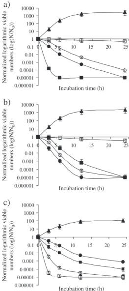
The antimicrobial activity is plotted inFig. 1— as determined for 10%(w/w) WPI edible films with 5%(w/w) glycerol, incorporated with LA and PRO, COS (3 kDa) and NA, against one Gram-negative (i.e. E. coli) and one Gram-positive (i.e. S. aureus) bacteria, and one yeast (i.e. Y. lipolytica) over 24 h of contact. Once again, it was possi-ble to observe that control WPIfilm disks did not entail any antimi-crobial activity: each bacterium grew ca. 3 log cycles, while the yeast grew ca. 2 log cycles during 24 h.
WPIfilms incorporated with both organic acids exhibited — as al-ready shown via the agar diffusion assay, the highest antimicrobial activity against Gram-positive bacteria, when compared with that exhibited by the other antimicrobial compounds; statistically signi fi-cant differences (pb0.05) were indeed obtained. Moreover, LA pro-duced a higher antimicrobial activity than that displayed by PRO against both bacteria; however, statistically significant differences were observed (pb0.05) in this assay among the two acids after 3 h (Fig. 1).
LA displayed a bacteriocidal effect (i.e. a reduction by 99.9% of the initial viable numbers) against both bacteria, which was statistically higher (pb0.05) against S. aureus than E. coli. Significant differences (pb0.05) were observed within 6 h of contact with regard to the total reduction of viable cells attained within 12 h for the former, and within 24 h for the latter bacterium. On the other hand, LA exhib-ited the lowest activity against the yeast— with a reduction of ca. 3 log cycles within 24 h; statistically significant differences (pb0.05) were found relative to the other antimicrobial compounds, as soon as after 3 h— seeFig. 1. The lower effectiveness of LA against yeasts was somehow expected, due to the strong response capacity of yeasts to the mode of action of such a compound— arising from the different structures and chemical composition of their cell wall relative to that of bacteria (Dibner & Butin, 2002; Ray, 2004).
Unlike observed in the agar diffusion assay, this analytical meth-odology appears more accurate and precise— as it showed statistical-ly significant differences (pb0.05) that could not be observed in the
Table 2
Antimicrobial activity, expressed as inhibition zone (mm) (average ± standard
devia-tion, n = 3) of 10%(w/w) WPI ediblefilms with 5%(w/w) glycerol, incorporated with
6 g L− 1LA and 10 g L− 1PRO, 20 g L− 1COS 3 kDa or 0.25 g L− 1NA (as appropriate),
against model microorganisms. Antimicrobial
agent
Model microorganism
Escherichia coli Staphylococcus aureus Yarrowia lipolytica
None 0.0 ± 0.0a 0.0 ± 0.0a 0.0 ± 0.0a LA 11.4 ± 0.5b 16.2 ± 0.8d 9.3 ± 0.4e PRO 10.6 ± 0.4b 15.0 ± 0.7d 18.5 ± 0.4f COS 23.2 ± 1.3c 11.9 ± 1.0b 9.6 ± 0.3e NA 0.0 ± 0.0a 0.0 ± 0.0a 21.5 ± 0.8c
a,b,c,d,e,fMeans within the same column, labeled with the same letter, do not statistically
previous assay, e.g. between LA and PRO against bacteria, and be-tween LA and COS against Y. lipolytica (Fig. 1c).
PRO exhibited a statistically higher activity (pb0.05) against the Gram-positive than the -negative bacteria. That compound displayed a bacteriocidal effect against S. aureus, and produced a reduction of ca. 4 log cycles against E. coli over 24 h. Unlike happened with LA and COS, PRO produced a statistically higher activity (pb0.05) against the yeast— which could be observed as soon as after 3 h of contact, with a bacteriocidal effect attained by 24 h (seeFig. 1). This result came not as a surprise, since said compound is known to be an effec-tive agent in preventing growth of yeasts on the surface of food prod-ucts (Ray, 2004).
The antimicrobial properties of the two weak organic acids tested here has been attributed to their undissociated form, which can easily penetrate the lipid membrane of the microbial cell; once in the cyto-plasm, it dissociates into anions and protons, thus leading to decrease of intracellular pH, coupled with disruption of the transmembrane proton motive force via changing the permeability of the cell mem-brane (Lind et al., 2005). Hence, the energy that would otherwise be used for microbial growth is wasted in sustaining the homeostatic pH value, which will directly hamper viability (Eswaranandam, Hettiarachchy, & Johnson, 2004; Ray, 2004).
Furthermore, the statistically higher antibacterial activity (pb0.05) of LA than PRO, only distinguishable via the viable cell count assay, may be explained by their different dissociation con-stants (pKa)— i.e. 3.86 and 4.87, for LA and PRO, respectively (Campos et al., 2011; Lind et al., 2005; Ray, 2004). According toSundberg and Jonsson (2005), the effectiveness in inhibition brought about by weak organic acids is higher as their pKa values are lower, since the fraction of dissociated molecules (anions and protons) inside the microbial cells will accordingly be higher at any given pH.
On the other hand, the higher antimicrobial activity associated with both organic acids against the Gram-positive bacterium (using the two different assays) is consistent with that reported byRay and Sandine (1992)— according to whom Gram-negative bacteria possess an extra resistance mechanism relative to Gram-positive ones, arising from the outer membrane in the former that acts as an extra barrier to the action of such compounds upon the cytoplasmic membrane (Montville & Bruno, 1994). However, the antimicrobial ac-tivity attained by both organic acids against Gram-negative bacteria is not unexpected at all, if one considers that a lower amount of its water-soluble molecules gain access to the periplasm through the water-filled channels formed by transmembrane proteins of the outer membrane (Nikaido, 2003).
COS exhibited the highest antimicrobial activity against the Gram-negative bacterium— which was statistically higher (pb0.05) already by 3 h than that against the Gram-positive bacterium and the yeast, or when compared with those exhibited by the other antimicrobial com-pounds tested— seeFig. 1. Moreover, this compound exhibited a bac-teriocidal effect against both bacteria, which was attained by 6 and 24 h, against E. coli and S. aureus, respectively. In the case of Y. lipoly-tica, COS produced a reduction of 4 log-cycles over 24 h. These behav-iors are consistent withFernandes et al. (2008) and Xia et al. (2011), who showed a higher antimicrobial activity of COS (3 kDa) and (5– 10 kDa), respectively, in M-H broth against Gram-negative than -positive bacteria.
The antimicrobial activity of COS has been attributed especially to its positive charge— which allows a strong binding to the negatively charged surfaces of microorganisms (as mentioned above). Conse-quently, COS restrains the movement of microbiological substances and penetrates into microbial cells, thus preventing growth by avoid-ing translation of DNA into RNA (Fernandes et al., 2008). The highest effectiveness against Gram-negative bacteria may derive from the surface characteristics of the cell wall— which holds larger negative charges in the case of Gram-negative bacteria, hence allowing stron-ger binding to the positively charged COS, and consequently a higher extent of penetration of this compound (Chung et al., 2004). Note, once again, that poor information regarding the antimicrobial activity of COS upon incorporation into ediblefilms, and no information at all regarding incorporation into whey proteinfilms is available to date, so more extensive conclusions cannot be formulated at this stage.
NA held the highest effectiveness against the yeast; statistically significant differences (pb0.05) were achieved when compared with the other agents. This compound led Y. lipolytica to depletion within 3 h; however, only a bacteriostatic effect was observed against Gram-negative and -positive bacteria over 24 h of contact. This result is consistent with that obtained via the disk diffusion method— and was expected since NA does not inhibit microbial cells by permeabi-lizing their plasma membrane: instead, it blocks microorganism growth by binding to their cell membrane sterols (primarily ergosterol), the principal (and almost exclusively) sterol present in membranes of yeasts and molds (Welscher et al., 2008).
3.3. Film characterization 3.3.1. Film appearance
Allfilms formulated were flexible, homogeneous and transparent — except for those incorporated with COS, which exhibited a slight 0.000001 0.00001 0.0001 0.001 0.01 0.1 1 10 100 1000 10000 0.000001 0.00001 0.0001 0.001 0.01 0.1 1 10 100 1000 10000 0.000001 0.00001 0.0001 0.001 0.01 0.1 1 10 100 1000 10000 0 5 10 15 20 25 0 5 10 15 20 25 0 5 10 15 20 25 Incubation time (h) Incubation time (h) Incubation time (h)
b)
c)
Normalized logarithmic viable
numbers (log(N/N
0
))
Normalized logarithmic viable
numbers (log(N/N
0
))
Normalized logarithmic viable
numbers (log(N/N
0
))
a)
Fig. 1. Effect (average ± standard deviation, n = 3) of antimicrobial agent, viz. 6 g L− 1
LA (●), 10 g L− 1PRO (○), 20 g L− 1COS 3 kDa (■) or 0.25 g L− 1NA (□) (as
appropri-ate), and none (▲), upon survival of E. coli (a), S. aureus (b) and Y. lipolytica (c), at
105
yellow-brownish color. Their surfaces were smooth, without visible pores or cracks. Thefilms could easily be separated from their casting plates— except for those incorporated with PRO, which became rather sticky.
Appearance of the two sides of thefilm was different. The film side facing the casting plate was typically shiny, whereas the other was dull; this is likely an indication of some phase separation occurring in the solution during drying. Similar results were reported previously byFernández, Apodaca, Cebrián, Villarán, and Maté (2007), as well as
McHugh and Krochta (1994)in the case also of whey proteinfilms. 3.3.2. Moisture content, solubility, density, water activity, thickness and water vapor permeability
The data tabulated inTable 3pertain to the moisture content (MC), solubility (S), density (ρs), water activity (a
w), thickness and water
vapor permeability (WVP) of WPI ediblefilms (10%,w/w) containing glycerol (5%,w/w)— as film matrix base, as affected by incorporation of the various antimicrobial compounds tested.
Incorporation of LA and NA into WPIfilms did not significantly (pN0.05) affect the MC, S, WVP and ρsvalues, relative to plain WPI
films (control films). In turn, when PRO and COS were incorporated in said films, a statistically significant increase (pb0.05) of MC, S and WVP, together with a statistically significant decrease (pb0.05) ofρswere attained with regard to the controlfilm and those
incorpo-rated with other antimicrobial compounds— seeTable 3.
The statistically significant differences (pb0.05) in MC, S, WVP andρsvalues, when LA or PRO were added into WPIfilms, were
cor-roborated byManab et al. (2011), in particular regarding S and WVP. This is probably explained by the different pKa values of the two organic acids, as well as by the presence of two binding groups– carboxyl and hy-droxyl (i.e.\COOH and \OH, respectively) – which are characteristic of LA, instead of a single binding group (i.e.\COOH) — as typical of PRO. The higher dissociation of LA (as a result of its lower pKa), coupled with the existence of two binding groups in dissociated form will contribute to establishment of a higher density network with the NH3+groups
on the whey protein backbone via hydrogen bonding/hydrophobic interactions— which will, in turn, lead to the significantly lower values of MC, S and WVP, and the significantly higher values of ρs
when LA is present (Manab et al., 2011).
On the other hand, the highest differences in MC, S, WVP andρs
values (pb0.05), relative to control films, were observed upon incor-poration of COS. This observation may be rationalized by the hydro-philic nature of this compound, attributable to the large fraction of free amino groups inD-glucosamine units. The high reactivity of the NH3+group of COS (which is a positively charged molecule) will likely
contribute to destabilization of the protein structure, thereby chang-ing the molecular organization of thefilm network and eventually in-creasing the number of free amino or hydroxyl groups of proteins. In addition, the amino groups of COS and of non-crosslinked proteins are expected to form hydrogen bonds with\OH groups of water molecules, thus increasing the susceptibility to hydration— and so leading to increases in moisture content, solubility and permeability
(Gontard, Duchez, Cuq, & Guilbert, 1993; McHugh et al., 1994).Sorbal, Menegalli, Hubinger, and Roques (2001)reported that the hydrophilic-ity of the antimicrobial additives considered will increase the water content of thefilm, which would thus affect the solubility therein. How-ever, the WVP values exhibited by our WPIfilms incorporated with the various antimicrobial compounds (Table 3) appeared lower than those reported elsewhere for ediblefilms manufactured from other materials: e.g.Chana-Thaworn, Chanthachum, and Wittaya (2011)found WVP values of 15.1 g mm m− 2d−1kPa− 1 using 1%(w/w) hydroxypropyl methylcellulosefilms incorporated with 0.3 g L−1kiam wood extract,
whereas Pranoto et al. (2005) obtained WVP values of 18.7 and 14.9 g mm m−2d− 1kPa−1forfilms obtained from 1%(w/w) alginate and 1%(w/w) chitosan, respectively, after incorporation of 0.1%(w/w) garlic oil.
The statistically nonsignificant (pN0.05) changes in MC, S, WVP and ρs values (p
N0.05) of WPI films when NA was incorporated (using the controlfilms as reference) were also observed byFajardo et al. (2010)and byTüre, Eroğlu, Özen, and Soyer (2009)for WVP — when the same compound was incorporated into chitosan, or into wheat gluten and methyl cellulosefilms, respectively. These results probably arise from the low hydrophilic nature of the NA molecule (Fajardo et al., 2010).
The partial insolubility of WPIfilms, as observed here, has been reported elsewhere (Fairley, Monahan, German, & Krochta, 1996a; McHugh & Krochta, 1994)— and may be rationalized by the presence of stronger intermolecular bonds (e.g. disulfide bonds, as a result of heat treatment) between the protein molecules within the matrix of WPIfilms (McHugh, Avena-Bustillos, & Krochta, 1993; McHugh & Krochta, 1994). The incorporation of PRO and COS increases solubility significantly (pb0.05) — seeTable 3; this may indicate that those compounds interfere significantly with the protein polymeric net-work. In addition, the significant decrease (pb0.05) observed in ρs
of suchfilms corroborates the fact that PRO and COS likely decrease networking within thosefilms, thus producing films with lower den-sity (Hart, Craine, & Hart, 2003; Yoshida & Antunes, 2004). In general, a higher solubility of ediblefilms indicates a lower water resistance, and thus a higher WVP. However, a high solubility of ediblefilms may appear as an advantage for specific applications (Stuchell & Krochta, 1994).
Regarding aw, incorporation of LA did not produce significant
(pN0.05) changes relative to the control films. On the other hand, when PRO, COS and NA were incorporated in the WPIfilms, no signif-icant differences arose (pN0.05) relative to those films incorporated with LA; however, a statistically significant increase (pb0.05) was ap-parent relative to controlfilms.
On the other hand, incorporation of the antimicrobial compounds tested did not significantly (pN0.05) affect the thickness of the WPI films. These results are similar to those reported by Kokoszka, Debeaufort, Lenart, and Voilley (2010), Osés et al. (2009), and Simelane and Ustunol (2005) — i.e. 0.12±0.08, 0.13±0.01 and 0.14 ± 0.02 mm, respectively, for WPIfilms with similar protein and glycerol concentrations.
Table 3
Physical properties (average ± standard deviation), viz. moisture content (MC), solubility (S), density (ρs
), water activity (aw), thickness and water vapor permeability (WVP), of
10%(w/w) WPI ediblefilms with 5%(w/w) glycerol, incorporated with 6 g L− 1LA, 10 g L− 1PRO, 20 g L− 1COS 3 kDa or 0.25 g L− 1NA (as appropriate).
Antimicrobial agent MC (%, n=3) S (%, n=3) ρs (g cm− 3, n=5) aw (n=4) Thickness (mm, n=5) WVP (g mm m− 2d− 1kPa− 1, n=3) None 16.8 ± 0.25a 67.6 ± 0.44a 1.29 ± 0.02a 0.47 ± 0.00a 0.13 ± 0.01a 10.1 ± 0.20a LA 17.4 ± 0.71a 69.0 ± 1.30a 1.27 ± 0.02a 0.49 ± 0.02a,b 0.14 ± 0.02a 10.9 ± 0.75a PRO 22.2 ± 0.70b 78.6 ± 1.24b 1.19 ± 0.02b 0.53 ± 0.02b 0.17 ± 0.03a 12.8 ± 0.22b COS 23.4 ± 0.65b 80.2 ± 1.34b 1.16 ± 0.03b 0.54 ± 0.03b 0.19 ± 0.04a 13.4 ± 0.41b NA 18.2 ± 1.30a 71.0 ± 3.14a 1.24 ± 0.03a 0.50 ± 0.01b 0.15 ± 0.02a 11.1 ± 1.04a a,b
3.3.3. Tensile properties
Results of the tensile testing of 10%(w/w) WPIfilms containing 5% (w/w) glycerol and several antimicrobial compounds are shown in
Fig. 2. Incorporation of said antimicrobial compounds produced sta-tistically significant differences (pb0.05) in tensile strength (TS), elongation at break (EB) and Young's modulus (YM) relative to the controlfilms; the magnitude of such differences was dependent on the compound added. Incorporation of such antimicrobial com-pounds into the WPIfilms significantly (pb0.05) reduced their TS (mechanical resistance), thus resulting in weakerfilms. These results are in agreement with those conveyed byCagri et al. (2004), who stated that incorporation of additives other than cross-linking agents generally lowers TS of ediblefilms.
The incorporation of LA and NA produced the lowest reduction in TS; however, statistically significant (pb0.05) differences were obtained with regard to controlfilms. On the other hand, the incorpo-ration of such compounds did not significantly (pN0.05) change the EB (or extensibility) and YM (or stiffness) properties of the WPI films. This result may be rationalized by the fact that those com-pounds, when incorporated into suchfilms, do not destabilize the otherwise stable structure of the proteinaceous network— so they did not increase the free volume and mobility of the protein chains (Hart et al., 2003).
The incorporation of PRO demonstrated, in turn, to produce the highest variation in tensile properties of WPI films, being highest (pb0.05) for EB and lowest (pb0.05) for TS and YM; this led to ex-tremely fragilefilms (seeFig. 2). This result is consistent with our finding reported above, and based on visual appearance; such a ference between the two organic acids may be attributed to their dif-ferent pKa values, as well as to the presence of one versus two binding groups. Therefore, the lower dissociation of PRO associated with the presence of a single binding group may support establish-ment of a lower density network with the protein polymer, so higher intermolecular spacing within, and mobility of the polymer chains themselves will lead to more fragile films (Bodnár, Alting, & Verschueren, 2007; Krochta & de Mulder-Johnston, 1997; Manab et al., 2011).
WPIfilms incorporated with COS showed values of YM statistically similar (pN0.05) to those obtained for the control films, and for films incorporated with LA and NA; however, a significantly (pb0.05)
lower TS and a significantly (pb0.05) higher EB was observed (see
Fig. 2). The aforementioned result may be accounted for by the high reactivity of the NH3+groups of the COS molecule— which probably
interfere with the cross-linked network of native proteins, thus lead-ing to molecular reorganization of the interactions in the film matrix, and increasing the intermolecular spacing, and consequently the intrinsic chain mobility (Bodnár et al., 2007; Krochta & de Mulder-Johnston, 1997).
The reduction in TS, as affected by incorporation of additives, has previously been investigated in various hydrocolloid-based films (Gontard et al., 1993; Park & Chinnan, 1990); changes in tensile prop-erties, characterized by decreases in density and reversibility of inter-molecular interactions, have also been reported byYang and Paulson (2000). These phenomena increase the mean free volume between polymer chains (Gontard et al., 1993). The effect of adding spice ex-tracts tofilms has been also tackled — and, in all cases, significant de-creases in TS and YM were observed (Chana-Thaworn et al., 2011; Fang, Tung, Britt, Yada, & Dalgleish, 2002; Rojas-Graü et al., 2007).
3.3.4. Optical properties
3.3.4.1. Light transmission andfilm transparency. Light transmission (T) in the UV–vis range and transparency values — of 10%(w/w) WPI films containing 5%(w/w) glycerol and incorporated with different antimicrobial compounds, are presented inTable 4.
No values of T were noted in the UV light range (at 200 nm), for all WPIfilms; however, at 280 nm, such values ranged from 1.3±0.0% to 2.3 ± 0.2%, depending on the antimicrobial compound considered. Statistically significant differences (pb0.05) in T were not recorded, relative to the controlfilms, when LA, PRO and NA were incorporated — unlike what happened when COS was added (seeTable 4). In any case, these results are low when compared with those exhibited by some synthetic polymerfilms at 280 nm — i.e. 67.5, 80.0 and 79.1%, for LDPE, oriented polypropylene (OPP) and PVDC, respectively (Shiku, Hamaguchi, & Tanaka, 2003). The aforementioned results suggest that WPIfilms possess excellent barrier properties in the 200–280 nm UV light region, probably owing to the high content of aromatic amino acids in their protein-based structure that can ab-sorb UV-light (Limpan, Prodpran, Benjakul, & Prasarpran, 2010).
On the other hand, T ranged from 10.9 ± 0.2 to 58.9 ± 0.5% in the visible range (350–800 nm) — once again depending on the antimi-crobial compound incorporated into the WPIfilm (seeTable 4). Sta-tistically significant differences (pN0.05) were not obtained in terms of T values for WPIfilms incorporated with LA, PRO or NA, relative to control films; however, a statistically significant increase (pb0.05) in T was observed upon COS incorporation. The aforemen-tioned difference may be associated with the yellow-brownish color exhibited by WPIfilms containing COS (as mentioned before).
Nevertheless, the T values obtained here for WPIfilms upon incor-poration with the antimicrobial compounds were significantly lower than those reported byGounga et al. (2007) — for 7%(w/w) WPI with 20%(w/w) glycerol upon addition with pullulan, and byFang et al. (2002) — for 12%(w/w) WPI with 40%(w/w) glycerol and 10 mM Ca2+; this means that our WPIfilms blocked passage of visible
light in a more effective way. These differences may arise from the distinct formulations of thefilm solution, or from the differences in thefilm-forming WPI product itself.
Finally, the transparency of the WPIfilms incorporated with anti-microbial compounds ranged from 1.35 to 3.09% (seeTable 4). Statis-tically significant differences (pN0.05) were not recorded when LA or NA were added relative to the controlfilms, whereas statistically sig-nificant differences (pb0.05) were observed with PRO and COS. Moreover, LA produced the lowest change in WPIfilm transparency, whereas COS displayed the highest one. In addition, WPIfilms with LA showed a slightly higher transparency than LDPE films — i.e. 0.0 0.1 0.2 0.3 0.4 0.5 0.6 0.7 0 10 20 30 40 50 60 70 80 90 100 EB (%) TS (MPa) andYM (10 -2 MPa) LA PRO None COS NA a e f g h TS YM EB
d
e h h i c e b b hFig. 2. Tensile properties (average ± standard deviation, n = 10), viz. tensile strength (TS), elongation at break (EB) and Young's modulus (YM), of 10%(w/w) WPI edible
films with 5%(w/w) glycerol, incorporated with 6 g L− 1LA, 10 g L− 1PRO, 20 g L− 1
COS 3 kDa or 0.25 g L− 1NA (as appropriate). Means with the same color labeled
3.05, whereas PRO and NA led to higher transparency than OPP and PVDCfilms — i.e. 1.67 and 1.51, respectively (Shiku et al., 2003). 3.3.4.2. Color. The color measurements using L, a, b andΔE factors, pertaining to WPIfilms incorporated with LA, PRO, COS and NA, are shown inTable 5— for the upper and lower surfaces. WPI films incor-porated with LA and PRO, as well as with NA were significantly (pb0.05) clearer and brighter (i.e. with a higher mean L value) than controlfilms. On the other hand, WPI films incorporated with COS appeared to be significantly (pb0.05) darker (i.e. with a lower mean L value), more red (i.e. with a greater mean positive a value) and more yellow (i.e. with a greater mean positive b value) than the other four types offilms — seeTable 5. This result is consistent with the yellow-brownish color exhibited by thesefilms; and was antici-pated since the natural color of COS (3 kDa) in solution is yellow-brownish.
The total color difference was expressed viaΔE values; incorpora-tion of LA produced statistically significant (pb0.05) lower values of ΔE as compared with control films, so color changed less when this agent was added to WPIfilms. This result is not surprising, since LA is often used as acidulant to reduce variation in color (Cagri et al., 2004). When NA and PRO were incorporated, statistically significant differences (pb0.05) were not found relative to the control films. On the other hand, WPI films incorporated with COS showed the highest color change (pb0.05). Therefore, incorporation of LA, PRO and NA in WPIfilms will not likely affect appearance of the food prod-uct, unlike will happen if COS is used. This is consistent with the transparency values obtained before (Table 4), showing lower trans-parency of thefilms incorporated with COS. However, addition of COS would provide an advantage in terms of optical properties to WPI films, if the main purpose were to cover defects that certain products may typically develop on their surface.
Finally, significant differences (pN0.05) were not observed in ΔE values among the lower and upper surfaces of WPIfilms, under all conditions.
4. Conclusions
This study demonstrated that WPI, following incorporation of distinct antimicrobial compounds, exhibits different degrees of effectiveness against several target microorganisms. Organic acids, and LA in particular, lead to the highest antimicrobial activity against the Gram-positive bacte-rium, whereas COS was strongest against its Gram-negative counterpart. NA could not inhibit bacteria, but displayed the highest effectiveness against the yeast.
The complementary utilization of two antimicrobial assays— one more qualitative and one more quantitative in nature, provides a more complete picture of the antimicrobial effectiveness of each ac-tive compound. The viable cell count assay was successfully adapted to evaluate the antimicrobial activity of active ediblefilms; it demon-strated a high sensitivity and a good reproducibility, and allowed a better differentiation between the various antimicrobial ediblefilms than the agar diffusion assay.
Incorporation of LA and NA produced the lowest change in all physical properties measured. Conversely, incorporation of COS and PRO led to the highest change (pb0.05) in optical and tensile properties, respectively.
The overall results of our antimicrobial assays and physical tests back-up the following formulation for an active ediblefilm: 10%(w/w) WPI with 5%(w/w) glycerol (as base matrix), incorporated with 6 g L−1LA and 0.25 g L−1NA. This formulation is tentatively suggested for applica-tion in dairy products, namely cheese wrapping; however, specific tests are to be done to confirm its effectiveness in common practice. Selection of these two antimicrobial compounds stems from their good synergistic performance against microorganisms commonly found on cheese sur-faces— i.e. bacteria and yeasts, without significantly compromising the tensile, barrier and optical properties of the resulting WPIfilms. Acknowledgments
Partial funding for this research work was provided by project Milkfilm, administered by Agência de Inovação — POCTI: Programa
Table 4
Optical properties (average ± standard deviation, n = 5), viz. light transmission (%) and transparency (A600/mm), of 10%(w/w) WPI ediblefilms with 5%(w/w) glycerol,
incorporat-ed with 6 g L− 1LA, 10 g L− 1PRO, 20 g L− 1COS 3 kDa or 0.25 g L− 1NA (as appropriate).
Antimicrobial agent Wavelength (nm) Transparency 200 280 350 400 500 600 700 800 None 0.0 ± 0.0a 1.3 ± 0.0a 10.9 ± 0.2a 24.4 ± 0.4a 31.4 ± 0.5a 35.5 ± 0.6a 37.3 ± 0.9a 38.9 ± 1.1a 3.43±0.38a LA 0.0 ± 0.0a 1.4 ± 0.2a 11.5 ± 0.5a 25.3 ± 0.7a 32.6 ± 1.5a 36.9 ± 1.3a 38.1 ± 1.7a 40.6 ± 0.6a 3.09±0.16a PRO 0.0 ± 0.0a 1.6 ± 0.3a 14.0 ± 3.1a 28.0 ± 3.4a 36.3 ± 4.6a 39.1 ± 3.1a 41.1 ± 3.0a 44.1 ± 3.0a 2.40±0.14b COS 0.0 ± 0.0a 2.3 ± 0.2b 27.8 ± 0.5b 45.9 ± 2.2b 52.0 ± 1.5b 55.4 ± 0.6b 56.7 ± 0.7b 58.9 ± 0.5b 1.35±0.31c NA 0.0 ± 0.0a 1.5 ± 0.2a 12.6 ± 1.7a 26.3 ± 1.7a 35.0 ± 3.3a 37.9 ± 2.0a 39.1 ± 1.9a 42.4 ± 2.6a 2.81±0.25a
a,bMeans within the same column, labeled with the same letter, do not statistically differ from each other (pN0.05).
Table 5
Color properties (average ± standard deviation, n = 4), viz. L (black–white), a (green–red), b (blue–yellow) and ΔE (color difference) for upper and lower surface, of 10%(w/w) WPI
ediblefilms with 5%(w/w) glycerol, incorporated with 6 g L− 1LA, 10 g L− 1PRO, 20 g L− 1COS 3 kDa or 0.25 g L− 1NA (as appropriate).
Antimicrobial agent Surface L a b ΔE
None Upper 87.84 ± 0.41a 0.73 ± 0.04a 8.56 ± 0.96a 12.10 ± 0.24a Lower 87.94 ± 0.36a 0.80 ± 0.09a 8.82 ± 0.88a 12.00 ± 0.35a LA Upper 91.72 ± 0.12b 0.12 ± 0.01b 3.01 ± 0.06b 6.39 ± 0.05b Lower 91.71 ± 0.17b 0.11 ± 0.03b 2.91 ± 0.17b 6.30 ± 0.08b PRO Upper 90.16 ± 0.49b −0.05±0.01c 5.69 ± 0.28ª,b 9.44 ± 0.14ª,b Lower 90.70 ± 0.24b 0.00 ± 0.03c 5.48 ± 0.11ª,b 9.24 ± 0.18ª,b COS Upper 50.50 ± 1.12c 27.19 ± 0.78d 49.40 ± 1.92c 72.70 ± 0.56c Lower 50.86 ± 1.34c 26.89 ± 0.73d 48.28 ± 2.26c 71.60 ± 0.63c
NA Upper 90.93 ± 0.07b −0.03±0.02c 4.80 ± 0.05a,b 8.34 ± 0.14a,b
Lower 91.31 ± 0.11b
−0.04±0.02c
4.72 ± 0.09a,b
8.15 ± 0.08a,b
a,b,c