UNIVERSIDADE DA BEIRA INTERIOR
Ciências da Saúde
A novel strategy to produce a drug delivery system
for skin regeneration
Diana Patrícia Rodrigues Gaspar
Master degree thesis in
Biomedical Sciences
(2
ndcycle of studies)
Supervisor: Ilídio Joaquim Sobreira Correia, Ph.D.
UNIVERSIDADE DA BEIRA INTERIOR
Ciências da Saúde
Uma nova estratégia para produzir um dispositivo
para entrega de fármacos que será usado na
regeneração da pele
Diana Patrícia Rodrigues Gaspar
Dissertação para obtenção do Grau de Mestre em
Ciências Biomédicas
(2º ciclo de estudos)
Orientador: Prof. Doutor Ilídio Joaquim Sobreira Correia
IV Those whom I owe my life and who I love forever,
VI
Acknowledgments
After all these months, I have got quite a list of people who contributed in some way to this thesis and it is a pleasure to thank the many people who made this work possible.
In first, I would like to express my sincere gratitude to my supervisor Professor Ilídio Correia, for his guidance, patience and support during all my master’s degree. I thank him also for providing me an opportunity to grow as a student and investigator in the unique research environment that he creates.
I also would like to thank to Eng. Ana Paula from the Optical Center of Universidade da Beira Interior for the help in acquiring scanning electron microscopy images of the different microparticles and nanoparticles produced. I would also like to thank to Dr. Catarina Ferreira for the help in the acquisition of confocal laser microscopy images.
In my daily work, I have been blessed with a friendly and cheerful group of fellow colleagues. So, I wish to thank them for helping me to get through the difficult times, and for all the emotional support, comraderie, entertainment and caring that they provided me.
To my closest friends that have always accompanied me in these last 5 years and whose presence helped me to make the completion of my master’s thesis possible. They always stood beside me and encouraged me. For all this, a big thanks to all of them.
I also want to thank my grandparents, uncles and cousins who while away have always been present in my heart and who gently offer counsel and unconditional support at each turn of the road.
Lastly, and most importantly, I would also like to express my sincere thanks to my parents, Lucinda and Joaquim Gaspar. Thanks for the unconditional support and for always believing in me when I doubted myself. To them I dedicate this thesis.
VIII
Abstract
Skin lesions are traumatic events that lead to the increase of fluid loss, infections, scarring and locally immunocompromised regions. These injuries can be caused by genetic disorders, acute trauma or even surgical interventions. In these situations, a substantial area of skin can be damaged, often without the possibility of being regenerated. Scientists have put a lot of effort in the development of suitable drug delivery systems suitable to release therapeutic molecules that are required for the initials phases of the wound healing process. Cell microencapsulation arises as an alternative approach for sustained in situ cell delivery. This technology is based on the immobilization of cells within a polymeric matrix, surrounded by a semi-permeable membrane, that isolate the encapsulated cells from the host immune system. Nonstanding, the microparticulate matrix still allows the exchange of nutrients, gases, waste and releasing of bioactive molecules, such as extracellular matrix components and growth factors secreted by cells. Nevertheless, the optimization of cell-based therapy demands the development of alternative strategies to improve cell administration. Alginate has been used for cell microencapsulation, due to its simple gelling process, excellent biocompatibility, biodegradability properties and its stability under in vivo conditions. On the other hand, nanoparticulate systems have been widely used in the biomedical field, as drug delivery devices that can improve the efficiency and widening the applications of the microencapsulation systems. Therefore, the present study aimed to develop biodegradable alginate microparticles that were used for human fibroblasts cells and chitosan nanoparticles encapsulation, in order to improve the wound healing process. To do so, two types of microparticles were firstly produced with alginate and a mixture of alginate and collagen. Subsequently, these carriers were characterized according to their size and geometry by scanning electron microscopy. Confocal images were also acquired to confirm cell encapsulation in microparticles. The cytotoxic profile of the carriers was assessed. Cell release from microparticles was observed over time after encapsulation through optical microscopic analysis. In second part of the work, chitosan nanoparticles loaded with a model protein (bovine serum albumin) were produced and were incorporated in microparticles. The encapsulation efficiency of this protein in nanoparticles was determined. Then, both the morphology and size of these nanoparticles were characterized. The results herein obtained showed that the developed microparticles and nanoparticles can be used as systems tailored for sustainable cells and drug release.
Keywords
X
Resumo
As lesões na pele são acontecimentos traumáticos que levam ao aumento da perda de fluidos, a infecções, à formação de cicatrizes e ao aparecimento de regiões imunocomprometidas. Estas feridas podem ser causadas por desordens de origem genética, traumas ou mesmo devido a cirurgias. Deste modo, uma área substancial da pele pode ser danificada, muitas vezes sem a possibilidade de regeneração. Os investigadores têm procurado desenvolver novos sistemas de entrega de drogas, de forma a acelerar o processo de cicatrização. O microencapsulamento celular surgiu recentemente como uma nova abordagem, para entrega controlada e de longa duração de agentes terapêuticos produzidos e secretados pelas próprias células, tais como componentes da matriz extracelular e factores de crescimento, os quais são essenciais para a regeneração. Esta tecnologia tem por base a imobilização de células, dentro de uma matriz polimérica rodeada por uma membrana semi-permeável. Assim, as células não são reconhecidas pelo sistema imunitário do hospedeiro e a membrana permite a difusão de nutrientes e gases para o interior da matriz e a saída das moléculas bioactivas secretadas pelas células e dos resíduos resultantes do metabolismo celular. No entanto, a terapia celular necessita ainda de ser optimizada. O alginato é um polímero que tem sido usado para o encapsulamento celular, devido ao seu fácil processo de gelificação, excelente biocompatibilidade, biodegradabilidade e estabilidade in vivo. Por outro lado, os sistemas nanoparticulados têm sido amplamente utilizados em aplicações biomédicas, por exemplo na produção de dispositivos de entrega direcionada de moléculas bioactivas, uma vez que permitem obter um perfil de libertação controlado. O presente trabalho teve como objectivo o desenvolvimento de micropartículas de alginato para encapsular fibroblastos humanos e nanopartículas de quitosano, com o intuito de futuramente serem usadas como agentes promotores da cicatrização de feridas. Inicialmente, foram produzidos dois tipos de micropartículas, um à base de alginato e outro de alginato com colagénio. As micropartículas produzidas foram caracterizadas quanto ao seu tamanho e geometria por microscopia electrónica de varrimento. Posteriormente, foram também adquiridas imagens de confocal para confirmar o encapsulamento de células nas micropartículas. O perfil citotóxico dos transportadores foi caracterizado através de testes de viabilidade celular, os quais confirmaram a biocompatibilidade dos transportadores. O perfil de libertação das células foi observado por análise microscópica ao longo dos dias. Numa segunda parte do trabalho foram produzidas nanopartículas de quitosano com o objetivo de serem incorporadas nas micropartículas como transportadores de factores de crescimento e, assim, favorecer a cicatrização das feridas. A eficiência de encapsulação das nanopartículas foi avaliada através da incorporação de uma proteína modelo, albumina de soro bovino. Posteriormente fez-se a caracterização da morfologia e do tamanho destas nanopartículas. Os estudos efectuados demonstraram que o sistema desenvolvido é adequado para a libertação de células e moléculas bioativas de forma controlada, prolongada e em concentrações fisiológicas.
XI
Palavras-Chave
XIII
Index
Chapter I: Introduction
1.1. Skin ... 2
1.1.1. Functions and Structure ... 2
1.1.2. Epidermis ... 3
1.1.3. Dermis ... 3
1.1.4. Hypodermis ... 4
1.2. Wounds... 5
1.2.1. The Wound Healing Process ... 6
1.2.1.1. Haemostasis and Inflammation ... 6
1.2.1.2. Migratory Phase ... 7
1.2.1.3. Proliferative Phase ... 7
1.2.1.4. Maturation Phase ... 8
1.3. Tissue Engineering: Skin Substitutes... 9
1.3.1. Cell Encapsulation as a Strategy for Skin Regeneration ... 14
1.3.1.1. Hydrogels used for Cell Encapsulation ... 17
1.3.2. Alginate as a Material for Cell Encapsulation ... 19
1.4. Nanoparticles ... 21
1.4.1. Nanoparticles for Growth Factors Delivery ... 22
1.4.2. Chitosan as a Polymer for Drug Delivery ... 23
1.5. Objectives ... 25
Chapter II: Materials and Methods
2.1. Materials ... 272.2. Methods ... 27
2.2.1. Encapsulation of Human Fibroblasts Cells in Alginate ... 27
2.2.2. Optical Microscopy Analysis of the Microencapsulation System ... 28
2.2.3. Scanning Electron Microscopy Analysis of the Microparticles ... 28
XIV
2.2.5. Characterization of the Cytotoxic Profile of the Microparticles ... 29
2.2.6. Statistical Analysis of the Cytotoxic Profile Results ... 30
2.2.7. Synthesis of Deacetilated Chitosan ... 30
2.2.8. Production of Chitosan Nanoparticles loaded with BSA ... 31
2.2.9. Determination of BSA Encapsulation Efficiency in Chitosan Nanoparticles ... 31
2.2.10. Characterization of the Morphology of the Chitosan Nanoparticles ... 31
Chapter III: Results and Discussion
3.1. Morphology and Optical Properties of the Microparticles ... 333.2. Microscopy Analysis of the Microencapsulation System ... 37
3.3. Fluorescence Studies of Encapsulated Cells in Microparticles with Hoechst 33342 ... 39
3.4. Characterization of the Cytotoxic Profile of the Microparticles ... 42
3.5. Characterization of BSA loaded Chitosan Nanoparticles ... 44
3.6. Encapsulation Efficiency of BSA in Chitosan Nanoparticles... 46
Chapter IV: Conclusions and Future Perspectives
4. Conclusions and Future Perspectives ... 48Chapter V: References
5. References ... 51XVI
List of Figures
Chapter I: Introduction
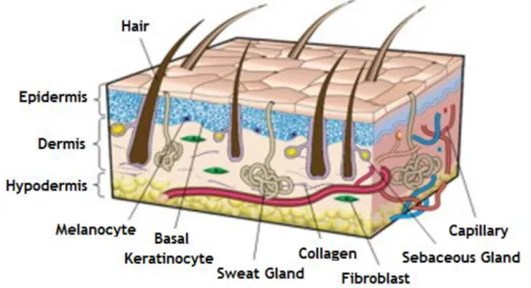
Figure 1: Representation of the structure of the human skin ……… 2
Figure 2: Phases of the wound healing process ……… 6
Figure 3: Representation of inflammatory phase of the wound healing process ……… 7
Figure 4: Illustration of migratory and proliferative phases of the wound healing process ………… 8
Figure 5: Representation of the maturation phase of the wound healing process ……… 8
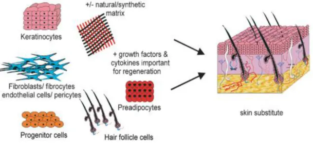
Figure 6: A schematic of the requirements to create a fully functional skin substitute ……… 13
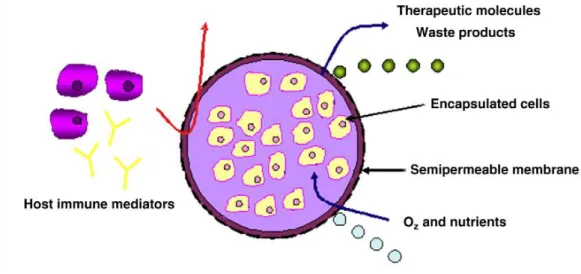
Figure 7: Representation of an encapsulation system ……… 14
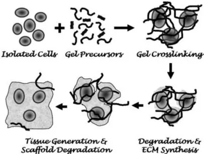
Figure 8: Representation of the process of cell encapsulation ……… 17
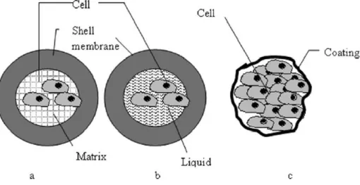
Figure 9: Classification of the different microencapsulation devices ……… 18
Figure 10: Chemical structure of sodium alginate ……… 19
Figure 11: Structure of chitosan ……… 24
Chapter II: Materials and Methods
Figure 12: Representation of the process of human fibroblasts cells encapsulation into alginate microparticles with and without collagen ……… 28Figure 13: Reduction of the MTS into formazan by viable cells ……… 30
Chapter III: Results and Discussion
Figure 14: Macroscopic and microscopic photographs of produced microparticles ……… 33Figure 15: SEM images of the two types of microparticles without cells after 3 days of being produced ……… 34
Figure 16: SEM images of human fibroblasts cells inside of the different microparticles after 3 days of being encapsulated ……… 35
Figure 17: SEM images of human fibroblasts cells inside of the different microparticles after 10 days of being encapsulated ……… 36
Figure 18: SEM images of human fibroblasts cells inside of the different microparticles after 17 days of being encapsulated ……… 36
XVII
Figure 19: Microscopic photographs of human fibroblasts cells inside of the microparticles after 1 day of being encapsulated ……… 37
Figure 20: Microscopic photographs of human fibroblasts cells inside and outside of the
microparticles ……… 38
Figure 21: CLSM images of human fibroblasts cells inside alginate microparticles after 3, 10 and 17 days of being encapsulated ……… 40
Figure 22: CLSM images of human fibroblasts cells inside alginate and collagen microparticles after 3, 10 and 17 days of being encapsulated ……… 41
Figure 23: Evaluation of cell viability by a MTS assay after 3 and 10 days of being encapsulated
into microparticles ……… 43
Figure 24: Characterization of the morphology of the nanoparticles produced with high molecular weight deacetilated chitosan by SEM ……… 45 Figure 25: Calibration line and its respective equation used to calculate the encapsulation efficiency of BSA in chitosan nanoparticles ……… 46
XIX
List of Tables
Chapter I: Introduction
Table 1: Skin Substitutes Available for Clinical Use ……… 12 Table 2: Materials used for cell encapsulation ……… 16
XXI
List of Abbreviations
2D Two-dimensional
3D Three-dimensional
aFGF Acid fibroblast growth factor
BCA Bicinchoninic acid
bFGF Basic fibroblast growth factor
BSA Bovine serum albumin
CaCl2 Calcium chloride
CH3COOH Acetic acid
CLSM Confocal laser microscopy
DD Degree of deacetylation
DMEM Dulbecco’s modified Eagle’s medium
ECM Extracellular matrix
EDTA Ethylenediaminetetraacetic acid
EGF Epidermal growth factor
EtOH Ethanol
FBS Fetal bovine serum
FGF Fibroblast growth factor
GAGs Glycosaminoglycans
GF(s) Growth factor(s)
HA Hyaluronic Acid
HMW High molecular weight
K- Negative control
K+ Positive control
KGF Keratinocyte growth factor
MMP Metalloproteinases
MTS 3-(4,5-dimethylthiazol-2-yl)-2,5-diphenyl-2H-tetrazolium
NaOH Sodium hydroxide
XXII
PEG Polyethylene glycol
PGA Polyglycol acid
PHEMA Poly (hydroxylethyl methacrylate)
pI Isoelectric point
PLA Polylactic acid
PLGA Poly (lactic-co-glycol) acid
PMS Phenazine methosulfate
PVA Poly (vinyl alcohol)
SEM Scanning electron microscopy
TGF-β Transforming growth factor beta TIMP Tissue inhibitors of metalloproteinase
TPP Pentasodium tripolyphosphate
UV-VIS Ultraviolet-visible
Chapter I:
Chapter I:Introduction
2
1.1. Skin
1.1.1. Functions and Structure
Skin is considered the largest organ of the human body, covering about 1.7m2 and comprising approximately 8% of the total body mass 1-4.
Skin serves, primarily, as a protective and defensive barrier against the external environment and allows the maintenance of the body temperature. This organ is also involved in defensive mechanisms, which confer physical, immunological, metabolic and UV radiation-protective barriers against microbes and toxic chemicals. Moreover, skin can also be used as a entry point for therapeutic substances such as vaccines 1,3,5,6. Skin importance is demonstrated by the fact that when large areas of this tissue are lost, caused by diseases or injuries (as in the case of extensive burns), result in significant morbidity and mortality 3,7.
Skin is organized in three anatomical distinct layers known as the epidermis, dermis and hypodermis 3,8,9 (figure 1). These layers have different thickness, strength and flexibility 8. The epidermis is the most superficial layer of the skin and provides the first barrier to avoid invasion of foreign substances into the body. Keratinocytes and melanocytes are prominent cell types of this layer. The main function of the dermis is to support the structural integrity of the skin giving it durability and elasticity. Such properties are confered by the major cellular components of the dermis, the fibroblasts. The hypodermis contains mostly adipose tissue, which functions as a thermal insulator and also helps to protect the underlying structures 3. Skin appendages like hair follicles, sebaceous and sweat glands are founded in the dermis and epidermis 9.
Chapter I:Introduction
3
1.1.2. Epidermis
The epidermis is the superficial layer of the skin and serves as an impermeable boundary between the environment and the body. This sheet is a multilayered cellular structure with little or no extracellular matrix (ECM) and do not present blood vessels 4,8.
Besides keratinocytes, which account for about 80% of epidermal cells, the epidermis is also composed of the pigment-producing cells, the melanocytes and specialized dendritic Langerhans cells, that have an essential role in the skin immune defense system 8.
The epidermis may be subdivided, from the inside to the outside, into five layers: the strata basale, spinosum, granulosum, lucidum and corneum 4.
The continuous proliferation, differentiation and, ultimately, cell death allows compartmentalization into a number of strata representing different stages of keratinocyte maturation 8. The keratinocyte cell division starts in the innermost strata basale and pushes daughter cells apically towards the next spinosum strata, where there are more cells. As they progress toward the skin surface, they move into the granulosum strata, where they accumulate lipid granules, critical for the maintenance of the water barrier 8.
Pigmentation is imparted by melanin, which is produced by melanocytes and transferred to keratinocytes in the final sublayer of the strata lucidum. The most external layer, known as the stratum corneum is formed by completely differentiated dead keratinocytes, interspersed with intercellular lipids (mainly ceramides and sphingolipids) 8.
The basement membrane allows the separation of the epidermis from the dermis 3. This membrane has many functions, among which it is responsible for epidermis binding to the dermis. It also determines polarity of the epidermis and provides a barrier against epidermal migration, which prevents the direct contact of epidermal cells with the dermis 10.
1.1.3. Dermis
The dermis is the layer between epidermis and hypodermis. It contains a high number of fibroblast cells that produce collagen and elastin, which are the main constituents of the ECM and give support and elasticity to the skin, respectively 3,11.
This layer is divided in two zones: a lower layer called reticular and a superficial one nominated papillary. The deeper layer, which is the main component of the dermis, is continuous with hypodermis and consists of dense irregular connective tissue. This tissue gives strength and elasticity to skin 3,12,13. On the other hand, the papillary layer contains extensions nominated papillae, which are projected to the epidermis 13.
The dermis contains blood capillaries, which supply the epidermis and help to regulate body temperature, smooth muscle fibers, sweat and sebaceous glands and their ducts, hair follicles and sensory nerve endings 13.
Chapter I:Introduction
4
1.1.4. Hypodermis
The hypodermis is the bottom layer of the skin that is located beneath the dermis and it is involved in the connection between skin, bones and adjacent muscles 14.
This layer is well vascularized and is composed mainly of adipose tissue, where about half of the total stored fat in the body is localized 13,14. However, their amount vary with age, sex and feeding 13. Thus, hypodermis confers insulation, acts as an energy source and also contributes to the mechanical properties of the skin 9,14.
Nevertheless, some authors do not contemplate hypodermis as being part of the skin, considering it as a subcutaneous tissue 13.
Chapter I:Introduction
5
1.2. Wounds
Wounds affect millions of people worldwide, rendering this pathology one of the major issues in modern health care 15,16. A wound can be described as a disruption of the structural or functional integrity of the skin, that is caused by physical or a thermal damage. Moreover, they can also result from an underlying medical or physiological condition 3,17,18.
Based on the type of repair process, wounds can be classified as acute or chronic wounds. Acute wounds are usually characterized by a complete healing, with minimal scar formation. The primary causes of acute wounds include mechanical injuries due to external factors, such as abrasions and tears, that are caused by frictional contact between the skin and hard surfaces. Other acute wounds include burns and chemical injuries, which arise from a variety of sources such as radiation, electricity, corrosive chemicals and thermal sources 17. The chronic wounds arise from tissue injuries that heal slowly or fail to heal due to repeated tissue insults or underlying physiological conditions, like diabetes, persistent infections and poor primary treatment. Such is responsible for the disruption of the events sequences, that occur during the wound healing process 17.
Wounds are also classified based on the number of layers and area of skin affected. Therefore, they can be divided into epidermal, superficial thickness, deep partial-thickness and full-partial-thickness, with increasing depth of the injury 17,19. Epidermal injuries caused by sunburns, light scalds or grazing are characterized by erythema and minor pain. These injuries do not require specific surgical treatment, since only the epidermis is affected and it regenerates rapidly without scarring 19.
Superficial partial-thickness wounds affect the epidermis and superficial parts of the dermis. This type of wounds is characterized by epidermal blistering and severe pain, especially in the case of thermal trauma. The blood vessels, sweat glands and hair follicles are affected 17,19.
Deep partial-thickness injuries involve a great dermal damage that results in fewer skin appendages (such as hair follicles, sweat and sebaceous glands) and therefore they take longer to heal 19.
Full-thickness injuries are characterized by the complete destruction of the underlying subcutaneous fat or deeper tissues, in addition to the epidermis and dermal layers 17,19.
Except for very superficial wounds and in early fetal life, the regenerative capacity of the skin is limited and in the majority of the wounds occurs the formation of high amounts of scar tissue 3.
Chapter I:Introduction
6
1.2.1. The Wound Healing Process
The wound healing is a complex biological process that involves growth and tissue regeneration, in order to prevent organism deregulation. After an injury, multiple biological pathways are immediately activated in a variety of cellular and matrix components that act together to reestablish the integrity of the damaged tissue and replace the injured tissue 15,17,18,20-23.
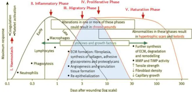
Wound healing comprises five overlapping stages that involve complex biochemical and cellular processes (see figure 2). These are described as haemostasis, inflammation, migration, proliferation and maturation phases 15,17,20-23.
Figure 2: Phases of the wound healing process. This procedure occurs in a cascade of events that is
correlated with the appearance of different cell types in the wound, during various stages of the healing process (ECM: Extracellular matrix; MMP: Metalloproteinases; TIMP: Tissue inhibitors of metalloproteinases) (adapted from 18).
1.2.1.1. Haemostasis and Inflammation
Haemostasis occurs immediately after tissue damage due to the onset of bleeding 3,17. Components of the coagulation cascade, inflammatory pathways and immune system (e.g. neutrophils, monocytes and lymphocytes) are needed at the wound site to prevent ongoing fluid losses 18,24. Haemostasis is achieved initially by the formation of a platelet plug, followed by the formation of a fibrin matrix, that becomes the scaffold for infiltrating cells. The clot dries to form a scab and provides strength and support to the injured tissue 15,17,22.
Inflammatory phase occurs simultaneously, involving both cellular and vascular responses. The main purpose of this phase is to clean the wound by removing dead tissues and to
Chapter I:Introduction
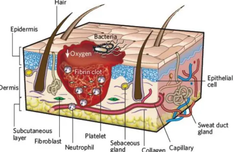
7 prevent the occurrence of infection 3. Thus, in this phase, neutrophils and platelets are abundant at the injured site 15. The release of protein-rich exudate into the wound causes vasodilation through the release of histamine and serotonin, and allows phagocytes to enter to the wound, surrounding the dead cells. Platelets released from damaged blood vessels become activated as they enter in contact with mature collagen and form aggregates as part of the clotting mechanism (figure 3) 15,17,22.
Figure 3: Representation of inflammatory phase of the wound healing process (adapted from 15).
1.2.1.2. Migratory Phase
The migration phase involves the movement of epithelial cells (such as keratinocytes) and fibroblasts to the injured area, in order to replace damaged or lost tissue. These cells regenerate from the margins (figure 4), rapidly growing over the wound under the clot accompanied by epithelial thickening 15,17.
1.2.1.3. Proliferative Phase
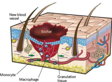
The proliferative phase occurs almost simultaneously or just after the migration phase and is characterized by fibroblast migration, deposition of ECM and formation of granulation tissue. This tissue is formed by the in-growth of capillaries and lymphatic vessels into the wound. In this phase occurs also the formation of blood vessels, which is known as angiogenesis, as depicted in figure 4. The fibroblast proliferation and collagen synthesis occurs each time edema recedes 3,15,17,18,22,24.
Chapter I:Introduction
8
Figure 4: Illustration of migratory and proliferative phases of the wound healing process. A scab was
formed on the surface of the wound (adapted from 15).
1.2.1.4. Maturation Phase
During the maturation or remodeling phase, all the processes that are activated after the injury ceases (figure 5). The majority of endothelial cells, macrophages and myofibroblasts undergo apoptosis or exit from the wound, leaving a mass that contains few cells and consists mainly of collagen and other ECM proteins. This stage involves the formation of cellular connective tissue and strengthening of the new epithelium. Such determines the nature of the final scar. Moreover, the acellular matrix is mainly composed of collagen type III that will be replaced by collagen type I, later on. This process is carried out by matrix metalloproteinases that are secreted by fibroblasts, macrophages and endothelial cells 3,15,17,24. The re-epithelialized wound surface is slightly higher than the surrounding surface, and the healed region does not presents any skin appendages 15.
Chapter I:Introduction
9
1.3. Tissue Engineering: Skin Substitutes
There are a number of pathologies in which skin loss or damage occurs. Among them, burns and chronic ulcers are responsible for the loss of superficial epidermis and dermis, abnormal wound healing and the failure of (or delay associated with) wound healing process 9,16. The healing of a skin wound is a complicated phenomenon that includes a wide range of cellular, molecular, physiological and biological processes. The immediate coverage of the wound with a dressing is a cornerstone of wound management 6.
In the past, the application of dressing materials on top of a wound was aimed for bleeding inhibition, protection of the wound from the pathogens (e.g. bacterias), as well as avoid water and electrolyte disturbances 17,20. However, traditional dressings do not restrict moisture evaporation and may cause dehydration of the wound bed 25.
With the purpose to overcome such problems, grafts have been used for deep partial- and full-thickness wounds treatment 6. Tissues for skin grafting can be obtained from the self-patient (autografts), from a different person of the same specie (allografts) or from animals of different species (xenografts) 26. The characteristics of autografts are ideal, since the patient does not present any immunological rejection. Although autografts exhibit the best clinical outcome, they suffer from several disadvantages such as the limited quantity, donor site morbidity and the requirement of surgery at multiple sites to harvest donor tissue from the patient 7,27,28. Allografts and xenografts have distinct advantages for the treatment of skin defects, especially due to the unlimited tissue quantity availability. However, these type of grafts are associated with the risk of disease transmission and require the use of immunosuppressants with associated side effects, which can lead to graft rejection 2,23,27,29,30. Therefore, these practices have significant limitations for the patient thus, not accomplish the purpose of the skin recovery 23,29,31.
In order to abolish the limitations of autografts, allografts and xenografts, different studies have been performed in the area of tissue engineering. This is a multidisciplinary area of knowledge, in which new three-dimensional (3D) scaffolds with adequate properties are developed to allow cell attachment, migration, proliferation and differentiation until the newly formed tissue is structurally stabilized 32. These biomaterials support tissue regeneration that had suffered some type of injury. Therefore, its purpose is to recreate living, healthy and functional tissues. 32,33. These scaffolds are also usually employed to create an immune-protected environment that allows nutrient, gases and waste products diffusion required for cells survival 34.
Therefore, a huge effort has been done in the area of tissue engineering to improve the wound healing mechanism. The different skin substitutes have been developed in order to avoid
Chapter I:Introduction
10 the need to remove the skin’s patient from one part to another and decrease the patients’ risk of infection and sepsis. Also, they contribute to reduce the mortality and morbidity caused by scarring, changes in pigmentation, the total number of surgical procedures and hospitalization period 35,36. The concern for the development of skin substitutes is also suitable for extensive wounds that cannot heal spontaneously, since they affect a high percentage of total body surface area or for injuries that cannot heal due to inflammation or a deficiency wound healing process. These substitutes can also be used to accelerate the healing of wounds that would heal by themselves, however very slowly 36.
Skin substitutes should act as scaffolds for cells that are used to replace lost tissue rather than just facilitate the process of wound healing 6,17,35,37. These skin substitutes try to mimic the normal physiologic responses, during the wound healing and function as coating to protect the wound against infection, while allowing gaseous exchanges 17,38. Consequently, an ideal skin substitute must also protect the injury from fluid and proteins loss, be easy to handle and apply to the wound site, undergo controlled degradation, enable exudates remotion, inhibit exogenous microorganism invasion (antimicrobial properties) and improve aesthetic appearance of the wound site 6,27,39-41. It should also be non-toxic, non-allergenic, be easily removed without trauma, and it should be made from a readily cheap and available biomaterial, that requires minimal processing.
Presently there are a lot of skin substitutes that have been developed. They can be classified based on: 1) the anatomical structure that they reproduce (dermo-epidermal (composite), epidermal and dermal); 2) the period among which they are used (permanent, semi-permanent or temporary); 3) the origin of the material used to produce the biomaterial (biological, synthetic and semi-synthetic) and 4) cellular or acellular components 28.
Dermo-epidermal or composite skin substitutes aim to mimic the structure of normal skin where both epidermal and dermal layers are injured. Most of these skin substitutes have fibroblast and keratinocytes incorporated into a scaffold, in order to form a temporary covering. These systems act rather like temporary biologically active wound dressings, providing growth factors (GFs), cytokines and a ECM for host cells, while initiating and regulating wound healing process 9,19. The keratinocytes usually used in this type of skin substitute provide pain relief and accelerate wound healing 6, however they do not survive longer than few weeks when applied to the wound. AllograftTM, ApligraftTM and OrCelTM are examples of current commercially available dermo-epidermal skin substitutes 9,19.
Epidermal substitutes usually contain autologous keratinocytes that are isolated by skin biopsy. The use of autologous keratinocytes in this type of skin substitute is advantageous, since the risk of rejection is very low. However, these skin substitutes present also some disadvantages, such as long preparation time, variable engraftment rates, difficult handling due to their thickness, fragile cellular layers and high production costs. EpicelTM, MySkinTM and
Chapter I:Introduction
11 CellSprayTM are some of the commercially available epidermal substitutes that have been approved for clinical use 9,19.
Engineered dermal substitutes were designed and produced to restore dermal tissue by promoting new tissue growth and optimizing healing conditions. However, they need to be covered by a permanent epidermal surface. The majority of products for dermal substitutions are composed by acellular matrices. After their application, they are colonized and vascularized by the underlying cells, producing an autologous neo-dermis. Others skin substitutes have human cells incorporated and are applied as transient wound dressings that stimulate the healing mechanism. In DermagraftTM, fibroblasts are seeded in a mesh, where they secrete GFs, deposit dermal matrix proteins and thus improve the healing process. It is much easier to manufacture these types of skin substitutes when compared with cell-containing bilayered skin substitute constructs. AlloDermTM and KarodermTM are others examples of the commercially available dermal bioengineering constructs 9,19.
Some of the developed tissue engineered products and skin substitutes presently available are described in Table 1.
Chapter I:Introduction
12 Table 1: Skin Substitutes Available for Clinical Use.
Type DressingName of Major Components Refs
Epidermal and Dermal
Skin Substitutes
IntegraTM A bovine collagen based dermal analogue and a thin temporary epidermal silicone sheet 9,17,42
BiobraneTM
A nylon mesh that is bonded to a silicone membrane and
coated with collagen
17,42
TranCyteTM A polyglycolic acid (PGA)/polylactic acid (PLA) layer with ECM proteins 17
ApligraftTM Bovine type I collagen mixed with a suspension of allogeneic fibroblasts and keratinocytes 9,11,14,17
Dermal Skin Substitute
AllodermTM Allograft human dermis with all the cellular material removed 9,17
DermagraftTM Cultured human allogeneic fibroblasts on a biodegradable PGA or polyglactin mesh 9,14,17
Epidermal Skin Substitute
EpicelTM Cultured autologous human keratinocytes which are grown to stratified cell sheets 9,17
MySkinTM Cultured autologous human keratinocytes on a silicone polymer substrate 9,11,14,17
Hyalograft 3-DTM Autologous human fibroblasts incorporated on a laser-microperforated hyaluronate 17
LaserskinTM Autologous human keratinocytes cultured on a membrane of benzyl hyaluronate 14,17
Chapter I:Introduction
13 Scientists have been developed other types of engineering skin substitutes with others natural and synthetic materials. Examples of natural materials include polypeptides, hydroxyapatites, hyaluronic acid (HA), glycosaminoglycans (GAGs), fibronectin, collagen, chitosan and alginates. Such materials have the advantage of presenting low toxicity and a low inflammatory response 43. Examples of synthetic materials include polyglycolide, polylactide and polylactide coglycolide, which are used for sutures and meshes. However, there are other synthetic materials like polytetrafluoroethylene and polyethylene terephthalate that are also used to fabricate skin substitutes 43. Nevertheless, there is still a remaining gap to be filled, since no engineering skin substitute used until now, presents all the criteria needed for fully restore the functional skin 23,43.
The ultimate goal of tissue engineering of the skin is to rapidly create a construct that offers the complete regeneration of functional skin, including all the skin appendages (such as hair follicles, sweat glands and sensory organs) and layers (epidermis, dermis and fatty subcutus) with rapid vascularization and the establishment of a functional vascular and nerve network and scar-free integration with the surrounding host tissue 43. These advances are depicted in figure 6.
Figure 6: A schematic of the requirements to create a fully functional skin substitute (adapted from 14).
In this new generation of skin substitutes, engineered scaffolds either produced with natural or synthetic materials will also be potentially useful for the delivery of additional signal molecules, such as GFs and nucleic acids to the wound site. This procedure could help in speeding up cell migration, adhesion, proliferation and differentiation, allowing the formation of granulation tissue and re-epithelization in a shorter period 6,17. The use of a combination of these signaling molecules not only improve the tissue repair, but also regulates and promotes the in
Chapter I:Introduction
14 vitro tissue growth 6,7,38. Other approach to design these skin substitutes are to mimic the functions of the ECM components naturally found in tissues 32.
1.3.1. Cell Encapsulation as a Strategy for Skin Regeneration
Cell therapy is one of the most exciting fields in regenerative medicine. It aims to replace, repair or enhance the function of damaged tissues or organs 44. Recently, cell therapy emerged as a promising strategy in skin tissue engineering, since it is a viable alternative for improving and restoring biological function of tissues 32,45.
Encapsulation of living cells in spherical-shaped devices (microbeads or microparticles) has been used for the continuous delivery of drugs and cells to treat various health disorders, such as endocrine (diabetes, hypoparathyroidism), central-nervous diseases (Parkinson's and Alzheimer's) and cancer 12,46-53.
This technology is based upon the entrapment of cells within a polymeric matrix surrounded by a semi-permeable membrane for the long-term release of therapeutics molecules 54. The cells produce and secrete the bioactive agents continuously to the outside matrix, while the semi-permeable membrane isolate the cells from the host immune system preventing the recognition of the immobilization cells as a foreign material (e.g., antibodies and cytokines), and allowing the exchange of nutrients, gases and waste 44,47,48,50,54-56. These features are depicted in figure 7. On the other hand, the polymeric matrix structure and chemistry must be suitable to allow cell survival and tissue formation, while its degradation rate should be similar to that of tissue growth. Finally, the degradation products must not be toxic for the encapsulated cells 33.
Figure 7: Representation of an encapsulation system. It is expected to allow nutrient, gases and GFs diffusion, processes that are essential for cell survival (adapted from 44).
Chapter I:Introduction
15 Cell immobilization shows important advantages compared with the encapsulation of proteins or other bioactive molecules, since it allows a continuous release of the therapeutic molecules in physiological concentrations 44,54. Furthermore, it also allows the delivery of cells into the defect area and to support the cellular functions during the early phases of the regeneration process 57. Therefore, cell encapsulation is one of the primary techniques of tissue engineering to delivery cells to the injured site, without being dependent on cells’ ability to migrate to the defect area 58.
One of the most important considerations for the design and production of a cell microencapsulation device is the material biocompatibility that is used. Moreover, these materials can not affect the surrounding host tissue, neither interfere with cell homeostasis 59. Table 2 presents several examples of recent materials that have been used for cell encapsulation 33.
Chapter I:Introduction
16 Table 2: Materials used for cell encapsulation.
Material Degradation Mechanism Cells Encapsulated Refs Chitosan Enzymatic (lysozyme) Chondrocytes or cardiomyocites 33
Alginate-co-Gelatin Hydrolytic Hepatocytes 12,33
HA Enzymatic (hyaluronidase) Chondrocytes, fibroblasts, human mesenchymal stem cells
12,33
Chondroitin Sulfate Enzymatic (chondroitinase) Chondrocytes 33
PEGylated Fibrinogen Enzymatic (plasmin) Bone marrow stromal cells 33
Self-assembled
Peptide Gels Dissolution
Human mesenchymal stem cells, preosteoblasts, endothelial cells, embryonic
stem cells
33
Elastin-like
Polypeptide Hydrolytic Chondrocytes, osteoblasts, adipose-derived stem cells 12,33
Poly (ethylene
glycol) (PEG) based Enzymatic (lipase) or hydrolytic Chondrocytes 33
Polyfumarate based Hydrolytic Bone marrow stromal cells 33
Chapter I:Introduction
17
1.3.1.1. Hydrogels used for Cell Encapsulation
Hydrogels are a class of hydrophilic biomaterials that have been used for cell encapsulation, with therapeutic purposes. The hydrogel structure allows cell encapsulation within a 3D environment, similar to that of the natural ECM of soft tissues, thus allowing good mass transport to maintain cell viability 12,60,61. Hydrogels can absorb the wound exudates and have cross-linked networks that have high water contents and tissue-like elastic properties. These attributes make them ideal candidates to be used in tissue engineering, especially for wound healing 21,33.
The design and production of a suitable hydrogel for cell encapsulation, must meet several criteria. In a typical cell microencapsulation process (figure 8), cells are suspended in a liquid precursor solution prior to encapsulation. The process by which hydrogel gelation or solidification occurs must be mild and cell friendly. The hydrogel structure and chemistry must be suitable for cell survival and tissue formation, while its degradation must occur in a timescale comparable to that of tissue regeneration 62. Finally, the degradation products of the hydrogel must not have adverse effects for the encapsulated cells 33. Generally, the encapsulation process can occur through a change in temperature for thermal sensitive gels (e.g., agarose, collagen, or gelatin), or via ion-gelation (e.g., alginate) and photo-based cross-linking mechanism (e.g., PEG) 63.
Figure 8: Representation of the process of cell encapsulation. This strategy involves the mixture of cells
with precursors, in a liquid solution followed by a gelation process (adapted from 33).
Hydrogels used for cell microencapsulation are commonly processed into spherical beads, which in size range from 100 to 2000 µm 64. Hydrogel microparticles have been developed for cell encapsulation, due to their spherical shape, which is considered advantageous from a mass transport perspective, offering an optimal surface-to-volume ratio for protein, nutrient and cells
Chapter I:Introduction
18 diffusion 44,62. Spherical appearance is increasingly recognized as a valuable tool in tissue engineering, since the microparticles appear to mimic the morphology and physiology of the cells in living tissues and organs better than conventional, two-dimensional (2D) monolayer cultures 65.
They also provide a highly hydrated microenvironment for embedded cells that can present biochemical, cellular, and physical stimuli that guide cellular processes such as differentiation, proliferation, and migration 44. They can also induce cell–cell and cell-ECM interactions within the bulk of the material 66. Moreover, these microcarriers provide a high degree of permeability for low-molecular mass nutrients and metabolites 21,44,62.
The versatility of these systems is associated with the variables that can be modeled to obtain an optimal system for a specific application, such as the particle composition, size, shape, existence of porosity, cell culture conditions, surface topography and chemistry or incorporation of bioactive agents 67.
Cell microencapsulation devices can be classified into 3 categories (figure 9): 1) matrix-core/shell microcapsules; 2) liquid-matrix-core/shell microcapsules and 3) cells-matrix-core/shell microcapsules.
In matrix-core/shell microcapsules, the cells are hydrogel-embedded, which has several advantages as the cells are kept in an aqueous environment, in contact with a soft and biocompatible material. Liquid-core/shell microcapsules generally seem to allow better cell growth and protein production, although their mechanical resistance is lower than that of the matrix-core capsules. In cell-core/shell microcapsules, the objective is to produce thinner membranes, which optimize cell diffusion 62.
Figure 9: Classification of the different microencapsulation devices. (a) Matrix-core/shell microcapsules
(cells embeded in hydrogel matrix); (b) Liquid-core/shell microcapsules (cells suspended in liquid core); (c) Cells-core/shell microcapsules (direct cell coating) 62.
Chapter I:Introduction
19 Hydrogels can be produced with natural materials, that can replicate the physiological characteristics of native tissue 66. Polysaccharides (e.g., agarose, gelatin and HA) and natural ECM proteins (e.g., collagen, gelatin and fibrin) have been used for the production of hydrogels 45,66.
Synthetic polymers have also been used in regenerative medicine to produce hydrogels using various molecules such as polycaprolactone, poly (lactic-co-glycolic acid) (PLGA), PEG and its derivatives, poly (vinyl alcohol) (PVA), polyurethane, poly (hydroxylethyl methacrylate) (PHEMA) and others. The mechanical and chemical properties of synthetic polymers can be tailored for different applications without having the immunogenicity-related concerns of some naturally occurring polymers, which constitutes the main advantage of this type of materials over the natural ones 45,66.
1.3.2. Alginate as a Material for Cell Encapsulation
As previously described in the text, different materials have been employed for cell encapsulation. Among them, alginate is nowadays one of the most studied and characterized materials due to its excellent biocompatibility and stability under in vivo conditions 68.
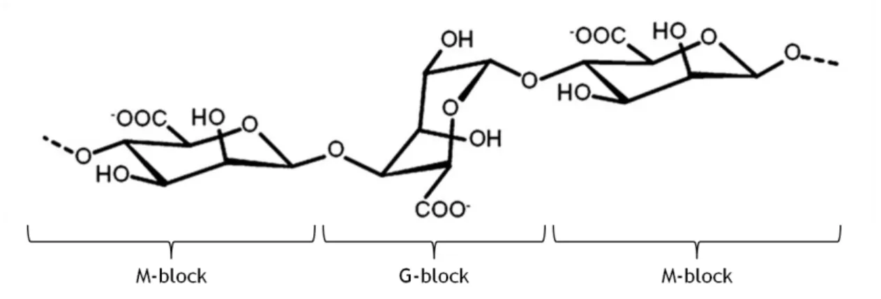
Alginate is a linear copolymer of (1–4)-linked β-D-mannuronic acid (M-block) and α-L-guluronic acid (G-block) (figure 10). It is a naturally occurring polysaccharide extracted from brown seaweed and bacterium and its composition vary depending upon the source from which it is isolated. Both cell adhesion and hydrogel stiffness can be influenced by these two uronic acid salts ratio 7,12,31,60,69,70. However, only the G-block of alginate is believed to participate in intermolecular cross-linking with divalent cations (e.g., calcium, barium, and strontium) to form 3D structures. Calcium chloride (CaCl2) is the most frequently used cross-linking agent to reticulate alginate 54,68,70-72.
Chapter I:Introduction
20 Because of its low toxicity, abundance, mechanical integrity/stability and easy gelling properties, alginate hydrogel has been investigated for a variety of tissue engineering applications, particularly in cell encapsulation 7,31,60,71. In addition, it does not interfere with the
cellular function of the immobilized cells in alginate gels and finally the encapsulation procedure can be conducted under very mild conditions, such as body temperature and at physiological pH 34,72.
The biocompatibility presented by this polymer is essential for the production of devices that protect the enclosed cells from the host immune system 44,73. Other important feature is
that its hydrated 3D network allows cells to adhere, spread, migrate and interact with each other. Moreover, cell encapsulation in alginate is a flexible technology and some parameters may be adjusted (i.e., alginate concentration, alginate composition, microparticle size and cell seeding density) to meet specific cell culture parameters 74.
However, the gelling reaction of alginate can be reversed by removing the calcium cross-linker through the exposure of the carrier to a number of other ions like sodium. Such mechanism leads to the scaffold degradation in vivo 31,60,71. Therefore, under in vivo conditions and in order to have a successful tissue engineering approach, the rate of scaffold degradation should be similar to that of tissue regeneration 31.
Alginate has also been used for wound dressings production, and it is known to promote wound healing mechanism 71. When in contact with the lesion bed, alginate, as a gel, is easily removed from the injury, decreasing pain and trauma associated with the dressing change procedure 1. Furthermore, it provides a moist environment that accelerates tissue granulation formation, re-epithelialization 1, and minimize bacterial infection at the wound site 71. Alginate interferes with the normal pro-inflammatory response to bacterial infection given by macrophages, decreasing the time of clearance of pathogenic organisms from the organism 75.
Chapter I:Introduction
21
1.4. Nanoparticles
An appropriate delivery carrier of biomolecules is very critical in the area of drug delivery, pharmaceutics, tissue engineering and even stem cell research 76. For drug delivery purposes, nano-systems, ranging in size from 10-1000nm, have been developed to act as a reservoir of therapeutic agents, with spatial and temporal control release profiles of the drug, leading to desirable therapeutic outcomes 77. In recent years, a significant effort has been devoted to develop drug delivery nanodevices for treating various diseases, such as cancer 77.
Different types of nano-sized carriers, such as polymeric nanoparticles, solid lipid nanoparticles, ceramic nanoparticles, magnetic nanoparticles, polymeric micelles, polymer-drug conjugates, nanotubes, nanowires, nanocages and dendrimers have been developed for various drug delivery applications 78.
Both natural and synthetic materials can be used to produce these different nanoparticulate delivery systems 77. During this last decade many of biodegradable polymers such as polysaccharides (e.g., alginate, chitosan), polyesters (PLA, PGA and polycaprolactone) have been used to produce devices for protein and drug delivery. Among them, the polysaccharides and their derivatives have been used in different studies 79,80. Nanoencapsulation of drugs, proteins, or other molecules within these biodegradable polymers has been recognized as an effective way for producing a nanoparticle that can hold target biomolecules and do its delivery 76.
Despite the variety of nanoparticles, these nanodevices must have some general characteristics, such as the ability to incorporate the drug without loss of activity, tunable release kinetics, stability when applied in vivo, biocompatibility, lack of immunogenicity, degradability and the potential to reach specific organs and tissues 77. Nanocarriers may also protect a drug from modify drug tissue distribution profile and/or improve intracellular penetration and distribution 78.
The nanoparticles, due to their small size, can penetrate through smaller capillaries and be taken by cells, allowing an efficient drug accumulation at the target sites 53.
These nanosystems can be prepared by various methods such as chemical cross-linking, ionic cross-linking, emulsion droplet coalescence, reverse micellar and self assembly chemical modification 81,82. Drug loading into nanoparticulate systems can be done during the preparation of particles (incorporation) and after the formation of particles by incubating drug into the solution 82-84. In these systems, drug is physically embedded into the matrix or adsorbed onto its surface 82. On the other hand, drug release from nanoparticles is determined by the diffusion of drug molecules through the polymer network and/or material degradation 77.
Recently, it has emerged the need to control and to prolong the release profile of the drugs incorporated into the nanoparticles. In some cases (as pulmonary administration),
Chapter I:Introduction
22 nanoparticles are also impractical for management owed to the fact that their direct delivery poses many challenges, including formulation instability due to particle-particle interactions and poor delivery efficiency due to low-inertia nanoparticles 85. To overcome such obstacles, it has arisen the hypothesis to incorporate these nanodevices in microparticles. Thus, this microencapsulation technique allows a better retention of bioactive molecules in the system preventing them from being released and increasing the period among which they are released 77,85.
1.4.1. Nanoparticles for Growth Factors Delivery
The immobilization of GFs in 3D matrices has already been studied by several investigators 86. GFs are proteins that induce a change in cellular function by transducing proliferation or differentiation signals and are involved in the modulation of tissue growth and development 87,88.
Topical administration of GF is still a challenging mission, since proteins present a short half-life in vivo, side effects caused by multiple or high doses applied and protein denaturation, which are some of the barriers for a successful GF based therapy. Most, if not all, GFs that are administered in their native form and without any protection are susceptible to be degraded or be rapidly eliminated from the blood circulation, resulting in insufficient amounts of these proteins to play their function in the body 88. Different methods have been tested to overcome these disadvantages. The encapsulation of a GF in a delivery system has been demonstrated to be a good strategy for GFs-based therapeutics 88.
Owing to their ultra-small size, nanoparticle formulations have many advantages over the traditional dosage forms used for GFs delivery. They are able to enhance the action of these active agents at the treatment sites for a sufficient period of time, in order to allow the cells to migrate, proliferate and differentiate at the injury. They also increase the systemic circulation lifetime and then release them in a sustained and controlled manner 53,89. These delivery systems also minimize the release of GFs to non-target sites, and support tissue regeneration that normally occurs over long periods of time. Therefore, this direct GF delivery technology is crucial to deliver key exogenous signalling proteins needed for the developing of new tissues 88. Encapsulating GFs within or attaching them to a polymer nanocarrier can remarkably improve its safety, efficacy and enable the development of new therapies 88.
In skin, GFs are involved in the maintenance of homeostasis during wound healing 90, because they stimulate endogenous repair mechanisms by providing the right signals to cells and thereby leading to an accelerated functional skin restoration 88. In wounds, the topical application of several GFs to stimulate fibroblast and endothelial cell proliferation has been used
Chapter I:Introduction
23 for heal the impaired wound, in order to increase the granulation tissue and capillary formation, and thus stimulate the wound healing mechanism 91.
Among the different GFs, particularly important factors for skin regeneration include fibroblast GF (FGF) family and epidermal GF (EGF), keratinocyte GF (KGF), vascular endothelial GF (VEGF) and transforming GF beta (TGF-β). VEGF functions as an endothelial cell mitogen, chemotactic agent and inducer of vascular permeability 92. VEGF is also important due to its effects on multiple components of the wound-healing cascade, including angiogenesis and recently has been shown its role in epithelialization and collagen deposition 92. Other GFs that positively regulate re-epithelialization include members of the FGF family such as basic FGF (bFGF), EGF and TGF-β 15. The clinical use of EGF has also been extensively investigated in wound healing processes, especially in the treatment of diabetic ulcers 53. Both bFGF and acidic FGF (aFGF) have many biological activities that stimulate the proliferation of fibroblasts and capillary endothelial cells, thus promoting angiogenesis and wound healing 91.
1.4.2. Chitosan as a Polymer for Drug Delivery
Chitosan is a natural biodegradable, biocompatible, bioadhesive and bacteriostatic polymer and it gained a lot of attention in the pharmaceutical field, since it has been used for the production of a wide range of controlled drug delivery systems 81,93. This polymer is extracted and purified from the shells of shrimp, crab and other crustaceans, and from some of the fungi cell walls 94.
Chitosan is a copolymer of glucosamine and N-acetyl glucosamine linked by β(1–4) glucosidic bonds obtained by N-deacetylation of chitin 81,93,94. The content of glucosamine is called as the degree of deacetylation (DD). Depending on the source and preparation procedure, its molecular weight may range from 300 to over 1000kD with a DD from 30% to 95% 6. The molecular weight and DD can be modified during its preparation to obtain different properties 81,93,94. Highly deacetilated forms (DD>85%) exhibit a relatively low degradation rate and may last for several months in vivo, whereas the forms with lower DD are degraded more rapidly 6. These basic molecular parameters of chitosan influence the protein loading and delivery 93. Different DD and molecular weight of chitosan have been used for the production of micro/nanoparticles 82.
In its crystalline form, chitosan is normally insoluble in aqueous solution above pH 7, however, in dilute acids (pH<6.5), the protonated free amino groups on glucosamine facilitate solubility of the molecule 6. Chitosan is polycationic in acidic media (pKa 6.5) and can interact with negatively charged species, such as pentasodium tripolyphosphate (TPP) and sodium sulfate, as depicted in figure 11. This characteristic can be employed to prepare cross-linked chitosan nanoparticles and microparticles, that can be used for protein and vaccine delivery 93.
Chapter I:Introduction
24 Figure 11: Structure of chitosan (adapted from 95).
Several studies reported the use of chitosan for the production of skin substitutes 17,23. This polymer presents different properties that promote the wound healing mechanism, since it has an homeostatic effect and stimulates the synthesis of collagen by fibroblast. Furthermore, it also promotes the infiltration of polymorphonuclear cells at the wound site, which is an essential event for wound healing takes place. Recently, it was also reported that chitosan was used for the incorporation of different GFs, such as bFGF, to improve the rate of healing 6.
Chapter I:Introduction
25
1.5. Objectives
In the present study, a new microencapsulation system for future application in skin regeneration had been developed. The main objectives of this study were:
Development of alginate microparticles with human fibroblasts cells encapsulated;
Evaluation and characterization of the physical and biological properties of the produced vehicles through spectroscopic techniques;
Development of chitosan nanoparticles loaded with a model protein, bovine serum albumin (BSA);
Characterization of the produced nanoparticles through scanning electron microscopy (SEM) analysis.
Chapter II:
Chapter II:Materials and Methods
27
2.1. Materials
Acetic acid (CH3COOH), amphotericin B, BSA, CaCl2, high molecular weight (HMW) chitosan, collagen solution from calf skin, Dulbecco’s modified Eagle’s medium (DMEM-F12), ethylenediaminetetraacetic acid (EDTA), L-glutamine, penicillin G, TPP, phosphate-buffered saline (PBS), sodium alginate, sodium hydroxide (NaOH), streptomycin and trypsin were purchased from Sigma-Aldrich (Portugal). (3-(4,5-dimethylthiazol-2-yl)-5-(3-carboxymethoxyphenyl)-2-(4-sulphofenyl)-2H-tetrazolium) (MTS) was obtained from Promega (Madison,WI, USA). Pierce BCA® protein assay reagent A and B were purchased from Thermo Scientific (Portugal). Human Fibroblast Cells (Normal Human Dermal Fibroblasts adult, criopreserved cells) were purchased from PromoCell (Spain). Fetal bovine serum (FBS) was purchased from Biochrom AG (Germany). Hoechst 33342® was obtained from Invitrogen (Carlsbad, CA, USA).
2.2. Methods
2.2.1. Encapsulation of Human Fibroblasts Cells in Alginate
Human fibroblasts cells were encapsulated into microparticles by gelifying alginate microcarriers with calcium ions 79. For this purpose, a solution of sodium alginate was prepared with milli-Q water. Subsequently, human fibroblasts cells were harvested, counted and resuspended in the alginate solution to a final ratio of 71.8×10⁵ cells/mL. The final moisture with
a concentration of 1.5% (w/v) was vortexed to ensure a complete mixing of the cells within the solution. The suspension was loaded into a syringe and extruded using a syringe pump (KdScientific, KDS, Sigma), through a 25G needle into a 10mL HEPES buffered CaCl2 (5% (w/v),
pH 7.4) solution, under magnetic stirring, for 10 min, at room temperature. This gelling agent was prepared by dissolving CaCl2 in milli-Q water. Droplets produced immediately microparticles
due to the ionic cross-linking of alginate matrix with divalent cations. The produced microparticles were then recovered by filtration using a 0.22µm filter paper and then washed three times with DMEM-F12 31,47,48,60,76,96. Other type of microparticles composed of collagen and alginate were also produced under the same procedure. The process is represented in figure 12.