Ema Luís Pereira Gomes Alves
NEUROTOXICITY OF
METHYLENEDIOXYMETHAMPHETAMINE
(MDMA; ECSTASY) ON RAT BRAIN MITOCHONDRIA
Ema Luis Pereira Gomes Alves
Neurotoxicity of
methylenedioxymethamphetamine
(MDMA;”ecstasy”) on rat brain mitochondria
Neurotoxicity of methylenedioxymethamphetamine
(MDMA;”ecstasy”) on rat brain mitochondria
Ema Luis Pereira Gomes Alves
Dissertação apresentada à Faculdade de Farmácia da Universidade do Porto
para obtenção do grau de doutor em toxicologia.
Dissertation presented to the Faculty of Pharmacy of the University of Porto to
to obtain a PhD degree in toxicology.
Supervisor
Professor Doutor Félix Dias Carvalho
Professor Associado com Agregação da Faculdade de Farmácia da
Universidade do Porto, Porto, Portugal.
Co-Supervisor
Doutora Maria Teresa Burnay Summavielle
Instituto de Biologia Molecular e Celular (IBMC), Universidade do Porto, Porto
Portugal
Departamento de Ciências Biomédicas, Escola Superior de Tecnologias da
Saúde (ESTSP), Instituto Politécnico do Porto (IPP), Porto, Portugal
Co-Supervisor
Professor Doutor José Barata Cutódio
“Para ser grande, sê inteiro: nada
teu exagera ou exclui.
Sê todo em cada coisa. Põe quanto és
no mínimo que fazes.
Assim em cada lago a lua toda
brilha, porque alta vive.”
Ao(s) Professore(s) Doutore(s)
Félix Dias Carvalho e
Maria Teresa Burnay Summavielle.
Aos meus pais e irmãos.
O Decreto de Lei 288/70, art. 8, § 2,refere:
“ É admitido na elaboração da dissertação o aproveitamento, total ou parcial, do
resultado de trabalhos já publicados, mesmo em colaboração, devendo, neste caso, o
candidato esclarecer qual a sua contribuição pessoal”
Neste sentido, integram esta dissertação resultados publicados nos trabalhos
seguidamente enumerados, para os quais contribuiu pessoalmente, preparando todo o
material experimental, procedendo às determinações analíticas, organização e análise
dos resultados e redacção do texto, em colaboração com os restantes autores.
Artigos originais e comunicações apresentadas em congressos cujos
resultados integram este trabalho:
Artigos publicados em revistas internacionais:
Alves E, Summavielle T, Alves CJ, Gomes-da-Silva J, Custódio JB, Fernandes E,
Bastos ML, Tavares MA, Carvalho F (2007) Monoamine oxidase-B mediates
ecstasy-induced neurotoxic effects to adolescent rat brain mitochondria. J Neurosci.
27:10203-10210.
Alves E, Summavielle T, Alves CJ, Custódio JB, Fernandes E, Bastos ML, Tavares
MA, Carvalho F (2007) Ecstasy-induced oxidative stress to adolescent rat brain
mitochondria in vivo: influence of monoamine oxidase type A. (submitted).
Alves E, Binienda Z, Carvalho F, Fernandes E, Bastos ML, Tavares MA, Summavielle
T (2007) Acetyl-L-carnitine provides effective in vivo neuroprotection over
MDMA-induced neurotoxicity in adolescent rat brain mitochondria (in preparation)
Alves E, Custódio JB, Summavielle T, Melo P, Bastos ML, Carvalho F (2007)
Comunicações orais apresentadas em congressos nacionais:
Alves, E., Summavielle, T., Gomes-da-Silva, J., Custódio, J.B., Fernandes, E., Bastos,
M.L., Carvalho, F. “Long-Term” effects of MDMA exposure in adolescent male rats:
abnormal thermoregulation, oxidative stress and mtDNA deletions in isolated whole
brain mitochondria. Annual Meeting of the Portuguese Society of Pharmacology.
Lisbon, December 5-7, 2005, Portugal.
Alves, E., Summavielle, T., Gomes-da-Silva, J., Custódio, J.B., Fernandes, E., Bastos,
M.L., Carvalho, F. Effects of MDMA exposure in adolescent male rats. IBMC. PhD
Training seminars. Porto, March 22, 2006, Portugal.
Alves, E., Summavielle, T., Alves, C., Gomes-da-Silva, J., Monteiro, P., Fernandes, E.,
Bastos, M.L., Carvalho, F. Monoamine oxidase B mediates ecstasy-induced neurotoxic
effects in adolescent’s rat brain mitochondria. Annual Meeting of the Portuguese
Society of Pharmacology. Porto, December 5-7, 2006, Portugal.
Alves, E., Summavielle, T., Alves, C., Gomes-da-Silva, J., Monteiro, P., Fernades, E.,
Bastos, M.L., Carvalho, F. Monoamine oxidase B mediates ecstasy-induced neurotoxic
effects in adolescent’s rat brain mitochondria. Annual meeting of the Portuguese
Society of Pharmacology. Coimbra, December 5-7, 2007, Portugal.
Comunicações em painel apresentadas em congressos internacionais:
Alves, E., Summavielle, T., Faria, R., Gomes-da-Silva, J., Custódio, J.B., Fernandes,
E., Bastos, M.L., Carvalho, F. MDMA exposure in adolescent male rats induces
long-term abnormal thermoregulation and oxidative stress in whole-brain mitochondria.
Satellite Meeting of the International Society for Neurochemistry/European Society for
Neurochemistr : Cellular and Molecular Mechanisms of Drugs of Abuse and
Neurotoxicity: Cocaine, GHB and substituted amphetamines. Venice, August 16 -19,
Alves, E., Summavielle, T., Alves, C., Gomes-da-Silva, J., Monteiro, P., Fernandes, E.,
Bastos, M.L., Carvalho, F.. Monoamine-oxidase B mediates ecstasy-induced
neurotoxic effects in adolescent’s rat brain mitochondria. Neuroscience 2006. Atlanta, 1
October 14-18, 2006, EUA.
Alves, E., Binienda, Z., Gonçalves, A., Fernandes, E., Carvalho, F., Summavielle, T.
Acetyl-L-carnitine provides neuroprotection against MDMA-induced neurotoxicity
through prevention of mitochondrial dysfunction. First Annual International Drug Abuse
Research Society (IDARS) Meeting, pre-satellite meeting of the International Society
for Neurochemistry and American Society for Neurochemistry. Merida, August 14-17,
2007, Mexico.
Alves, E., Binienda, Z., Gonçalves, A., Fernandes, E., Carvalho, F., Summavielle, T.
Methylenedioxymethamphetamine-induced mitochondrial damage in CNS: protective
effects of acetyl-L-carnitine. Neuroscience 2007. San Diego, November 3-7, 2007,
EUA.
Comunicações em painel apresentadas em congressos nacionais:
Neurobehaviour Unit, IBMC, University of Porto and Toxicology Department of the
Faculty of Pharmacy, University of Porto. “Ecstasy, um tóxico cerebral”. Brain
Awarness Week (BAW) . Lisbon, December 13, 2005,Pavilhão do Conhecimento,
Portugal.
Alves, E., Summavielle, T., Faria, R., Gomes-da-Silva, J., Custódio, J.B., Fernandes,
E., Bastos, M.L., Carvalho, F. MDMA exposure in adolescent male rats induces
long-term abnormal thermoregulation and oxidative stress in isolated whole brain
mitochondria. REQUIMTE 4th Annual Meeting. Fatima, March 31 to April 1st, 2006,
Portugal.
Alves, E., Summavielle, T., Alves, C., Gomes-da-Silva, J., Monteiro, P., Fernandes, E.,
Bastos, M.L., Carvalho, F. Ecstasy-induced neurotoxic effects in adolescent’s rat brain
mitochondria. Protective effect of L-deprenyl. 2nd Symposium of Neurobiology and Drug
Alves, E., Binienda, Z., Gonçalves, A., Faria, R., Fernandes, E., Carvalho, F.,
Summavielle, T. In vivo neuroprotective effects of acetyl-L-carnitine against MDMA
induced neurotoxicity at the mitochondrial level. 10th Meeting of the Portuguese Society
for Neurosciences. Ofir, May 23-26, 2007, Portugal.
Alves, E., Summavielle, T., Alves, C., Fernandes, E., Bastos, M.L., Tavares, M.A.,
Carvalho, F. Persistent mitochondrial oxidative stress in the adolescent rat brain after
ecstasy administration. Link age meeting. Porto, November 28 to December 1, 2007,
Agradecimentos
Thankwords
Ao Professor Doutor Félix Dias de Carvalho: O Professor Félix foi um orientador fora de série.
A ele agradeço o entusiasmo, a amizade, e principalmente a forma excelente como orientou todo o trabalho que integra esta tese. Agradeço ainda o apoio com que sempre pude contar nas decisões que fui tomando e os conselhos sabios e experientes sempre no sentido de me fazer ver e seguir os caminhos correctos. Não posso deixar de mencionar ainda a imensa disponibilidade que sempre demonstrou para ajudar a resolver todos os tipos de problemas com que me fui deparando ao longo deste percurso. Todos os obrigadas serão poucos para agradecer tudo o que fez por mim.
À Professora Doutora Maria Teresa Burnay Summavielle:
A Teresa foi a pessoa com quem mais proximamente pude contar durante estes três anos de trabalho. Um muito obrigada pela tua paciência, compreensão, ensinamento, apoio e sobretudo pelo ombro amigo que foste em situações bastante dificeis. Obrigada também pelos momentos agradáveis que proprorcionaste a todo o grupo, pela tua boa disposição e por todo o esforço que tens feito para gerir da melhor forma o Grupo do Neurocomportamento.
Ao Professor Doutor José Barata Custódio:
Por me ter ensinado a base de todos os procedimentos experimentais de que precisei para a realização desta dissertação. Um obrigada muito sincero.
À Professora Doutora Maria Amélia Tavares:
Foi, sem dúvida uma ajuda preciosa em tempos bastante dificéis e sem a qual a realização deste trabalho não seria possível. Muito obrigada por me ter acolhido no seu grupo de trabalho.
À Professora Doutora Maria Joana da Costa Gomes da Silva:
À Professora Doutora Eduarda Fernades:
Pelo trabalho que teve com a extracção da ecstasy e sem a qual a realização deste
trabalho não seria possível. Muito obrigada.
Ao Pedro Melo:
Pela grande ajuda que disponibilizou na execução da parte gráfica desta tese, pela
amizade sincera, e, sobretudo pela boa disposição.
À Ana Magalhães:
Pelos conselhos sábios, pela amizade e companheirismo.
À Juliana:
Por me ter ensinado técnicas essenciais à execução desta dissertação e pela sua
amizade
À Ana:
Por me ter ensinado a fazer análise estatistica, pela amizade e boa disposição.
A todos os membros do Neurocomportamento:
Por me terem ajudado a rir dos meus problemas;
Por me ensinarem a ser mais calma e paciente;
Por serem um grupo de trabalho excepcional e, acima de tudo,
Por terem sido os melhores amigos e companheiros de trabalho que alguma vez tive!
Muito obrigada
Ao Professor Doutor Alejandro Santos:
Por me ter ensinado uma das técnicas com as quais mais gostei de ter trabalhado, por
me ajudar a interpretar e a apresentar da melhor forma os resultados obtidos. Muito
obrigada.
A todos os membros do laboratório FIV:
Ana do Vale
Marta
Diana
Daniela
Carolina
Pela vossa amizade sincera. Pelos vossos conselhos sábios. Pela vossa ajuda
técnica. Pela vossa boa disposição à hora de almoço. E…por tudo aquilo que só vocês
sabem.
Um muito obrigada sincero!
À Dr. Maria João Santos Marques:
Pelo trabalho que teve com as quantificações do ATP e sobretudo pela amizade e
companheirismo. Obrigada
A todo o Grupo de Toxicologia:
Ao Professor Doutor Fernando Remião:
Pela sua boa disposição e pela grande ajuda que prestou numa das últimas fases
deste trabalho. Um obrigada muito sincero
À Renata:
Pela enorme paciência e ajuda em alturas bastante complicadas. Um muito obrigada.
À Professora Doutora Maria de Lurdes Bastos.
Pela gestão excelente que faz do grupo de Toxicologia da Faculdade de Farmácia,
pelas inúmeras correcções que fez dos trabalhos que fui realizando e, finalmente, por
me ter recebido no seu grupo de trabalho.
Muito obrigada
À Helena Pontes:
Pela amizade, pelo apoio e por todos os jantares agradáveis que partilhamos ao longo
destes três anos de trabalho.
Muito obrigada
Ao Ricardo:
Pela boa disposição com que sempre brindou todas as ocasiões tanto de convivio
como de trabalho.
Muito obrigada
Ao João:
Pela troca de ideias, pela ajuda na interpretação dos resultados e principalmente pela
amizade.
À Vera:
Pela amizade sincera e apoio em momento menos bons.
Muito obrigada
A todo o restante Grupo da Toxicologia:
Professora Doutora Helena do Carmo
Engenheira Maria Elisa
Obrigada pela vossa amizade.
À Doutora Maria do Rosário:
Muito obrigada pelo animo, ajuda preciosa e palavras sábias.
Ao meu pai:
Obrigada do fundo do coração. Por todas as vezes (tantas) que desanimei e que me
deste aquele empurrão, com palavras duras mas que sabias serem as mais acertadas.
Espero que um dia tenhas em mim apenas um bocadinho de todo o orgulho que eu
tenho em ti. Um dia escreveste: “…o meu incentivo para que continues com a mesma
determinação….”É esse incentivo, pai, que mais agradeço. Sem o teu apoio, com
certeza que não teria chegado até aqui. Espero dar-te um dia mais alegrias…e,
sinceramente …espero que me continues a acompanhar.
Adoro-te pai!!! Obrigada por tudo!
À minha mãe:
Antes de mais, desculpa… por tão de perto teres vivido todos os percalços, más
disposições (tão frequentes…), choros…, desistências, as minhas maluqueiras (como
tão carinhosamente lhes chamas). .…Obrigada, por tão subtilmente me teres ensinado
o caminho…por nunca me teres deixado desistir, e….por me manteres presa à terra e
à realidade.
Obrigada Mãe.
Ao meu irmão Luís Miguel: …
.”Sê tudo em cada coisa, Põe quanto és no mínimo que fazes”. Estes foram os teus
votos na altura da minha última queima das fitas em Coimbra…, esta é a frase
introdutória desta dissertação…. Querido irmão, quero que saibas que estas foram
talvez as palavras que me deram mais força durante a última parte do meu
doutoramento, talvez a mais difícil…Obrigada também pelos “empurrões”, pelas
“estaladinhas”, pelos teus bons conselhos, pelas imagens….e por todo o teu
Ao meu irmão Luís Duarte:
Obrigada pela tua amizade. Obrigada pelas longas conversas e por todo o conforto
emocional que me proporcionaste! Não há palavras que descrevam o quanto te estou
grata.
Ao meu irmão Manuel Luís:
Obrigada meu querido. Pela alegria que me tem dado ver-te crescer. Pela alegria que
tens dado a toda a família, pelas gargalhadas que tantas vezes me fizeste soltar
quando a única coisa que me apetecia era chorar. Espero continuar a poder contar
com essa tua boa disposição!
Obrigada Nequinhas.
À minha avó Maria Manuela e ao meu avô Luis:
Muito obrigada por tudo.
Ao meu tio Romeu e à minha tia Filomena
Obrigada por saber que sempre pude contar convosco.
À memória da minha avó
Ao meu Tio Fernando:
Muito obrigada pelo apoio e entusiasmo.
Ao meu avô:
Um muito obrigada da tua: “Zefa velha”. Os teus ensinamentos acompanhar-me-ão
pela vida. Obrigada pelos valores morais e espírito de sacrifício que me incutiste!
Ao meu tio Alexandre:
Nem sei que dizer….Apenas que sem ti nunca teria levado a cabo o meu
doutoramento. Obrigada pelos bocadinhos agradáveis que fomos tendo durante esta
fase em que tantas vezes me tentaste chamar à razão. Vamos ver se “apanho aquele
cavalo quando o vir passar…” Prometo que vou tentar estar tão atenta quanto tu
estiveste!
Foste sem dúvida uma das pessoas mais importantes nesta fase da minha vida.
Obrigada.
À Dialina:
À Carina:
Foste sempre uma amiga especial. Obrigada por toda a força que me transmitiste!
Ao Mário:
Obrigada por me teres socorrido em alturas de stress extremo. Obrigada pelos cafés
agradáveis que me ajudaram a ganhar animo para nova batalha na escrita…
Ana Rita
Vanessa
Angélica
Carla
Mariana
Filipa
Joana Maria
Claudia
Obrigada pela vossa amizade
À Nita:
Apesar de te ter conhecido nesta última fase do meu doutoramento foste sem dúvida
uma verdadeira amiga. Sem a tua ajuda na parte das figuras a tese ficaria a perder
bastante. Obrigada pela paciência que tiveste e por todo o trabalho!
À Fundação para a Ciência e Tecnologia:
Muito obrigada pela bolsa de doutoramento concedida sem a qual a realização deste
Abstract
Repeated administration of 3,4 methylenedioxymethamphetamine (MDMA; “ecstasy”)
results in long-lasting decreases of serotonin (5-HT) levels in most serotonergic nerve
terminals. This effect may result in permanent neurological deficits, like sleep
disorders, depressed mood, persistent elevation of anxiety, impulsiveness and hostility,
and selective impairment of episodic memory, working memory and attention. Although
several factors may contribute to MDMA-induced neurotoxicity, namely MDMA
metabolism, sustained receptor stimulation, hyperthermia, enzymatic and
non-enzymatic oxidation of neurotransmitters, inhibition of neurotransmitters synthesis,
inflammation, and oxidative stress, the relative contribution of these factors for the
toxicological outcome is still to be clarified.
Despite MDMA associated neurotoxicity is well documented, the majority of the
scientific approaches are performed mainly in adult animal models, using chronic or
short-term acute exposure. However, the greatest percentage of consumers are
adolescents whose nervous system is still under development and may be more
susceptible to neurotoxic insults. This reality renders important the use of an
appropriate animal model that closely simulates what happens in humans. The present
work aims to contribute for a better understanding of the consequences of MDMA
exposure in adolescents, in what concerns to their brain mitochondrial integrity and
function.
In the present dissertation we have tested the following main hypothesis: A possible
contribution for the long-lasting neurotoxicity of MDMA on the serotonergic system may
result from the fact that MDMA uses the 5-HT transporter (SERT) to enter the nerve
terminals and release the vesicular 5-HT into the cell cytoplasm by reverting the
functioning of the vesicular monoamine transporters (VMAT). Part of the released
serotonin is metabolized by monoamine oxidase (MAO), with the consequent
production of (hydrogen peroxide) H2O2. Since MAO is bounded to the external
membrane of mitochondria, H2O2 may diffuse into this organelle and lead to formation
of other reactive oxygen species (ROS) that could attack mitochondrial lipids, proteins
and DNA. To test the formulated hypothesis, an adolescent animal model of exposure
to MDMA was used. Adolescent male Wistar rats, aged 45 days, were divided in three
series of four experimental groups. MDMA group of animals was administered four
doses of 10 mg/Kg intraperitoneally (i.p.), two hours apart of each other and controls
were given isovolumetric doses of saline. The contribution of MAO-B for
MDMA-induced damage was assessed by including two experimental groups that consisted of
the MAO-B selective inhibitior selegiline, plus MDMA (2mg/Kg of selegiline i.p. 30 min
contribution of MAO-A for MDMA-induced damage was accessed by including two
groups consisting of the selective MAO-A inhibitor clorgyline, plus MDMA (1mg/Kg of
clorgyline i.p. 30 min before exposure to MDMA) and clorgyline (1mg/Kg i.p.). The
putative protective effect of the drug acetyl-L-carnitine (ALC) against MDMA-induced
damage was also tested. For this purpose, additional groups of animals treated with
ALC plus MDMA (100mg/Kg ALC i.p. 30 min before exposure to MDMA) and ALC
(100mg/Kg i.p.) were used.
One of the most prominent acute physiological effects of MDMA is the elevation of
body temperature. Rat’s body temperature was monitored every 15 minutes on the day
of injections for a period of nine hours and then every day until the day of sacrifice
(PND 59). Special attention was given to mitochondria and the biochemical evaluations
were made either by the analysis of isolated whole brain mitochondria or dissection of
specific brain areas. Whole brain mitochondria were analysed for lipid peroxidation,
protein carbonylation and western blot analysis of subunit II of NADH dehydrogenase
(NDII) and subunit I of cytochrome c oxidase (COXI). Dissected brain areas were
analysed to detect a mitochondrial DNA (mtDNA) deletion comprising subunit I of
NADH dehydrogenase (NDI), NDII and COXI mitochondrial genes by polymerase chain
reaction (PCR). Specific brain slices were stained with 2,3,5-triphenyltetrazolium
chloride (TTC) in order to determine the mitochondrial metabolic activity of tissues.
The results obtained in this dissertation confirmed the raised hypothesis, since the
exposure of adolescent rats to a binge neurotoxic dose of MDMA resulted, in fact, in
alterations at the brain mitochondrial level that could be prevented at a great extent by
either MAO-B inhibition or ALC supplementation. In particular, the following alterations
were observed:
- MDMA administration induced increased oxidation of lipids and proteins of brain
mitochondria, observable at long-term after exposure. This effect was extensively
prevented by MAO-B but not by MAO-A inhibition. ALC supplementation prevented this
effect only at the level of protein oxidation.
- MDMA administration induced a deletion of the mitochondrial genome in a region that
encompasses NDI, NDII and COXI mitochondrial genes and decreased the expression
of the correspondent protein subunits NDII and COXI in several areas of the rat brain.
MAO-B inhibition prevented this effect. However, MAO-A inhibition decreased even
more the expression of COXI. ALC supplementation was able to diminish the effects
but its protective activity is less pronounced than that observed with MAO-B inhibition.
This was the first time that the deletion of mitochondrial genes, induced by MDMA,
were described.
- MDMA administration induced dual effects in the mitochondrial metabolic
on the studied brain regions. Brain regions like caudate putamens (CPU), nucleus
accumbens (NAccb), ventral tegmental area (VTA), hippocampus (hip) and cerebellum
(cereb) had its mitochondrial metabolism diminished while others, namely substancia
nigra (SN) and raphe nuclei (RN), presented increased metabolic activity. MAO-B
inhibition afforded protection against this effect in the majority of the brain regions
analysed. The effect of ALC supplementation was less clear. In some of the regions,
namely CPU, Naccb and cereb a protective effect was achieved, but in other (VTA, RN,
SN and hip), an increased metabolic dysfunction was observed.
- MDMA is capable of inducing a hyperthermic response on the day of exposure and
abnormal thermoregulation thereafter. This effect is not prevented by MAO inhibition or
ALC supplementation;
MDMA administration was followed by a loss of body weight gain throughout the period
of experiment. MAO-B inhibition was not able to significantly prevent. ALC
administration, conversely, aggravated this effect, although in a non significant way.
In conclusion, MDMA-induced neurotoxic effects are associated with oxidative damage
to brain mitochondria and these effects are dependent on MAO-B activity. MAO-A is
also related with MDMA inflicted damage, but, its inhibition is not able to prevent the
toxic effects. ALC administration prevented a great part of MDMA-induced damage,
emphasising the central role played by ROS in the overall process and evidencing the
importance of antioxidant neuroprotection for the prevention of the MDMA neurotoxic
effects.
Resumo
A administração repetida de 3,4 metilenedioximetamfetamina (MDMA, “ecstasy”)
resulta em decréscimos prolongados dos níveis de serotonina (5-HT) na maioria dos
terminais serotonergicos. Este efeito pode resultar em deficits neurológicos
permanentes, tais como alterações dos padrões de sono, alterações na fala, níveis de
ansiedade elevados e persistentes, impulsividade, hostilidade e descoordenação
selectiva da memória episódica, memória de trabalho e atenção. Embora vários
factores possam contribuir para a toxicidade induzida pela MDMA, nomeadamente o
seu metabolismo, a estimulação prolongada dos receptores, a hipertermia, a oxidação
enzimática e não enzimática de neurotransmissores, a inibição da síntese de
neurotransmissores, a ocorrência de processos inflamatórios e stress oxidativo, a
contribuição relativa destes factores para os efeitos toxicológicos carece de
clarificação.
Apesar da neurotoxicidade associada à MDMA estar bem documentada, a maioria dos
estudos científicos são levados a cabo em modelos animais adultos, usando
exposições crónicas ou curtas e agudas. Contudo, parte relevante dos consumidores
encontra-se na adolescência, altura em que o sistema nervoso continua em
desenvolvimento, podendo, desta forma, estar mais susceptivel a insultos
neurotóxicos. Esta realidade torna importante o uso de um modelo animal apropriado
que simule proximamente aquilo que se passa nos humanos. O trabalho aqui
apresentado pretende contribuir para uma melhor compreensão das consequências da
exposição de adolescentes a MDMA no que diz respeito à função e integridade das
mitocondrias cerebrais.
Na presente dissertação foi testada a seguinte hipótese: Uma possível contribuição
para a neurotoxicidade a longo prazo da MDMA no sistema serotonérgico pode ser
explicada pelo facto da MDMA usar o transportador de 5-HT (SERT) para entrar nos
terminais nervosos e induzir a libertação da 5-HT vesicular no citoplasma por reversão
do funcionamento dos transportadores vesiculares de monoaminas (VMAT). Parte da
serotonina libertada é metabolizada pela monoamina oxidase (MAO), com a
consequente formação de peroxido de hidrogénio (H2O2). Uma vez que a MAO está
ancorada à membrana externa da mitocondria, o H2O2 pode difundir para o interior
deste organelo e originar outras espécies reactivas de oxigénio (ROS), as quais
podem atacar lipidos, proteinas e DNA mitocondrial. Para testar a hipótese formulada
usou-se um modelo animal adolescente de exposição a MDMA. Ratos Wistar
adolescentes, com 45 dias, foram distribuidos por 3 séries de quatro grupos
experimentais. Ao grupo de animais tratados com MDMA foram administradas
animais controlo foram administradas doses isovolumétricas de solução salina. A
contribuição da MAO-B para os danos induzidos pela MDMA foi estudada através da
inclusão de mais dois grupos experimentais: um grupo previamente tratado com
selegilina (um inibidor selectivo da MAO-B), numa dose de 2mg/Kg de selegilina
administrados i.p., 30 min antes da exposição à MDMA e um grupo de animais
tratados apenas com selegilina (2mg/Kg i.p.). A contribuição da MAO-A para os danos
induzidos pela MDMA foi explorada através do uso de um inibidor selectivo da MAO-A,
clorigilina, administrado também previamente numa dose de 1mg/Kg de clorigilina i.p.,
30 minutos antes da exposição à MDMA. O possível efeito protector da
acetil-L-carnitina (ALC) contra os danos induzidos pela MDMA foi também testado. Para este
propósito, grupos adicionais de animais tratados com ALC e MDMA (100mg/Kg ALC
i.p. 30 min antes da exposição à MDMA) e ALC (100mg/Kg i.p.) foram usados.
Um dos mais proeminentes efeitos fisiológicos da exposição aguda a MDMA é o
aumento da temperatura corporal. A temperatura corporal dos ratos foi monitorizada a
cada 15 minutos no dia de exposição, por um período de nove horas, e depois
diáriamente até ao dia do sacrificio (PND 59). Foi dada especial atenção à mitocondria
e as avaliações bioquimícas foram feitas por análise das mitocondrias isoladas do
cérebro total ou por dissecção de áreas cerebrais específicas. As mitocondrias do
cérebro total foram analisadas para determinação da peroxidação lipidica,
carbonilação proteica e análise da subunidade II da NADH desidrogenase (NDII) e da
subunidade I da citocromo c oxidase (COXI) por western blot. As áreas cerebrais
dissecadas foram analisadas para detecção de uma delecção no DNA mitocondrial
(DNAmt) que compreende os genes mitocondriais correspondentes à subunidade I da
NADH desidrogenase (NDI), NDII e COXI por reacção de polimerase em cadeia
(PCR).
A coloração de fatias epecíficas do cérebro com cloreto de 2,3,5-trifeniltetrazólio (TTC)
foi feita para determinar a actividade metabolica mitocondrial dos tecidos.
Os resultados obtidos nesta dissertação confirmaram a hipótese formulada, uma vez
que a exposição de ratos adolescentes a uma dose neurotoxica de MDMA resultou, de
facto, em alterações a nivel das mitocondrias do cérebro sendo que este efeito foi
prevenido em grande extensão tanto pela inibição da MAO-B como por administração
de ALC. Em particular, as seguintes alterações foram observadas:
- A administração de MDMA provocou, a longo prazo, um aumento da oxidação de
lipidos e proteinas nas mitocondrias cerebrais. Este efeito foi extensivamente
prevenido pela inibiçao da MAO-B mas não da MAO-A. O suplemento de ALC
preveniu este efeito a nível da oxidação proteica.
- A administração da MDMA induziu uma delecção no genoma mitocondrial numa
da expressão das subunidades proteicas correspondentes NDII e COXI em diversas
áreas do cérebro de rato. A inibição da MAO-B preveniu este efeito. Contudo, a
inibição pela MAO-A acentuou o decrécsimo na expressão de COXI. A administração
de ALC foi capaz de diminuir os efeitos mas a sua actividade protectora é menos
pronunciada do que aquela que foi observada pela inibição da MAO-B. Esta foi a
primeira vez que se decreveram delecções de genes mitocondriais provocados pela
exposição a MDMA.
- A administração de MDMA induziu um efeito bilateral na performance metabolica
mitocondrial que, de acordo com os ensaios realizados com o cloreto de
trifeniltetrazólio (TTC), sofreu variações dependendo das regiões cerebrais estudadas.
Áreas cerebrais como o caudado putamens, núcleo acumbens, area tegmental ventral,
hipocampo e cerebelo revelaram um metabolismo mitocondrial diminuido, enquanto
que outras, nomeadamente a substancia negra e o núcleo da rafe, apresentaram um
aumento na actividade metabólica. A inibição da MAO-B conferiu protecção contra o
efeito na maioria das regiões analisadas. A administração de ALC foi menos clara. Em
algumas regiões, nomeadamente no caudado putamens, nucleo acumbens e cerebelo,
alcançou-se um efeito protector, mas noutras (area tegmental ventral, nucleo da rafe,
substancia negra e hipocampo) foi observado um aumento da disfunção metabólica..
-A MDMA é capaz de induzir uma resposta hipertérmica no dia de exposição e
termoregulação anormal nos dias subsequentes. Este efeito não é prevenido por
inibição da MAO-B nem por administração de ALC;
A administração de MDMA resultou numa diminuição do ganho de peso corporal ao
longo do período das experiências. A inibição da MAO-B não foi capaz de prevenir
este efeito de forma significativa. A administração de ALC, de forma oposta, agravou o
efeito, embora de forma não significativa.
Em conclusão, os efeitos neurotóxicos induzidos pela MDMA estão associados a
danos oxidativos nas mitocondrias do cérebro e estes efeitos são dependentes da
actividade da MAO-B. A MAO-A também está relacionada com o dano induzido pela
MDMA, mas, de forma contrária, a sua inibição não é capaz de previnir os efeitos
tóxicos indicando, desta forma, uma acção específica da droga nas estruturas ricas em
MAO-B (terminais serotonergicos) e descartando a inibição da MAO-A como uma
estratégia protectora. A actividade preventiva da ALC sobre grande parte do dano
induzido pela MDMA dá enfase ao papel central das espécies reactivas de oxigénio no
processo global da toxicidade induzida por esta droga e evidencia a importância da
neuroprotecção antioxidante para a prevenção dos danos oxidativos observados a
Abbreviations list
AD
Alzheimer diseaseADH Aldehyde dehydrogenase
ALC Acetyl-L-carnitine
ALS Amyotrophic lateral sclerosis
bp Base pairs
Cereb Cerebellum
CNS Central Nervous System
COXI Subunit I of mitochondrial compex IV (cytochrome c oxidase)
CPU Caudate putamens
DA Dopamine
DNPH Dinitrophenylhydrazine
DOPAC 3,4-Dihydroxy-phenylacetic acid
eNOS endothelial nitric oxide synthase
ETC Electron Transport Chain
FCx Frontal cortex
FFA Free fatty acid
GABA Gamma-aminobutyric acid
GSH Reduced gutathione
GPx Glutathione Peroxidase
5-HIAA 5-Hydroxyindole acetic acid
5-HIAL 5-Hydroxyindole acetaldehyde
Hip Hippocampus
HSP-70 Heat shock protein-70
5-HT Serotonin
iNOS Inducible nitric oxide synthase
HO. Hydroxil radical
H2O2 Hydrogen peroxide
8-OHdG 8-Hydroxy-2’-deoxyguanosine
i.p. Intraperitonially
LC L-Carnitine
L-DOPA L-Dihydroxyphenyllamine
LHON Leber’s hereditary optic neuropathy
MAO Monoamine oxidase
MAOi Monoamine oxidase inhibitors
MDA 3,4-Methylenedioxyamphetamine
MDMA 3,4- Methylenedyoximethamphetamine
METH Methamphetamine
MPT Mitochondrial Permeability Transition
mtDNA Mitochondrial DNA
NAccb Nucleus accumbens
NDI Subunit I of mitochondrial complex I (NADH dehydrogenase)
NDII Subunit II of mitochondrial complex I (NADH dehydrogenase)
NA Noradrenaline
NMDA N-methyl-D-aspartate
N-ME-α-MeDa N-methyl-α-methyldopamine
NO Nitric Oxide
nNOS neuronal nitric oxide synthase
NOS Nitric Oxide Synthase
OXPHOS oxidative phosphorylation
O3 Ozone
PD Parkinson’s disease
ONOO- Peroxinitrite
PND Postnatal day
PCR Polymerase Chain Reaction
RN Raphe nuclei
RNS Reactive nitrogen species
ROS Reactive oxygen species
SS Serotonin syndrome
SERT Serotonin transporter
SN Substancia nigra
SOD Superoxide dismutase
O2.- Superoxide radical
TH Tyrosine hydroxilase
TNF Tumor necrosis factor
TPH Tryptophan hydroxilase
TRAIL TNF-related apoptosis inducing ligand receptors
rRNA Ribossomal RNA
tRNA Transference RNA
TBA Tiobarbituric acid
TTC Triphenyltetrazolium cholride
UCP3 Mitochondrial uncoupling protein 3
VMAT Vesicular monoamine transporters
VTA Ventral tegmental area
Este trabalho foi desenvolvido no grupo de Neurocomportamento do IBMC e
financiado pela Fundação para a Ciência e Tecnologia (BD/12176/2003), Fundação
Calouste Gulbenkian (FCG/65710) e pelo Programa Plurianual do IBMC .
This work was developed on the Neurobehaviour Group at the IBMC and supported by
the Portuguese Foundation for Science and Technology (BD/12176/2003), Foundation
Index
Table of contents
Publications IX
Thankwords XIII
Abstract XIX
Resumo XXIII
Abreviations list XXVII
Index XXXI
Index of figures XXXIV
Index of tables XXXVII
Outline of the dissertation XXXIX
Overview XLI
Chapter I
1.General Introduction 3
1.1 An overview of mitochondrial structure and function
3
1.2 The interactions of xenobiotics with the ETC
4
1.2.1 ETC inhibitors 4
1.2.2 OXPHOS uncouplers 6
1.3 The contribution of oxidative stress to mitochondrial function 8
1.3.1 Nitric oxide: damaging or protector? 9
1.3.2 Main targets of oxidative modifications inside mitochondria 9
1.3.3 Supplementation of antioxidants to mitochondria 11
1.3.4 Measurement of oxidative damage in mitochondria 12
1.4 Mitochondria and cell death 13
1.4.1 Apoptosis vs necrosis 13
1.5 Neurodegenerative diseases and mitochondria 16
1.5.1 Alzheimer disease (AD) 16
1.5.2 Parkinson’s disease (PD) 18
1.5.3 Myoclonic epilepsy associated with ragged-red muscle fibers
(MERF) 20
1.5.4 Amyotrophic lateral sclerosis (ALS) 20
1.5.5 Leigh Syndrome 21
1.5.6 Leber’s hereditary optic neuropathy (LHON) 21
1.5.7 Chronic Uremia 21
1.7 Monoamine Systems and dysfunctions 24
1.7.1 Brief description of monoamine oxidases 24
1.7.2 Selegiline and MAO-B inhibition 25
1.8 Carnitines 30
1.9 Involvment of mitochondrial damage in the neurotoxicity of drug’’s
of abuse
31
1.9.1 Mitochondria as targets of drug toxicity 31
1.9.1.1Cocaine 31
1.9.1.2Methamphetamine (METH) 32
1.9.1.3 3,4-methylenedioxymethamphetamine (MDMA) 35
1.10 Brief History of MDMA 38
1.10.1 General effects on synaptic neurotransmission under MDMA
exposure 38
1.10.2 MDMA metabolism 39
1.10.3 MDMA exposure variables 39
1.10.4 MDMA and hyperthermia 40
1.10.5 MDMA-induced toxicity to the serotonergic system 42
1.10.6 The “Serotonin Syndrome” 47
1.10.7 The Serotonin Dopamine continuum in MDMA exposure 47
1.10.8 MDMA and mitochondria 48
1.10.9 MDMA and apoptosis 49
1.10.10 MDMA administration and development 50
1.10.11 The reward system 53
2. The animal model 57
3.General and specific objectives of the dissertation 61
Chapter II
Manuscripts
Manuscript I: Monoamine oxidase- B mediates ecstasy-induced
neurotoxic effects to adolescent rat brain mitochondria. 65
Manuscript II: Ecstasy-induced oxidative stress to adolescent rat
brain mitochondria in vivo: influence of monoamine oxidase type A. 75
Manuscript III: Acetyl-L-carnitine provides effective in vivo
neuroprotection over MDMA-induced neurotoxicity in adolescent rat brain
Chapter III
Unpublished results 127
1.1Introduction 129
1.2 Methods 130
1.3 Results 132
Chapter IV
1. Integrated overview of the performed studies 141
2. Conclusions 151
Chapter V
Index of figures
Chapter I
Figure 1.1: Coupling of oxidative phosphorylation with respiration throughout
the mitochondrial electron transport chain
3
Figure 1.2: Formation of ROS and RNS inside mitochondria and
mitochondrial antioxidant system 8
Figure 1.3: Mammalian mtDNA genome 10
Figure 1.4: Injured brain areas in AD 17
Figure 1.5: Injured brain areas in PD 19
Figure 1.6: General reactions catalysed by MAO 25
Figure 1.7: Chemical structure of selegiline 27
Figure 1.8: Brain targets of cocaine-induced toxicity in the rat 32
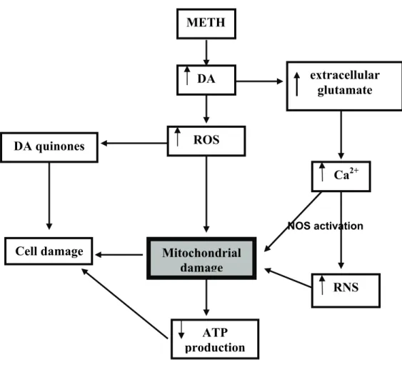
Figure 1.9: Brain targets of methamphetamine (METH)-induced toxicity in
the rat 33
Figure 1.10: Schematic representation of methamphetamine
(METH)-induced toxicity 35
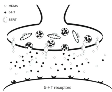
Figure 1.11: Schematic representation of
3,4-methylenedioxymethamphetamine (MDMA) primary action on serotonergic
cells
43
Figure 1.12: Monoamineoxidase (MAO) mediated 5-HT metabolism inside
pre-synaptic nerve endings 44
Chapter II
Manuscript I
Figure 1: MDMA induced hyperthermia in adolescent male Wistar rats.
Effect of selegiline
69
Figure 2: MDMA induced lipid peroxidation in whole-brain mitochondria and
the protective effect of selegiline
70
Figure 3: MDMA induced carbonyl formation in whole-brain mitochondria
and the protective effect of selegiline
70
Figure 4: Protective effect of selegiline over the deletion of the mitochondrial
gene sequences NDI, NDII and COXI in male Wistar rats exposed to a
neurotoxic dose of MDMA
71
Figure 5: MDMA induced decreased expression of the subunit NDII in whole
Figure 6: MDMA induced decreased expression of the subunit COXI in
whole-brain mitochondria and the protective effect of selegiline
72
Manuscript II
Figure 1.1: MDMA-induced hyperthermia in adolescent male Wistar rats.
Effect of clorgyline
86
Figure 1.2: Graphic representation of MDMA+clorgyline induced
hyperthermia in animals that died through the injection period
87
Figure 2: MDMA induced lipid peroxidation in whole brain mitochondria.
Effect of clorgyline
87
Figure 3: MDMA induced carbonyl formation in whole brain mitochondria.
Effect of clorgyline
88
Figure 4: MDMA induced decreased expression of the subunit NDII in whole
brain mitochondria. Effect of clorgyline
89
Figure 5: MDMA induced decreased expression of the subunit COXI in
whole brain mitochondria. Effect of clorgyline
90
Manuscript III
Figure 1: Body weight gain. Effect of ALC
110
Figure 2: MDMA induced hyperthermia in adolescent male Wistar rats.
Effect of ALC
111
Figure 3: MDMA induced lipid peroxidation in whole brain mitochondria.
Effect of ALC
112
Figure 4: MDMA induced carbonyl formation in whole brain mitochondria.
Effect of ALC
112
Figure 5: Protective effect of ALC over the deletion of the mitochondrial
gene sequences NDI, NDII and COXI in male Wistar rats exposed to a
neurotoxic dose of MDMA
113
Figure 6: MDMA induced decreased expression of the subunit COXI in
whole brain mitochondria. Effect of ALC
114
Figure 7: MDMA induced decreased expression of the subunit NDII in whole
Chapter III
Figure 1: Weight gain between the day of exposure and the day of sacrifice
measured in MDMA, selegiline, MDMA plus selegiline and control saline
treated animals
133
Figure 2: Body temperature measured subcutaneously throughout the
thirteen days following exposure within the four groups of animals under
analysis (MDMA, selegiline, MDMA plus selegiline and selegiline)
134
Figure 3: Amount of ATP per milligram of protein in brain areas of
adolescent male wistar rats 14 days after exposure
135
Figure 4: Percentage of white area analysed in brain slices of male Wistar
rats 14 days after exposure. Experiments with selegiline
136
Figure 5: Percentage of white area analysed in brain slices of adolescent
male Wistar rats 14 days after exposure. Experiments with ALC
137
Chapter IV
Index of tables
Table 1.1: ETC inhibitors 5
Table 1.2: OXPHOS uncouplers 6
Table 1.3: Mitochomdrial targeted antioxidants 12
Table 1.4: Methods commonly used to assess oxidative damage in
mitochondria 13
Table 1.5: Common AD related mitochondrial alterations 17
Table 1.6: Common PD related mitochondrial alterations 20
Table 1.7: Common age-related mitochondrial alterations 23
Table 1.8: Protective activities of acetyl-L-carnitine and selegiline in the
prevention of age related phenomena’s 24
Table 1.9: Selegiline improvement of neuronal functioning 26
Table 1.10: Common cocaine-associated biochemical alterations 32
Table 1.11: Common METH-associated biochemical alterations 34
Table 1.12: Common MDMA-associated biochemical alterations 36
Table 1.13: MDMA-induced effects on the serotonergic system 42
Table 1.14: Targets of MDMA-induced oxidation 46
Table 1.15: MDMA-induced mitochondrial damage 49
Table 1.16: MDMA-iinduced apoptotic-related events 50
Outline of the dissertation
The present thesis is structured in four main chapters:
Chapter I
Section 1: General Introduction
In this section, a general overview on the contribution of mitochondrial damage for
neurodegenerative diseases and xenobiotic toxicity is given. The most relevant insights
on mitochondrial related disorders like Parkinson and Alzheimer are presented and
discussed. The implications of prolonged and acute exposure to xenobiotics on
mitochondrial function and integrity is also reviewed, with particular emphasis on
MDMA.
Section 2: The animal model
In this section, an introduction to the animal model used is presented.
Section 3: General and specific objectives of the dissertation
The general and specific objectives of the dissertation are provided.
Chapter II
This chapter is divided in 3 sections, corresponding to the original manuscripts, and
presents the experimental work, results obtained and specific discussions to answer
the questions that derived from the general and specific objectives of this thesis.
Chapter III
In this chapter the unpublished results are presented.
Chapter IV
Section 1. Integrated overview of the performed studies.
The performed studies are integrated in a harmonized form.
Section 2. Conclusions
The conclusions that can be drawn from the present dissertation are summarized.
Chapter V
Overview
Mitochondria are gaining an important significance for the understanding of the
mechanisms involved in several brain disorders. Coupling of respiration with oxidative
phosphorilation (OXPHOS) performed in these organelles constitutes the major source
of energy to brain cells and implies a correct mitochondrial performance that when
disrupted can result in severe damage. Age-associated mechanisms and several
related brain disorders, particularly the neurodegenerative ones, like Parkinson and
Alzheimer are associated with severe mitochondrial dysfunction.
From what is at present known about MDMA-induced neurotoxicity, two main
aspects have to be retained in order to understand the overall results here presented:
(i) MDMA binds to the 5-HT re-uptake transporter and, inside axon terminals causes an
acute and powerful release of neurotransmitters; (ii) excess of serotonin in the
presynaptic nerve endings is metabolized by MAO flavoenzymes bounded to the
external mitochondrial membranes.
The first results obtained in the present work lead us to conclude that the
administration of a neurotoxic binge dose of MDMA to rats results in mitochondrial
oxidative damage in the central nervous system (CNS), namely lipid and protein
oxidation and mtDNA deletions, with subsequent diminished expression of the
correspondent proteic subunits that are important constitutive elements of the electron
transport chain (ETC).
When obtaining these results a theoretical hypothesis was designed. Increased
H2O2 formation as result of increased MAO function inside presynaptic nerve endings
lead to an overall increase of ROS inside mitochondria. Damage to several
mitochondrial constituents could therefore have resulted, compromising this way the
correct mitochondrial function and adequate energy supply.
To test the above mentioned hypothesis, additional experiments with MAO
inhibitors (MAOi) were performed. Two isoforms of MAO exist: MAO-A and MAO-B. In
brain, MAO-A is expressed predominantly in catecholaminergic neurons, whereas
MAO-B is expressed in serotonergic neurons, astrocytes and glia. Although
metabolism by MAO-B is only residual in the presence of MAO-A, it is fully effective in
the absence of the later, as it happens inside serotonergic nerves.
The first experiments with MAOi, performed with selegiline, a MAO-B inhibitor,
revealed, in accordance with the formulated hypothesis, a significant protection against
the overall effects on mitochondria.
With the first hypothesis proven, a second question arised. If MAO-B inhibition
what happens to rat brain mitochondria if MAO-A inhibition is performed before MDMA
administration? To answer this question, experiments with clorgyline, a specific MAO-A
inhibitor, were performed. In this case, the previous administration of clorgyline
revealed no protection against MDMA-induced neurotoxic effects. Moreover, toxicity
was increased and the majority of animals exposed died during the experiments.
With the two monoamine oxidases tested, we decided to examine the effects of
ALC supplementation on the MDMA-induced toxic effects at the brain mitochondrial
level. ALC was chosen because of its antioxidant properties and its active role in
mitochondria mainly by acting as a carrier of fatty acids across mitochondrial
membranes for energy production through β-oxidation. As expected, ALC exposure
protected at a great extent the mitochondrial damage associated with drug exposure.
Besides the new insights obtained from the above studies, further investigation
in order to confirm previous findings, or complete the new studies here presented, was
also performed. Measurement of body temperature of animals in the day of drug
administration confirmed previous reports of several authors that included hyperthermia
in the most pronounced effects of the drug. Failure of selegiline, clorgyline and ALC in
protecting against this effect states mitochondrial damage, at the levels here studied,
as unrelated with the characteristic deregulation of core body temperature.
ATP measurements on specific brain areas was also assessed in order to infer about
the extent of drug-induced mitochondrial damage on ATP production. The results
obtained confirmed a decrease of brain energy levels after exposure to MDMA and
lead us to hypothesize about a possible involvement of the deletion of mitochondrial
genes and deficient expression of the correspondent protein subunits with the
uncoupling of oxidative phosphorilation.
TTC staining of brain slices was performed in order to macroscopically confirm the
hypothesized mitochondrial metabolic dysfunction. The results achieved were in
accordance with the expected although no direct correlation with the results concerning
Chapter I
1. General Introduction
2. Animal Model
1. General Introduction
1.1 An overview of mitochondrial structure and functions
Mitochondria are double membrane cytoplasmic organelles found in eukaryotic cells,
structurally composed by two internal compartments, confined by an inner and an outer
membrane. The mitochondrial matrix is contained within the inner membrane and the
intermembrane space is comprised between the two membranes. The mitochondrial
matrix encloses the mtDNA, RNA, protein synthesizing and destoxifying systems,
constitutive mitochondrial proteins and various soluble enzymes involved in the
tricarboxylic acid cycle and β-oxidation pathways. The external mitochondrial
membrane is permeable to ions and solutes up to 14 KDa and has bounded enzymes
that interface with the cellular cytoplasm. The inner membrane contains the ETC which
is composed of five major complexes (I-V), each one with internal subunits. The
intermembrane space contains proteins encoded by the nuclear DNA that include
some that function as components of the ETC, apoptotic factors, transporters of
polypeptides, metal ions and hydrophobic precursor proteins and enzymes responsible
for metabolic processes (Koehler et al., 1998; Mesecke et al., 2005).
Coupling of OXPHOS with respiration through the ETC gives rise to energy production
(Cooper, 2000) (Fig. 1) that, together with regulation of apoptosis and redox state,
heme and iron sulphur center biosynthesis, amino acid and nitrogen metabolism, and
calcium homeostasis modulation, constitute the most important functions of
mitochondria(Murphy and Smith, 2000; Green and Kroemer, 2004).
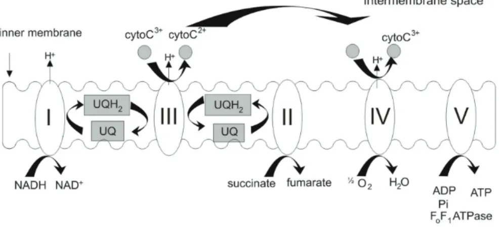
Figure 1.1: Coupling of oxidative phosphorylation with respiration throughout the mitochondrial electron
transport chain. The mitochondrial complex I (NADH:ubiquinone reductase) is responsible for the oxidation
of NADH and the energy produced in this step is further used to reduce ubiquinone (UQ). The
succinate to fumarate (Hagerhall, 1997). The ubiquinol:cytochrome c reductase complex (complex III) is
responsible for the oxidation of the reduced ubiquinol (UQH2) and for the reduction of cytochrome c.
Complex IV is finally responsible for the reduction of molecular oxygen to water. Throughout the entire
process, proton pumping is always coupled to electron flow, thus ensuring the efficiency of energy
production by the phosphorylation system, i.e., complex V, ADP/ATP and inorganic phosphate
transporters.
1.2 The interaction of xenobiotics with the ETC
The disruption of the electrochemical proton gradient may be attained by the use of
specific inhibitors of the ETC or by inhibiting the supply of reducing substrates to the
respiratory chain. Chemical substances with potential to disrupt the correct
mitochondrial performance include agents that increase membrane permeability and/or
induce the permeability transition or behave as alternate electron acceptors. In any
case, the correct mitochondrial function is compromised and severe mitochondrial
damage could result. These conditions would disable the mitochondrial capacity to
utilize the molecular oxygen and therein produce ATP.
1.2.1 ETC inhibitors
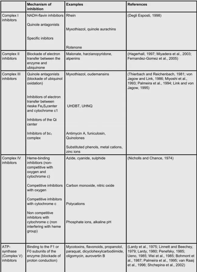
The mitochondrial complex I is the most vulnerable to chemical-induced damage.
Three types of inhibition are recognized, namely NADH-flavin inhibitors, quinole
antagonists and specific inhibitors (Table 1.1) and a large number of compounds with
capability to block the function of this important structure (Degli Esposti, 1998) (see
table1.1). Complex II inhibitors are the less potent and specific inhibitors of the ETC.
Nevertheless, there is a great diversity of compounds with ability to reduce the
enzymatic activity of this complex (Hagerhall, 1997; Miyadera et al., 2003). Complex III
inhibitors present a great variability of effect among species (Degli Esposti et al., 1990;
Degli Esposti et al., 1992; Ghelli et al., 1992; Vaidya et al., 1993; Kraiczy et al., 1996).
There are four different groups of compounds with specific characteristics of inhibition,
namely quinole antagonists, inhibitors of electron transfer between rieske Fe2S2 center
and cytochrome c1, inhibitors of the Qi center and inhibitors of the bc1 complex (Table
1.1). Finally, the inhibitors of complex IV, similarly to complex III are also divided in four
categories according to the specific targets of action, namely heme-binding inhibitors
(non-competitive with oxygen and cytochrome c), competitive inhibitors with oxygen,
competitive inhibitors with cytochrome c, and non-competitive inhibitors with
cytochrome c (Table 1.1). The inhibitors of complex V (ATPsynthase) act mainly by
blocking proton conduction through Fo fraction of complex V (ATPsynthase), although
the mycotoxins are the most potent ones, several other chemicals exist that share the
Table 1.1: ETC inhibitors
Mechanism of inhibition
Examples References
Complex I inhibitors NADH-flavin inhibitiors Quinole antagonists Specific inibitors Rhein
Myxothiazol, quinole aurachins
Rotenone
(Degli Esposti, 1998)
Complex II inhibitors
Blockade of electron transfer between the enzyme and ubiquinone
Malonate, harzianopyridone, atpenins
(Hagerhall, 1997; Miyadera et al., 2003; Fernandez-Gomez et al., 2005)
Complex III inhibitors
Quinole antagonists (blockade of ubiquinol oxidation)
Inhibitiors of electron transfer between rieske Fe2S2center
and cytochrome c1 Inhibitors of the Qi center
Inhibitors of bc1
complex
Myxothiazol, oudemansins
UHDBT, UHNQ
Antimycin A, funiculosin, Quinolones
Substituted phenols, metal cations, zinc ions
(Thierbach and Reichenbach, 1981; von Jagow and Link, 1986; Miyoshi et al., 1993; Palmeira et al., 1994; Link and von Jagow, 1995) Complex IV inhibitors Heme-binding inhibitiors (non-competitive with oxygen and cytochrome c) Competitive inhibitiors with oxygen Competitive inhibitors with cytochrome c Non competitive inhibitors with cytochrome c (non interfering with heme group)
Azide, cyanide, sulphide
Carbon monoxide, nitric oxide
Polycations
Phosphate ions, alkaline pH
(Nicholls and Chance, 1974)
ATP-synthase (Complex V) inhibitors
Binding to the F1 or F0 subunits of the enzyme (blockade of proton conduction)
Mycotoxins, flavonoids, propanolol, paraquat, dicyclohexylcarbodiimide, oligomycin, aurovertin B
(Lardy et al., 1975; Linnett and Beechey, 1979; Lardy, 1980; Penefsky, 1985; Ueno, 1985; Wei et al., 1985; Bohmont et al., 1987; Palmeira et al., 1995; van Raaij et al., 1996; Shchepina et al., 2002)
1.2.2 OXPHOS uncouplers
Uncoupling of OXPHOS leads necessarily to a decrease of ATP production. In the table below (Table 1.2), a summary of the most common mitochondrial uncouplers is presented.
Table 1.2: OXPHOS uncouplers
OXPHOS uncouplers
Properties Examples References
Proton carriers
Mobilization of protons across lipid bilayers
Carbonylcyanide-p-trifluoromethoxyphenylhydrazone (FCCP), carbonylcyanide m-chlorophenylhydrazone (CCCP), dichlorophenol
(Crane, 1977; Terada, 1981; Sun and Mauzerall, 1996; Beauvoit et al., 1999) Liophilic weak
acids
Increased permeability of lipid membranes to protons with dissipation of the
electrochemical proton gradient by the cycling movement of an uncoupler molecule
Substituted phenols, salicylanides, carbonyl cyanide
(McLaughlin and Dilger, 1980; Terada, 1981)
Free fatty acids
Protein mediated uncoupling by FFA (wasting of energy and inhibition of respiration by excessive FFA accumulation)
Sulfuramide, perfluorodecanoic acid (Langley, 1990;
Schnellmann and Manning, 1990; Wojtczak and Schonfeld, 1993; Hermesh et al., 1998) Chanel-type
ionophores
Chanels in the lipid membrane (increase of permeability with collapse of the proton electrochemical gradient)
Gramicidins (Katsu et al., 1987;
Luvisetto and Azzone, 1989)
Carrier-type ionophores
Formation of lipid-soluble complexes with ions (collapse of the electrochemical proton gradient)
Nigericin, valinomycin (Toro et al., 1976;
Felber and Brand, 1982; Kovac et al., 1982)
Cationic uncouplers
Increase in membrane permeability to ions
(Interference with the physical integrity of the membrane-induction of the MPT)
Tris-S-C4 , pentamidine (Degli Esposti, 1998;
Shinohara et al., 1998)
Membrane active compounds
Formation of chanells permeable to alkaline ions and/or protons or induction of the formation of large pores that lead to mitochondrial swelling
Alamethicin
tamoxifen, cyclosporine A, nafoxidine
(Takaishi et al., 1980; Mathew et al., 1981; Hoyt et al., 2000; Simpson et al., 2002)
Alternate electron acceptors
Competition with the electron natural acceptors of the carrier-dissipation of proton
electrochemical potential
Adriamycin, paraquat, substituted naphtoquinones,
(Doroshow and Davies, 1986; Bironaite et al., 1991; Henry and Wallace, 1995; Wallace, 1999)
In this thesis, a particular attention will be given to brain mitochondria
specifically focusing some aspects related with the ETC integrity and function,
mtDNA mutations and oxidative stress. Within this context, the important role of
MAO’S, enzymes responsible for brain monoamine neurotransmitter metabolism,
1.3 The contribution of oxidative stress to mitochondrial dysfunction
Oxidative stress is a disturbance in the pro-oxidant-antioxidant balance in favour of the
former, leading to potential damage. Both reactive nitrogen species (RNS) (Spanos
and Yamamoto, 1989b) and ROS (Jacobson and Duchen, 2002; Ott et al., 2007)
contribute to the development of oxidative stress conditions within cellular organisms. A
sustained oxidative stress status may ultimately affect cellular function and, within the
cell, mitochondria are particularly involved in this process, by being themselves
potential generators of these reactive species that although being essential to some
cellular functions could also contribute to cellular damage specially if produced in high
amounts. Both ROS and RNS are by-products of ATP production (Wiseman and
Halliwell, 1996) and their increased production generally arise from defects in
mitochondrial respiratory chain structure and function. These species usually include
oxygen radicals [(e.g. superoxide radical (O2.-) and hydroxyl radical (HO.)] and certain
non radicals [(e.g. hydrogen peroxide (H2O2), peroxynitrite (ONOO-), and ozone (O3)]
(Wiseman and Halliwell, 1996) that when overproduced constitute potential damaging
agents to mitochondria, its constituents and to the overall cellular function.
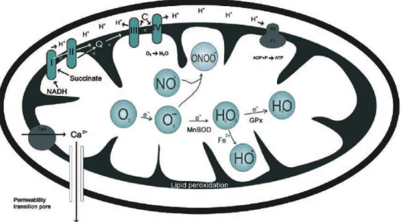
Figure 1.2: Formation of ROS and RNS inside mitochondria and mitochondrial antioxidant system.
In the ETC, the main producers of ROS are complexes I and III (Balaban et al., 2005),
whose primary product of oxidation is O2
.-. This radical alone is not very toxic but
further reaction with nitric oxide (NO) gives rise to the formation of the very reactive
specie ONOO- that is also quickly converted to H2O2, spontaneously or through the
transition metal ions generating HO.. Importantly, HO. is one of the strongest oxidizing
agents, producing severe damage in its vicinity, leading to oxidative damage of lipids,
proteins and DNA (Halliwell, 1996; Valko et al., 2004). Additional important
endogenous sources of oxidative stress are MAOs, flavoenzymes bounded to the
external mitochondrial membrane. The oxidation of monoamine neurotransmitters by
MAOs to its aldehyde derivatives gives rise to the production of H2O2 (Sandri et al.,
1990; Giorgio et al., 2005), which may subsequently be transformed into HO., as
mentioned before.
1.3.1 Nitric oxide: damaging or protector?
NO is synthesized mainly by three enzyme isoforms of the enzyme nitric oxide
synthase (NOS) that include the neuronal NOS (nNOS; typeI), inducible NOS (iNOS;
typeII) and endothelial NOS (eNOS, typeIII) (Knowles and Moncada, 1994; Yun et al.,
1996; Emerit et al., 2004). This RNS has a dual action in neuronal cells because
although playing essential roles in the modulation of several physiological activities like
the modulation of vascular tone (Palmer et al., 1987), neurotransmission (Garthwaite et
al., 2005) and immune system (Hibbs et al., 1988; Stuehr and Nathan, 1989), it is also
capable to induce cellular damage. NO binds to the mitochondrial cytochrome c
oxidase (Bolanos et al., 1994) and decreases its affinity for O2 thus affecting the basal
mitochondrial performance and decreasing the levels of energy production (Almeida
and Bolanos, 2001). Moreover, due to the increase in intracellular Ca2+ concentration
that follows N-methyl-D-aspartate (NMDA) activation, nNOS is activated (Knowles and
Moncada, 1994), resulting in the overproduction of NO, which is also able to cause the
opening of the mitochondrial permeability transition (MTP) structures located in the
mitochondrial membranes (Tatton and Olanow, 1999). This ultimate action has two
main consequences: the loss of membrane potential and the activation of mechanisms
involved in apoptotic cellular death by inducing the release of cytochrome c and other
intramembrane apoptotic factors (Tatton and Olanow, 1999).
1.3.2 Main targets of oxidative modifications inside mitochondria
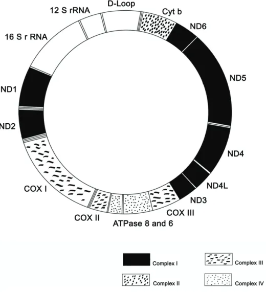
The mitochondrial genome is responsible for coding 13 proteins absolutely essential for
respiration and oxidative phosphorilation (Anderson et al., 1981). These proteins
include seven subunits of NADH dehydrogenase (NDI, II, III, IV, IVL, V and VI), three
subunits of cytochrome c oxidase (COXI, II and III) and two subunits of ATP synthase
of these proteins, might imply altered mitochondrial activity thus representing a
potential source of cellular dysfunction.
The mtDNA is one of the mitochondrial structures more prone to oxidative
modifications, the presence of free radical generating enzymes together with poor
repair mechanisms and the absence of protective histones, all converge to a high
sensitivity of mtDNA to the occurrence of mutations (Carew and Huang, 2002; Taylor
and Turnbull, 2005)
.
Figure 1.3: Mammalian mtDNA genome. mtDNA codes for two rRNA ((12S and 16S)-coding regions),
22tRNA (not signalled in the figure), and 13 subunits of the OXPHOS (oxidative phosphorylation) pathway
(13mRNA-coding regions). Adapted from (Suliman et al., 2003).
Mitochondrial proteins and lipids are also prone to oxidation. The high content in
polyunsaturated fatty acid residues in mitochondrial phospholipids gives rise to an
extended formation of lipid peroxides upon exposure to pro-oxidant conditions
(Esterbauer et al., 1991) and the direct oxidation of amino acids in the proteins of