Full paper published online: May 30, 2010 ISSN 1678-9199.
Biochemical comparison of venoms from young Colombian Crotalus durissus cumanensis and their parents
Céspedes N (1, 2), Castro F (3),Jiménez E (1), Montealegre L (1), Castellanos A (1), Cañas CA (4), Arévalo-Herrera M (1, 2), Herrera S (1, 2)
(1) International Center of Vaccines, Cali, Colombia; (2) Institute of Immunology,
University of Valle, Cali, Colombia; (3) Group of Herpetology, School of Sciences,
University of Valle, Cali, Colombia; (4) Valle de Lili Clinic Foundation, Cali, Colombia.
ABSTRACT: Crotalus durissus cumanensis, a rattlesnake endemic to Colombia and Venezuela, is considered one of the most lethal snake species in Latin America. The aim of the present study was to compare the protein content and biological activity of the venom obtained from eight specimens of C. durissus cumanensis, namely two adults from different localities of Colombia and six offspring born in captivity. Protein profiles of crude venoms were analyzed by SDS-PAGE and RP-HPLC, and biological activities were evaluated for lethality, edema, defibrination, hemolytic and coagulant activities to assess individual venoms of adults and a pool of young snake venoms. Transient edema appeared rapidly after venom inoculation, whereas hemorrhagic effect was not observed. Differences in protein profiles, lethality, hemolytic, coagulant and defibrinating activities between both adult snake venoms were observed; those from the mother snake exhibited higher activities. Venoms from young snakes were similar to the one obtained from the mother, but the coagulant effect was stronger in offspring venoms. Notably, biological effects of the father snake venom were not comparable to those previously described for C. durissus cumanensis from Venezuela and C. durissus terrificus from Brazil, confirming the high variability of the venom from Crotalus species.
KEY WORDS: snakes, Crotalus durissus cumanensis, rattlesnake, venom.
CONFLICTS OF INTEREST: There is no conflict.
FINANCIAL SOURCE: Colombian Institute for the Development of Science and Technology (Colciencias) grant n. 2304-13-17756 and International Center of Vaccines.
CORRESPONDENCE TO:
SÓCRATES HERRERA VALENCIA, Centro Internacional de Vacunas, AA 26020, Cali, Colombia. Phone: +57 2 521 6228. Fax: +57 2 557 0449. Email:
INTRODUCTION
Snakebites represent a medical emergency in many regions of the world, particularly
in tropical and subtropical areas where they are endemically distributed, affecting
mainly agricultural workers and children. Although the precise global incidence of
snakebites has proven difficult to estimate, it has been reported that there are at least
5 million snakebites per year worldwide, which results in 2.5 million clinical cases and
an estimated 125,000 deaths (1). The incidence of snakebite mortality is especially
high in Africa, Asia, New Guinea and Latin America. In Colombia, a total of about
3,000 snakebite accidents are reported yearly with an estimated mortality of 2 to 5%
(2).
Poisoning by snakebite provokes medical emergencies that affect different tissues
and organ systems depending on the species responsible for the bite. In the
Americas, the most severe cases result from bites by members of the Viperidae and
Elapidae families. Crotalus snakes (rattlesnakes), belonging to the Viperidae family,
are geographically distributed from Canada to Argentina. Crotalus durissus,
composed of 14 subspecies, has the widest geographical distribution (3). This
species has been associated with intraspecific variability in the chemical composition
of its venom and, consequently, in its clinical effects and related therapeutic
strategies (4-7). Although symptoms are usually variable, snake venom is highly toxic
and presents complex outcomes. In the case of Crotalus, local effects consist of
moderate pain, edema and local hemorrhage without necrosis, whereas systemic
manifestations are more severe and may cause considerable muscle damage
(myotoxicity), bleeding (hemostatic abnormalities), renal damage (nephrotoxicity) and
paralysis (neurotoxicity) (8).
Previous studies have shown great biochemical and pharmacological variation
among venoms of C. durissus subspecies from different regions of South America,
which appears more toxic than those of the same species from Central America
(9-11). Diversity in venom composition and biological activities has also been
associated with the age of snakes in C. durissus durissus (6, 12, 13). Moreover, in
this species toxicity appears to be induced in part by crotoxin, a potent neurotoxin
present in its venom in large quantities (14). Crotalus durissus cumanensis is a
subspecies endemic to Colombia and Venezuela, particularly abundant in the
Recent studies on the biochemical functional characterization of C. durissus
cumanensis venom from Venezuela have indicated high intraspecies variability
whereas different isoforms of crotoxin complex from this venom have been isolated,
purified and characterized by other researchers (16-21). In the current study, we
present preliminary chemical and functional characterization of venom obtained from
a snake family composed of adult C. durissus cumanensis (parents), captured in
different regions of Colombia, and from six young offspring born in captivity.
MATERIALS AND METHODS Snakes and Venoms
Eight specimens of C. durissus cumanensis were employed in the present study.
Two of them were adults – the male was captured in Guajira (northern Colombia),
while the female was from Tolima (central Colombia) – and the other six were their
offspring (both genders) born in captivity. Snakes were kept at room temperature
(23°C ± 5) in individual boxes (80 x 50 x 40 cm) at the snake holding facility in
Caucaseco Scientific Research Center (Cali, Colombia), and were fed live mice every
15 days. Venom samples were extracted manually by glandular compression,
collected in glass vessels, then centrifuged, lyophilized, and kept at –20°C until use.
Venoms from all snakes were subjected to chemical analysis to determine their
functional activities. Offspring were milked when they reached one year of age.
Mice
Male or female BALB/c mice weighing 18 to 22 g were used throughout the study for
functional assays of the venoms. Animals received food and water ad libitum. All the
procedures involving mice were conducted following national and international
recommendations and policies for animal use and were monitored by the Animal
Ethics Committee of the University of Valle.
Electrophoretic Analysis
Sodium dodecyl sulfate polyacrylamide gel electrophoresis (SDS-PAGE) of venom
samples was carried out according to the Laemmli method (22) using a
Mini-Protean® II system (Bio-Rad Laboratories, USA) as previously described (17).
Briefly, after dilution in run buffer, samples were loaded onto 12.5% acrylamide gels
8.3). Gels were stained with 0.1% Coomassie brilliant blue R250 and were analyzed
using the GeneSnap® software (v 6.07 Synoptics Ltd., UK) and the
GeneGenius/multigenius® equipment (Syngene, UK).
Chromatographic Analysis
Samples of C. durissus cumanensis venom were analyzed by reverse-phase
high-performance liquid chromatography (RP-HPLC) on a Waters HPLC system (USA)
consisting of 510 bombs, 717 PLUS autosampler and Waters 486 UV detector.
Venom samples (1 mg/mL) were dissolved in 1% acetic acid and loaded onto the
Zorbax 300SB-C8® (Agilent, USA) column (4.6 x 50 mm, 5 µm). The mobile phase
was a mixture of phase A [20:80:0.083 (v/v) acetonitrile/water/TFA] and phase B
[80:20:0.1 (v/v) acetonitrile/water/TFA], with a flow rate of 1.0 mL/minute and
monitored at 280 nm.
Determination of Lethality
Lethality was determined in BALB/c mice after intraperitoneal injections of venom
samples. Venoms were injected separately into groups of six mice each with doses
ranging from 0.125 to 5 mg/kg, serially diluted in 0.2 mL of saline solution. Survival
time of each animal was recorded for 48 hours and a LD50 value calculated according
to the Spearman-Karber method (23).
Edema-Inducing Activity
Edema formation induced by the venoms was assayed according to the method of
Yamakawa et al. (24). The time-course of edema was determined in mice injected
into the subplantar surface of the left hind paw with 50 µL of saline solution
containing 1.0, 2.5 or 5.0 µg of each venom. The other paw received the same
volume of saline solution without venom as a control. Edema was measured at
different intervals (30 minutes and 1, 2, 3, 4, 5, 6 and 24 hours). Results were
calculated as the difference between values obtained before and after venom
inoculation, compared with a control and plotted (expressed in millimeters) versus
Hemorrhagic Activity
The minimal hemorrhagic dose (MHD) was determined by the Instituto Clodomiro
Picado method (25), with crude venoms, and was defined as the amount of venom
protein resulting in a 10-mm diameter hemorrhagic area. A series of four doses of 20,
40, 60 and 80 µg were used, and 0.1 mL of each was inoculated subcutaneously into
the abdomen of each experimental BALB/c mouse. Mice were killed after one hour;
the skin was removed and the hemorrhagic diameter in the subcutaneous tissue was
measured to define the MHD.
Defibrinating Activity
Eight doses of venom (0.3, 0.6, 1.2, 2.5, 5.0, 10.0, 15.0 and 20.0 μg) were prepared
in 200 μL of saline solution and three female mice per dose group were injected
intravenously while three negative control mice were similarly injected with saline
solution only. One hour after the injections, animals were bled and 200 μL of blood
was collected in glass tubes and incubated for two hours at room temperature. The
minimal defibrinating dose (MDD) was defined as the minimal dose of venom able to
induce coagulation failure in blood samples of all injected mice.
Coagulant Activity
Coagulant activity of venom samples was determined using the method described by
Instituto Clodomiro Picado (25). Briefly, 0.2 mL of human citrated plasma was
incubated for five minutes at 37ºC and then doses ranging between 12 to 50 μg of
venom in a total volume of 0.1 mL of saline solution were added. Coagulant activity
was determined in triplicate preparations of these solutions and the mean clotting
time was calculated. Normal human citrated plasma plus saline solution was used as
coagulation control. The minimal coagulant dose (MCD) corresponds to the venom
concentration that induced clotting development in 60 seconds.
Hemolytic Activity
A gel containing a mixture of agar, blood and egg yolk was prepared in small Petri
dishes. Three-millimeter-diameter wells were punched in the gel and 15 μL of
different venom doses (1, 2, 4, 6 and 8 μg) prepared in saline solution were added in
triplicate to wells. Plates were incubated at 37°C for 24 hours, hemolytic zones
The indirect hemolytic activity was determined in terms of the minimal indirect
hemolytic dose (MiHD), considered to be the minimum amount of venom that
produced a 20-mm-diameter hemolytic zone (25).
RESULTS
Electrophoretic Analysis
Electrophoretic analysis demonstrated a high complexity in the venom composition
as indicated by protein characterization (Figure 1). All venoms exhibited proteins of
15, 25, 36 and 60 kDa together with other more variable proteins. A greater
complexity was observed in the venom of the snake captured in Guajira (Cd1, male)
than in the specimen from Tolima (Cd2, female). Similarly, a series of
high-molecular-weight proteins of 79, 100, 143 and 193 kDa were not detectable in Cd2. When the
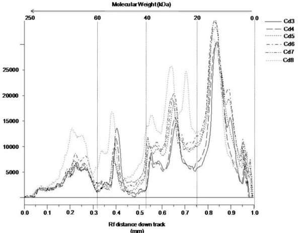
venoms of six offspring (Cd3-Cd8) were compared (Figure 1), they presented a
predominance of low-molecular-weight proteins with similar ranges to those shown in
the adult snakes. Analysis of these venoms using the GeneSnap® program showed
a distinct overlap of bands with similar molecular weights in five of the venoms,
whereas one presented some variations in the protein profile (Cd8), represented by
an additional protein with 20 kDa and several bands with molecular weight greater
than 25 kDa at high concentrations (Figure 2). A peak concentration revealed
proteins with lower molecular weight (15 kDa) (Figure 2) identified as phospholipases
A2 (crotoxin basic chain-1) trough tandem mass spectrometry (data not shown).
Additionally, another low-molecular-weight protein (approximately 12 kDa) was
Figure 1. Electrophoretic analysis of C. durissus cumanensis venoms. Venoms from the Guajira male (Cd1), the Tolima female (Cd2), and male (Cd3, Cd6, Cd7) and
female (Cd4, Cd5, Cd8) offspring were subjected to SDS-PAGE under non-reducing
Figure 2. Comparative electrophoretic profiles of venoms from the offspring. The analysis was carried out using the GeneSnap® program after gel documentation.
Cd3, Cd6, Cd7 were males and Cd4, Cd5, Cd8 were females.
Chromatographic Profile
Chromatographic profiles of both adult snakes (Figure 3) corroborated the variation
observed by SDS-PAGE in protein profiles. Analysis of offspring venoms showed
greater similarity with the Tolima snake (female) venom than with that of Guajira
snake (male). In the venoms from the mother and offspring, there was a compound
that eluted at 16 minutes (peak number 3) which corresponded to more than 50% of
the total protein content, whereas in the male venom, the most concentrated
Figure 3. Reverse phase chromatographic protein profile of Crotalus durissus venom. Guajira male (A), Tolima female (B) and offspring pool (C). The retention times were 12 (1), 14 (2), 16 (3), 18 (4), 21 (5) and 23 (6) minutes.
Lethal Activity
Female snake venom was the most lethal and potent [LD50 = 0.47 mg/kg mouse (CI
95% 0.45-0.49)] compared to the male venom [LD50 = 2.69 mg/kg mouse (CI 95%
2.63-2.70)]. Young snake venoms showed similar lethal activity to female snake
venom [LD50 = 0.41 mg/kg mouse (CI 95% 0.39-0.44)] (Table 1). In all cases, the
effect was observed in the first eight hours after venom inoculation. Animals
developed paraplegia, fasciculation and shortness of breath.
Table 1. Functional activitiesof C. durissus cumanensis venom
C. durissus cumanensis venom
Activity
Female Young Male
Coagulant activity (µg)a 27.7 ± 2.3 11.7 ± 0.6 29.3 ± 1.2
Hemolytic (µg)b 11.3 ± 0.7 14.7 ± 0.4 192 ± 0.7
Lethality (µg/g)c 0.47 (0.45 – 0.49) 0.41 (0.39-0.44) 2.69 (2.63 – 2.70)
Hemorrhagicd Absent Absent Absent
Defibrinating MDD
(µg)e 0.6 2.5 Absent
a
Minimum coagulant dose (MCD).
b
Minimum hemolytic dose (MiHD); each response was the mean of three observations.
c
Median lethal dose (LD50); data within parenthesis represent 95% confidence intervals.
d
Minimum hemorrhagic dose (MHD); each response was the mean of three observations; control:
Bothrops asper venom.
e
Edema-Inducing Activity
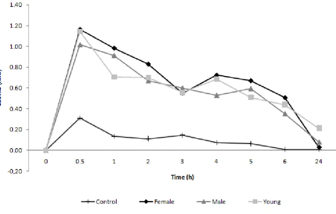
Edema developed rapidly after injection of both male and female venoms. The
maximum reaction was observed at 30 minutes, subsequently disappearing within 24
hours (Figure 4). Similar behavior was observed for venoms of young snakes and no
differences were noticed when different doses were employed.
Figure 4. Edematous activity of C. d. cumanensis venom. BALB/c mice were inoculated with 2.5 μg of each venom into the subplantar region of the hind paw.
Hemorrhagic and Hemolytic Activity
Minimal hemorrhagic dose (MHD) could not be estimated for any of the venoms as
none of them showed any hemorrhagic effect at the different tested doses. In terms
of hemolysis, female venom exhibited hemolytic activity with the venom dose of 11 ±
0.7 μg while young snake venom did the same with a 14.7 ± 0,4 μg dose, the male
venom, in turn, required significantly higher concentrations (192 ± 0.9 μg of venom)
Defibrinating and Coagulant Activities
As observed in Table 1, all venoms presented coagulant activity. The female venom
needed 27.7 μg to produce the effect whereas the male venom required 29.3 μg and
the offspring venoms, 11.7 μg. While the female venom showed defibrinating activity
in all tested doses down to 0.6 μg, venoms from young snakes required doses of at
least 2.5 μg to show coagulant activity and the male venom did not exhibit this
activity at any of the tested doses (Table 1).
DISCUSSION
In this study we confirmed the great variability previously observed in the protein
content and biological activities of adult snake venom from C. durissus cumanensis
(16, 17). Variability in venom composition has been attributed to environmental and
individual factors such as habitat, origin, seasonality, diet, age and gender (1). In the
current work, the environmental factors including habitat, seasonality and diet were
not considered because adult animals had been kept in captivity for more than ten
years under similar feeding and environmental conditions, and young snakes for at
least one year. Therefore, the observed differences between adult snakes may be
due to potential genetic diversity, possibly associated with their different geographic
origins. Additionally, the great similarity among protein profiles of male and female
offspring might also discard the gender factor. We cannot explain the similarities
among the venoms from the mother and from the offspring; therefore, further studies
are required to clarify how genes connected with venom proteins are transmitted
from parents to offspring.
The venoms from the female and from the young snakes were particularly similar in
their hemolytic and defibrinating activities and lethality, but they all differed from the
male adult venom as well as from those previously reported for C. durissus
cumanensis from Venezuela and for other Crotalus subspecies from Brazil, Costa
Rica and Guatemala, which suggests a geographic association in these effects (6,
10, 16, 17, 26). Besides the great similarities among mother and young snake
venoms, there were surprising differences since the adult female venom presented
greater coagulant activity and lower defibrinating property.
Although lethality is likely of multifactorial origin, both neurotoxicity and hemolytic
activities appear to play a preponderant role as lethality has been associated with the
lethality of C. durissus terrificus venom is mainly due to the action of a crotoxin
complex, which constitutes a major component (60%) of the venom (28, 29). Crotoxin
is a very potent neurotoxin that is composed of a PLA2 and a chaperone molecule
(crotapotine) and is capable of blocking neuromuscular transmission. In this study the
young and female venoms presented a higher concentration of this protein than the
male venom, as observed in the electrophoretic and chromatographic profiles,
therefore suggesting a higher lethal potential than the male venom. Indeed, in this
study, the female and offspring venoms were more fatal than male venom. The latter
was also less lethal than several other subspecies (6).
In general, it appears to exist a gradient in toxicity as female and young snake
venoms showed a lethality rate comparable to that reported for C. durissus
cumanensis from Venezuela, but were less toxic than venom from Brazilian snakes
and more lethal than those of Central America species (6, 7, 11, 16, 17).
Regarding the defibrinating activity it was significantly lower in the Colombian snakes
than in the C. durissus cumanensis from Venezuela (16, 17). In terms of coagulation
activity adult snakes appear to behave similarly in Colombian and Venezuela (2002)
Furtado et al (13), including the fact that newborn and young C. durissus durissus
and C. durissus terrificus have been significantly more potent than the adult snake
venoms (6, 14). It has been demonstrated that coagulant activity is due to the action
of thrombin-like enzymes (11) in others C. durissus species. It is likely that similar
components are present in C. durissus cumanensis venoms.
An interesting feature of this study is the fact that venoms tested herein induced a
pronounced edematous activity, but poor hemorrhagic activity. Edema activity has
been associated to metalloproteinases that also cause hemorrhage. Therefore, the
absence of hemorrhagic activity in these venoms may suggest that the edema is
induced by proteins different from these proteinases with edematous activity like
myotoxins and phospholipases A2 (19, 20, 30). In previous studies with C. durissus
venoms from other South American regions, no edematous or hemorrhagic activity
was observed (9-11, 31). This is in contrast to the positive results reported for C.
durissus snakes from Central and North America (11). Nevertheless, hemorrhagic
activity has been reported for C. durissus cumanensis venoms of Venezuela (16-17).
Finally, we have confirmed the presence of great variability in C. durissus
cumanensis venom that did not appear to be associated with age, gender, diet or
ongoing in an attempt to better understand the difference in protein profiles and
functional differences of venoms and their potential importance for envenoming
therapy in humans.
ACKNOWLEDGEMENTS
The authors are grateful to the Animal Biotechnology Center (CBA) for the
collaboration by supplying the animals. Thanks are also extended to Diego Alvarez
and Jorge Jimenez for their technical assistance. This work was supported by the
Colombian Institute for the Development of Science and Technology (Colciencias),
grant n. 2304-13-17756 and by the International Center of Vaccines.
REFERENCES
1. Chippaux JP. Snake-bites: appraisal of the global situation. Bull World Health
Organ. 1998;76(5):515-24.
2. Instituto Nacional de Salud. Protocolo de vigilancia del accidente ofídico. Bogotá:
Instituto Nacional de Salud Subdirección de Vigilancia y Control; 2007. p. 1-5.
3. Campbell JA, Lamar WW. The venomous reptiles of Latin America. Ithaca: Cornell
University Press; 1989. 962 p.
4. Chippaux JP, Williams V, White J. Snake venom variability: methods of study,
results and interpretation. Toxicon. 1991;29(11):1279-303.
5. Francischetti IM, Gombarovits ME, Valenzuela JG, Carlini CR, Guimaraes JA.
Intraspecific variation in the venoms of the South American rattlesnake (Crotalus
durissus terrificus). Comp Biochem Physiol C Toxicol Pharmacol. 2000;127(1):
23-36.
6. Saravia P, Rojas E, Arce V, Guevara C, López JC, Chaves E, et al. Geographic
and ontogenic variability in the venom of the neotropical rattlesnake Crotalus
durissus: pathophysiological and therapeutic implications. Rev Biol Trop.
2002;50(1):337-46.
7. Dos-Santos MC, Assis EB, Moreira TD, Pinheiro J, Fortes-Dias CL. Individual
venom variability in Crotalus durissus ruruima snakes, a subspecies of Crotalus
durissus from the Amazonian region. Toxicon. 2005;46(8):958-61.
8. Bolívar Mejia JA, Tabarez Morales JW, Orozco Cardona RE. Vigilancia de los
accidentes causados por animales ponzoñosos. Medellin: DSSA Salud Pública;
9. Gutiérrez JM, Dos Santos MC, Furtado MF, Rojas G. Biochemical and
pharmacological similarities between the venoms of newborn Crotalus durissus
durissus and adult Crotalus durissus terrificus rattlesnakes. Toxicon. 1991;29
(10):1273-7.
10. Dos Santos MC, Ferreira LC, Da Silva WD, Furtado MF. Characterization of the
biological activities of the 'yellow' and 'white' venoms from Crotalus durissus ruruima
compared with the Crotalus durissus terrificus venom. Neutralizing activity of Crotalus
durissus ruruima antivenins. Toxicon. 1993;31(11):1459-69.
11. Santoro ML, Sousa-e-Silva MC, Gonçalves LR, Almeida-Santos SM, Cardoso
DF, Laporta-Ferreira IL, et al. Comparison of the biological activities in venoms from
three subspecies of the South American rattlesnake (Crotalus durissus terrificus, C.
durissus cascavella and C. durissus collilineatus). Comp Biochem Physiol C
Pharmacol Toxicol Endocrinol. 1999;122(1):61-73.
12. Lomonte B, Gené JA, Gutierrez JM, Cerdas L. Estudio comparativo de los
venenos de serpiente cascabel (Crotalus durissus durissus) de ejemplares adultos y
recien nacidos. Toxicon. 1983;21(3):379-84.
13. Furtado MFD, Santos MC, Kamiguti AS. Age-related biological activity of South
American rattlesnake (Crotalus durissus terrificus). J Venom Anim Toxins incl Trop
Dis. 2003;9(2):186-201.
14. Brazil OV, Fariña R, Yoshida L, De Oliveira VA. Pharmacology of crystalline
crotoxin. 3. Cardiovascular and respiratory effects of crotoxin and Crotalus durissus
terrificus venom. Mem Inst Butantan. 1966;33(3):993-1000.
15. Botero D, Restrepo M. Parasitosis humanas. 3rd ed. Medellín: Corporación para
Investigaciones Biológicas; 1998. p. 5-6.
16. Pirela de Las Salas RC, López-Jonsthon JC, Hernández Rangel JL.
Caracterización toxinológica del veneno total de la serpiente de cascabel Crotalus
durissus cumanensis (VIPERIDAE), presente en la localidad de Porshoure, Guajira
venezolana. Rev Cient (Maracaibo). 2006;16(3):232-8.
17. Aguilar I, Guerrero B, Maria Salazar A, Girón ME, Pérez JC, Sánchez EE, et al.
Individual venom variability in the South American rattlesnake Crotalus durissus
18. Salazar AM, Aguilar I, Guerrero B, Girón ME, Lucena S, Sánchez EE, et al.
Intraspecies differences in hemostatic venom activities of the South American
rattlesnakes, Crotalus durissus cumanensis, as revealed by a range of protease
inhibitors. Blood Coagul Fibrinolysis. 2008;19(6):525-30.
19. Pereañez JA, Nuñez V, Huancahuire-Vega S, Marangoni S, Ponce-Soto LA.
Biochemical and biological characterization of a PLA2 from crotoxin complex of
Crotalus durissus cumanensis. Toxicon. 2009;53(5):534-42.
20. Romero-Vargas FF, Ponce-Soto LA, Martins-de-Souza D, Marangoni S.
Biological and biochemical characterization of two new PLA2 isoforms Cdc-9 and
Cdc-10 from Crotalus durissus cumanensis snake venom. Comp Biochem Physiol C
Toxicol Pharmacol. 2010;151(1):66-74.
21. Pereañez JA, Núñez V, Huancahuire-Vega S, Marangoni S, Ponce-Soto LA.
Biochemical and biological characterization of a PLAR (2R) from crotoxin complex of
Crotalus durissus cumanensis. Toxicon. 2009;53(5):534-42.
22. Laemmli UK. Cleavage of structural proteins during the assembly of the head of
bacteriophage T4. Nature. 1970;227(1):680-5.
23. Spearman-Karber R. Alternative methods of analysis for quantal responses. In:
Finney D, editor. Statistical method in biological assay. London: Charles Griffin;
1978. p.1-78.
24. Yamakawa M, Nozaki M, Hokama Z. Fractionation of Sakishima habu
(Trimeresurus elegans) venom and lethal hemorrhagic and edema forming activities
of the fractions. In: Ohsaka A, Hayashi K, Sawai Y, editors. Animal, plant and
microbial toxins. Biochemistry. New York: Plenum Press; 1976. vol. 1. p. 97-109.
25. Instituto Clodomiro Picado. Manual de procedimientos para la determinación de
actividades tóxicas de venenos y su neutralización por antivenenos. Costa Rica:
Publicaciones de la Universidad de Costa Rica, Facultad de Microbiología; 1988. 60
p.
26. Rojas G, Gutiérrez JM, Gené JA, Gómez M, Cerdas L. Neutralización de las
actividades tóxicas y enzimáticas de cuatro venenos de serpientes de Guatemala y
Honduras por el antiveneno polivalente producido en Costa Rica. Rev Biol Trop.
1987;35(1):59-67.
27. Bonilla C, Zavaleta A. Estudio bioquímico del veneno de la serpiente Bothrops
28. Gopalakrishnakone P, Dempster DW, Hawgood BJ, Elder HY. Cellular and
mitochondrial changes induced in the structure of murine skeletal muscle by crotoxin,
a neurotoxic phospholipase A2 complex. Toxicon 1984;22(1):85-98.
29. Bercovici D, Chudizinski AM, Dias VO, Esteves MI, Hiraich E, Oishi NY, et al. A
systematic fractionation of Crotalus durissus terrificus venom. Mem Inst Butantan.
1987;49(3):69-78.
30. Angulo Y, Chavez E, Alape A, Rucavado A, Gutiérrez JM, Lomonte B. Isolation
and characterization of a myotoxic phospholipase A2 from the venom of the arboreal
snake Bothriechis (Bothrops) schlegelii from Costa Rica. Arch Biochem Biophys.
1997;339(2):260-6.
31. Otero R, Osorio RG, Valderrama R, Giraldo CA. Efectos farmacológicos de los
venenos de serpientes de Antioquia y Chocó (Colombia). Toxicon. 1992;30(5-6):