RESUMO.- [Babesia bovis: expressão de moléculas de adesão em células endoteliais de cordão umbilical de bovinos estimuladas com plasma de bovinos infectados.] Dez bezerros machos, da raça Jersey, com 1 ano de idade com baços “in situ”, sorológica e parasitologicamente livres de Ba-besia, foram mantidos em baias individuais no isolamento a prova de artrópodes do Depto de Veterinária desde o nasci-mento e ao longo de toda a experimentação. Os animais fo-ram divididos aleatoriamente em dois grupos. Cinco animais (grupo A) foram inoculados por via intravenosa com 6,6 x10⁷
hemácias parasitados com amostra patogênica de Babesia bovis (BboUFV - 1 7ª passagem) , para a determinação subse-qüente “ex vivo” da expressão de moléculas de adesão . Cin-co animais não inoculados (Grupo B ) foram utilizados Cin-como controlo negativo . A expressão de moléculas de adesão ICAM - 1, VCAM , PECAM – 1, E - selectina e trombospondina ( TSP ) foi medida em células endoteliais da veia umbilical de bovinos (BUVECs). As células endoteliais estimuladas com um pool de plasma proveniente de animais infectados com BboUFV - 1 7ª passagem tinham uma imunocoloração muito mais intensa de ICAM - 1 , VCAM , PECAM - 1 de E - selectina e de TSP , em comparação com as células que não receberam o estímulo . Os resultados sugerem que as citocinas pró-inflamatórias li -berados na fase aguda da babesiose pode estar envolvida na expressão de moléculas de adesão , implicando , assim, elas na fisiopatologia da babesiose causada por B. bovis.
Babesia bovis
: expression of adhesion molecules in bovine
umbilical endothelial cells stimulated with plasma from
infected cattle
1Marlene I. Vargas2, Joaquín H. Patarroyo2*, Mayra Hernandez2, Ana P. Peconick2,
Adriana M. Patarroyo2, Gabriel A. Tafur2, Leandro S. Araújo2 and Fabrício Valente2
ABSTRACT.- Vargas M.I., Patarroyo J.H., Hernandez M., Peconick A.P., Patarroyo A.M., Ta-fur G.A., Araújo L.S. & Valente F. 2014. Babesia bovis: expression of adhesion molecules
in bovine umbilical endothelial cells stimulated with plasma from infected cattle. Pesquisa Veterinária Brasileira 34(10):937-941. Laboratório de Biologia e Controle de He-matozoários e Vetores, Departamento de Veterinária, Instituto de Biotecnologia Aplicada à Agropecuária, Universidade Federal de Viçosa, Campus Universitário, Viçosa, MG 36570-900, Brazil. E- mail: [email protected]
Ten male, 12-month-old Jersey with intact spleens, serologically and parasitologically free from Babesia were housed individually in an arthropod-free isolation system from bir-th and bir-throughout entire experiment. The animals were randomly divided into two groups. Five animals (group A) were intravenously inoculated with 6.6 X10⁷ red blood cells para-sitized with pathogenic sample of Babesia bovis (passage 7 BboUFV-1), for the subsequent “ex vivo” determination of the expression of adhesion molecules. Five non-inoculated ani-mals (group B) were used as the negative control. The expression of the adhesion mole-cules ICAM-1, VCAM, PECAM-1 E-selectin and thrombospondin (TSP) was measured in bovine umbilical vein endothelial cells (BUVECs). The endothelial cells stimulated with a pool of plasma from animals infected with the BboUFV-1 7th passage sample had a much more intense immunostaining of ICAM-1, VCAM, PECAM-1 E-selectin and TSP, compared to the cells which did not received the stimulus. The results suggest that proinflammatory cytokines released in the acute phase of babesiosis may be involved in the expression of adhesion molecules thereby implicating them in the pathophysiology of babesiosis caused by B. bovis.
INDEX TERMS: Babesia bovis, physiopathology, adhesion molecules, endothelial cells.
1 Received on April 1, 2014.
Accepted for publication on July 9, 2014.
TERMOS E INDEXAÇÃO: Babesia bovis, fisiopatologia, moléculas
de adesão, células endoteliais.
INTRODUCTION
Bovine babesiosis is one of the most important infections responsible for economic losses in cattle production around the world, with a greater importance in Australia, Latin America, and Africa. Eight species of Babesia that affect bo-vines was reported (Uilimber 2006). Nonetheless, In Latin America Babesia bovis and Babesia bigemina are species that occur, and Babesia bovis is considered the most pathogenic.
Babesiosis induced by B. bovis causes cerebral babesio-sis and multiple organ failure. In the microcirculation level could occurs the adhesion of parasitized or non-parasitized erythrocytes on endothelium as prerequisite to trigger syn-drome (Patarroyo et al. 1982; Caetano 2001). The similari-ties in the pathophysiology and clinical manifestations in babesiosis caused by B. bovis and malaria caused by Plas-modium falciparum were recognized by Clark & Jacobson (1998). In both pathologies were founded cytoadherence of erythrocytes in the capillary beds as more characteristic lesion (Wright et al. 1989).
The cytoadherence occurs due to the action of parasites, or their metabolic products. They induce structural and/ or antigenic changes in erythrocytes membrane, creating binding sites for receptors expressed in endothelial cells (Gohil et al. 2010).
Proteins involved in cytoadhesion have been described in P. falciparum and in B. bovis; the erythrocyte membrane protein 1 (PfEMP1) and 3 (PfEMP3) from the former and, the variable erythrocyte surface antigen 1 (VESA-1), from the later, these proteins are believed to cause adhesion al-though their endothelial ligands are unknown (Chen et al. 2000; O`Connor & Allred 2000).
Nonetheless, in P. falciparum infection was demons-trated several adhesion molecules, including ICAM-1, PE-CAM-1, thrombospondin (TSP), P-selectin, E-selectin and VCAM-1, expressed on the endothelium surface of capillary beds, playing an important role for P. falciparum cytoadhe-rence (Berendt et al. 1989, Treugiter et al. 1997, Ho et al. 1998).
Even though the pathophysiology similarities in both infections, few studies have focused on the adhesion mo-lecules occurring during babesiosis caused by B. bovis. A review by Cooke et al. (2005), only mentions TSP. In vitro adhesion experiments show that red blood cells infected by B. bovis adhere to surfaces treated with TSP and laminin (Parrodi et al. 1989)
Additionally, not all samples of B. bovis can cause cytoa-dherence or severe manifestations of the infection, inclusi-vely when the populations have been cloned from the same virulent isolate (Nevils et al. 2000).
Despite that no suitable model for B. bovis study exists, bovine cerebral microvascular endothelial cells are able to promote in vitro adhesion of parasitized erythrocytes. Regardless the application of its experimental procedures represented a new approach for the research of babesiosis caused by B. bovis, although there receptors remain undefi -ned (O`Connor et al. 1999).
The study and characterization of molecules expressed by the parasite and its ligands may bring innovative me-thods to prevent bovine babesiosis. For instance, the use of monoclonal antibodies against molecule or molecules expressed on the erythrocyte membrane with the purpo-se of blocking thepurpo-se proteins and preventing ligation with adhesion molecules.
The present work shows the expression, of the adhesion molecules PECAM-1, ICAM-1, E-selectin, TSP and VCAM, on bovine umbilical vein endothelial cells, stimulated with plas-ma from bovines infected with a virulent sample of B. bovis.
MATERIALS AND METHODS
Parasite. A strain of Babesia bovis, BbovUFV1 7th passage,
virulent, isolated from bovine infected in Viçosa/MG, Brazil was used in this study. The sample was cryopreserved in liquid ni-trogen. The passages of the strains were always made by needle inoculation in splenectomized calves (Bos taurus taurus) serologi-cally and parasitologiserologi-cally free from Babesia.
Experimental animals and infection. All animals used in the experiment received treatment in accordance with the ani-mal experimentation rules described in the International Guiding Principles for Biomedical Research Involving Animals. Ten male, 12-month-old Jersey with intact spleens, serologically and pa-rasitologically free from Babesia were housed individually in an arthropod-free isolation system from birth and throughout entire experiment. The animals were fed fodder and pellet (20% protein) and receiving water ad libitum. The animals were randomly divi-ded into two groups. Five animals (group A) were intravenously inoculated with 6.6 x10⁷ red blood cells parasitized with the
Bbo-vUFV1 7th passage sample. Five non-inoculated animals (group B)
were used as the negative control. The groups were clinically and parasitologically observed from one day before the inoculation to 18 days after the inoculation. Blood for obtain plasma was collec-ted every 48 hours to lessen the stress of infeccollec-ted animals.
Isolation and maintenance of bovine umbilical vein endothe-lial cells (BUVECs). Bovine umbilical vein endotheendothe-lial cells (BU-VEC) were isolated from fresh veins using collagenase type II, ac-cording to the protocol previously described by Jaffe et al. (1973)
with some modifications: cells were cultivated in complete DMEM
plus 10% inactivated fetal bovine serum, 2mM glutamine, 1mM CaCl2, 1mM MgCl2, 1%antibiotic/antimycotic, cultured in 75-cm
bottles and incubated at 37oC and 5% CO
2, with media changed
every 48 hours.
The identity of the endothelial cells was determined
morpho-logically, observing the polygonal appearance and confluent areas exhibiting the typical “cobblestone” appearance. To confirm the
identity, after seven passages of culture, a monolayer was incu-bated for four hours, with 10µg/ml of (Dil-Ac-LDL; Biomedical
Technologies, Stoughton, Mass) and observed under immunoflu -orescence microscopy.
Expression of adhesion molecules. After the formation of
the semi-confluent monolayer of BUVECs, the culture medium
was replaced, and 200µl of a pool of plasma from animals in acute phase (14 days after the inoculation) with BbovUFV1 7th passage
was added to the new medium. At that time inoculated animals showed clinical signs and parasitemia indicating that the
cytoki-nes and proinflammatory IL would be circulating in greater quan -tity, stimulating endotelial cells.
The cells were incubated for two hours. Endothelial cells with a pool of plasma from animals of group B were similarly incubated
and used as a negative control. The identification of the adhesion
descri-bed by Prudhommme et al. (1996), with some changes: PBS (pH 7.2, 2.9mMNa2HPO4 x 7H2O,154mM, NaCl,and 1mM, KH2PO4) was used instead of PBS with 0,1% BSA and IgG rabbit anti-mouse IgG or goat anti-rabbit IgG conjugated with peroxidase, were used ins-tead of biotinylated anti-mouse IgG and diaminobenzidine (DAB).
After stimulation, the cells were washed with PBS pH 7.2, fi -xed with 4% v/v paraformaldehyde for 20 minutes and washed twice with PBS pH 7.2. Cells were then placed in 0,1 M PBS-glycine for 10 minutes and washed twice with PBS pH 7.2. Endogenous peroxidase was blocked with a solution of 4% H2O2 in PBS pH 7.2
for 20 minutes. Nonspecific binding was blocked using 10% horse
serum in PBS for 30 minutes. Cells were then washed twice with PBS (pH 7.5). The cells were incubated over night at 4oC in a
humi-dity chamber, with the monoclonal antibodies (Moabs) anti ICAM-1, (CD54) clone 15.2; VCAM (CD106) clone B-K9; PECAM-1 (CD-31) clone JC/70A and thrombospondin (TSP) clone A6.1 and the
polyclonal monospecific anti-E-selectin (CD 62-E) all produced
by Neo Markers® and diluted 1:80 in PBS (pH 7.2). The adhesion
molecules were detected with rabbit anti-mouse IgG conjugated with horse radish peroxidase (Sigma® diluted at 1:200) and with
goat anti-rabbit IgG conjugated with peroxidase (Sigma® diluted
1:300), both specific for the heavy and light chains. DAB was used
as chromogen for the subsequent counter-staining with Harris Hematoxylin.
RESULTS
Animal infection
Ten days after inoculation were observed in one of the inoculated animals onset of some clinical signs of babe-siosis as increased rectal temperature (more than 39°C), reduced packet cell volume (PCV) (exceeding 30%) occur-red at same time of appearing of the parasite in circulating erythrocytes. In the rest of calves the clinical sings began on the 12th day post-inoculation. The maximum parasita-emia observed was 0.8% in day 14 post infection and ner-vous signs were observed in three calves on the same day.
Endothelial cells
After seven passages, the cultures were free from conta-minating cells; the confluent monolayer exhibited the typi -cal “cobblestone” appearance, and the uniformity to retain Dil-Ac-LDL. The fluorescence was present in the cytoplasm and cell membrane, but not in the nucleus. (Data not shown) Expression of adhesion molecules
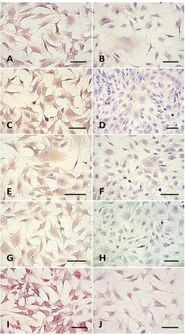
Figure 1 shows the results of the indirect immunopero-xidase staining using monoclonal antibodies against ICAM-1 (Fig.ICAM-1A), VCAM (Fig.ICAM-1C), PECAM-ICAM-1 (Fig.ICAM-1E), TSP (Fig.ICAM-1G), and polyclonal antibody against E-selectin (Fig.1I). All the stimulated BUVECs were positively immunostained, and the staining was evenly distributed on the cell surface. More intense immunostaining was found in the stimulated cells, compared with control cells which did not stained po-sitive for VCAM and TSP (Fig.1D,F). As observed in Figure 1I, the staining of E-selectin was more intense than that the other molecules.
DISCUSSION
The plasma used for stimulation was collected from ani-mals in the acute stage of babesiosis and possibly contai-ned high levels of proinflammatory cytokines. One of the
roll of cytokines is stimulate expression of several adhesion molecules on the surface of endothelial cells. Previous rese-arch showed an increase in cytokines TNF-α, IFN-γ and IL-12 in the plasma of animals inoculated with the BboUFV-1 7th passage sample, compared with control animals (De Freitas, 2001). These data agree with the works of Shoda et al. (2000) and East et al. (1997), who measured cytoki-nes expressed by macrophages in animals infected with B. bovis.
Likewise, the proinflammatory cytokines such as TNF-α and IFN-γ appear to be responsible for several biological activities on endothelial cells, as the stimulation of FCγ re
-Fig.1. Expression of adhesion molecules in bovine umbilical endo-thelial cells (BUVECS) after stimulation with a pool of plasma from infected animals with Babesia bovis strain BboUFV1 7th
ceptors (Pan et al. 1998). By other hand protein exoanti-gens can be found in the plasma of animals in the acute sta-ge of babesioses. These exoantista-gens are released either by the parasite or by rupture of red blood cells (Ristic 1988). B. bovis exoantigens are a heterogeneous group of proteins that could help to stimulate the endothelial cells.
Intense expression of ICAM-1, VCAM, PECAM-1, E-se-lectin and TSP was observed in Fig.1A,C,E,G,I when com-pared with the control treatment. Furthermore the in vitro studies performed by Shoda et al. (2000) showed that ma-crophages derived from monocytes activated by B. bovis ex-pressing high levels of proinflammatory cytokines 1β IL-12 and TNF-α. These facts suggest that the cytokines must also be present in infected animals of experiment, thereby allowing its circulation in the plasma.
The adhesion molecules ICAM-1 and VCAM were inten-sely expressed in BUVECs when the cells were stimulated with plasma, concurring with the results obtained by Bevi-lacqua (1993). They indicated that the expression of these molecules may be regulated by many types of cytokines, including TNF-α and other immunomodulators.
PECAM-1 was intensely expressed in the endothelial cells stimulated, whereas the control endothelial cells had little PECAM-1 expression. These observations agree with the results of Müller et al. (2002), which showed the oc-currence of homogeneous PECAM-1 expression in stimu-lated cultures of human umbilical vein endothelial cells (HUVEC) and human pulmonary microvascular endothelial cells (HPMEC) in pulmonary vascular disease. Apparently, the E-selectin expression in BUVECs was much higher than the expression of the other adhesion molecules studied. This may occur because E-selectin is the first adhesion mo -lecule to be induced after stimulation with TNF for 1 to 2 hours. The non-stimulated endothelial cells also expressed E-selectin, but at a lower intensity. Research carried out by Bischoff & Brasel (1995), proved that the E-selectin mRNA might be up-regulated by TNF-α in confluent monolayers of bovine endothelial cells.
The intense thrombospondin expression on the surfa-ce of the stimulated surfa-cells is divergensurfa-ce with the findings of Loganadane et al. (1997), who studied the expression of this molecule stimulated with the components of the extra-cellular matrix in HUBEC cultures. However, it is important to consider that this work did not deal with the correlation between concentration of the molecule in the sub-endothe-lial matrix and the culture density. These aspects affected the secretion of thrombospondin by endothelial cells in vi-tro.
The duration of the stimulus by the plasma used in our experiments was enough for the expression of adhesion molecules, considering Raab et al. (2002) findings. They demonstrated that mixture of cytokines, such as TNF-α, IL-1 or IFN-γ added to the incubation medium, can syner -gistically modulate the expression of adhesion molecules, such as VCAM-1, ICAM-1, PECAM-1 CD-34, E and P-selectin in HUVECs.
Despite that the role of adhesion molecules expressed during inflammation is well known, little is known about its function at vascular endothelium level on framework
of B. bovis infection. For Plasmodium falciparum, it is well known that molecules such as ICAM-1, PECAM-1, E-selectin and VCAM-1 act as receptors of ligands on the parasitized red blood cells (Cooke et al. 1994, Chen et al. 2000, Senezuk et al. 2001). These findings suggest that some of these mo -lecules could act as receptors for the adhesion of red blood cells to the vascular endothelium in animals infected by B. bovis. From all of that raises a hypothesis: if the both api-complexa are similar, will be the same molecules involve in the pathology of babesiosis as in malaria?
Based on Yipp et al. (2000), the CD36 is considered to be the main receptor for cytoadhesion in malaria. Never-theless, Hutchings et al. (2007) suggested that CD36 does not play a role in bovine babesiosis.
The in vivo expression of adhesion molecules is not always the same as in vitro. So, the limitations of in vitro models must be considered, with greater relevance the ab-sence of essential tissue micro-environment physiological factors.
The use of human monoclonal antibodies is based on the research of Van Kampen and Mallard (2001), who de-monstrated that the monoclonal human VCAM-1 anti-body is cross reactive to VCAM-1 expressed in bovine aortic endothelial cells (BAECs), which has expression kinetics si-milar to that of humans VCAM-1. The results of the present work clearly demonstrate that the human monoclonal an-tibodies recognize bovine molecules, such as ICAM-1, PE-CAM-1 and thrombospondin.
Unlike in malaria caused by P. falciparum, the identity of the receptors and/or adhesion molecules that mediate the adhesion in babesiosis caused by B. bovis is still not un-derstood.
CONCLUSION
The plasma from animals in acute phase of babesiosis cau-sed by Babesia bovis stimulates in vitro endothelial cells to produce adhesion molecules, which may be cause of red cells cytoadherence; these molecules possibly have an im-portant role in the pathophysiology of infection. Perhaps these molecules, as in malaria caused by Plasmodium falci-parum, have the same role in babesiosis.
Acknowledgements.- The authors thank FAPEMIG (Minas Gerais State
Research Foundation, Brazil) for providing financial support for the pro -ject. We thank Marcio A. Dias for technical assistance in the laboratory and Aloizio Carlos da Silva for assistance with animals experiment
REFERENCES
Berendt A.R., Simmonds D.L., Tansey J., Newbold C.I. & Marsh K. 1989. In-tercellular adhesion molecule-1 is an endothelial cell adhesion receptor for Plasmodium falciparum. Nature 341: 57-59.
Bevilacqua M. 1993. Endothelial-leukocyte adhesion molecules. Annu. Rev. Immunol. 11:767-804.
Bischoff J. & Brasel C. 1995. Regulation of P-selectin by tumor necrosis factor-a. Biomed. Biophys. Res. Commun. 210:174-180.
Caetano B.C. 2001. Estudos de citoaderência “in vitro” de eritrócitos de bovinos inoculados com Babesia bovis (Starcovici, 1893) em células en-doteliais de aorta bovina. Dissertação de Mestrado em Medicina Veteri-nária, Universidade Federal de Viçosa, Viçosa, MG. 70p.
2000. The semiconserved head structure of Plasmodium falciparum
erythrocyte membrane protein 1 mediates binding to multiple indepen-dent host receptors. J. Exp. Med. 192:1-10.
Clark I.A. & Jacobson L.S. 1998. Do babesiosis and malaria share a common disease process? Ann. Trop. Med. Parasitol. 92:483-488.
Cooke B.M., Berendt A.R., Craig A.G., Macgregor J., Newbold C.I. & Nash G.B. 1994. Rolling and stationary cytoadhesion of red blood cells parasitized by Plasmodium falciparum: separate roles for ICAM-1, CD36 and throm-bospondin. Brit. J. Haematol. 87:162-167.
Cooke B.M., Mohandas N., Cowman A.F. & Coppel R.L. 2005. Cellular adhe-sive phenomena in apicomplexan parasites of red blood cells. Vet. Para-sitol. 132:273-295.
De Freitas C.M. 2001. Avaliação da resposta imune de bovinos inoculados com amostras atenuada e virulenta de Babesia bovis (Starcovici, 1893). Dissertação de Mestrado em Medicina Veterinária, Universidade Fede-ral de Viçosa, Viçosa, MG. 74p.
East I.J., Zakrzewski H., Gale K.R., Leatch G., Dimmock C.M., Thomas M.B. & Waltisbuhl D.J. 1997. Vaccination against Babesia bovis: T cell from pro-tected and unpropro-tected animals show different cytokine profiles. Inter. J. Parasitol. 27:1537-1545.
Gohil S., Kats L.M., Sturm A. & Cooke B.M. 2010. Recent insights into altera-tion of red blood cells by Babesia bovis: moovin forward. Trends Para-sitol. 26:591-599.
Ho M., Schollaardt T., Niu X., Looareesuwan S., Patel K.D. & Kubes P. 1998. Characterization of Plasmodium falciparum infected erythrocyte and P-selectin interaction under flow conditions. Blood 91:4803-4809. Hutchings C.L., Li A., Fernandez K.M., Fletcher T., Jackson L.A., Molloy J.B.,
Jorgensen W.K., Lim Ch.T. & Cooke B.M. 2007. New insights into the al-tered adhesive and mechanical properties of red blood cells parasitized by Babesia bovis. Mol. Microbiol. 65:1092-1105.
Jaffe E.A., Nachman R.L., Becker C.G. & Minick C.R. 1973. Culture of hu-man endothelial cells derived from umbilical veins. Identification by morphological and immunologic criteria. J. Clin. Invest. 52:2745-2756. Loganadane L.D., Berge N., Legrand C. & Fauvel-Lafave F. 1997. Endotelial
cell proliferation regulated by citokines modulates trombospondim-1 secretion into the subendothelio. Cytokine 9:740-746.
Müller A., Hermanns M., Skrynski C., Nesslinger M., Müller K. & Khkpatrick J. 2002. Expression of the endothelial markers PECAM-1, vWf, and CD34 in vivo and in vitro. Exp. Mol. Pathol. 72:221-229.
Nevils MA., Figueroa J.V., Turk J.R., Canto G.J., Le V., Ellersieck M.R. & Car-son C.A. 2000. Cloned lines of Babesia bovis differ in their ability to in-duce cerebral babesiosis in cattle. Parasitol. Res. 86:437-443.
O’Connor R.M., Long J.A. & Allred D.R. 1999. Cytoadherence of Babesia bo-vis-infected erythrocytes to bovine brain capillary endothelial cells pro-vides an in vitro model for sequestration. Infect. Immun. 67:3921-3928.
O’ Connor R.M. & Allred D.R. 2000. Selection of Babesia bovis infected erythrocytes for adhesion to endothelial cells coselects for altered vari-ant erythrocyte surface vari-antigen isoforms. J. Immunol. 164:2037-2045. Pan L.F., Kreisle R.A. & Shi Y.D. 1998. Detection of Fcγ receptors on hu
-man endothelial cells stimulated with cytokines tumour necrosis fac-tor-alpha (TNF-α) and interferon-gamma (IFN-γ). Clin. Exp. Immunol. 112:533-538.
Parrodi F., Wright I.G., Bourne A.S. & Dobson C. 1989. In vitro adherence of bovine erythrocytes infected with Babesia bovis to trombospondina an laminin. Int. J. Parasitol. 19:567-569.
Patarroyo J.H., Vargas M.I. & Bicudo P. 1982. Description of lesions in cattle in a natural outbreak of Babesia bovis infection in Brazil. Vet. Parasitolol. 11:301-308.
Prudhomme J., Sherman I., Land K., Moses A., Stengleins S. & Nelsons J. 1996. Studies of Plasmodium falciparum cytoadherence using inmortal-ized human brain capillary endothelial cell. Int. J. Parasitol. 26:647-655. Raab M., Daxecker H., Markovic S., Karimi A., Griesmacher A. & Mueller M. 2002. Variation of adhesion molecule expressing on human umbilical vein endothelial cells upon multiple cytokine application. Clin. Chem. Acta. 321:11-16.
Ristic M. 1988. Babesiosis of Domestic Animals and Man. CRC Press, Flo-rida. 255p.
Senezuk A., Reeder J., Kosmala M. & Ho M. 2001. Plasmodium falciparum
erytrocyte membrane protein 1 functions as a ligand for P-selectin. Blood 98:3132-3135.
Shoda L.K.M., Palmer G.H., Florin-Christensen J., Florin-Christensen M., Godson D.L. & Wendy C.B. 2000. Babesia bovis-Stimulated macrophages express interleukin 1β, interleukin-12, tumor necrosis factor alpha and nitric oxide and inhibit parasite replication in vitro. Infect. Immun. 68:5139-5145.
Treugiter C.J., Heddini A., Fernandez V., Muller W.A. & Wahlgren M. 1997. PECAM-1/CD31, an endothelial receptor for binding Plasmodium falci-parum-infected erythrocytes. Nat. Med. 3:1405-1408.
Uilinber G. 2006. Babesia: a historical overview. Vet. Parasitol. 138:3 -10. Van Kampen C. & Mallard B. 2001. Regulation of bovine intercellular
adhe-sion molecule 1 (ICAM-1) and vascular cell adheadhe-sion molecule 1 (VCAM-1) on cultured aortic endothelial cells. Vet. Immunol. Immunopathol. 79:129-138.
Wright I.G., Goodger B.V., Buffington G.D., Clark I.A., Parrodi F. & Waltisbuhl D.J. 1989. Immunopathophysiology of babesial infections. Trans. R. Soc. Trop. Med. Hyg.83:11-13.