Mesenteric hypoperfusion and inflammation induced
by brain death are not affected by inhibition of the
autonomic storm in rats
Rafael Simas, Sueli G. Ferreira, Laura Menegat, Fernando L. Zanoni, Cristiano J. Correia, Isaac A. Silva, Paulina Sannomiya, Luiz F.P. Moreira*
Instituto do Corac¸a˜o (InCor) do Hospital das Clı´nicas da Faculdade de Medicina da Universidade de Sa˜o Paulo, Laboratory of Cardiovascular Surgery and Circulation Pathophysiology (LIM-11), Sa˜o Paulo/SP, Brazil.
OBJECTIVES: Brain death is typically followed by autonomic changes that lead to hemodynamic instability, which is likely associated with microcirculatory dysfunction and inflammation. We evaluated the role of the microcirculation in the hemodynamic and inflammatory events that occur after brain death and the effects of autonomic storm inhibition via thoracic epidural blockade on mesenteric microcirculatory changes and inflammatory responses.
METHODS: Male Wistar rats were anesthetized and mechanically ventilated. Brain death was induced via intracranial balloon inflation. Bupivacaine (brain death-thoracic epidural blockade group) or saline (brain death group) infusion via an epidural catheter was initiated immediately before brain death induction. Sham-operated animals were used as controls (SH group). The mesenteric microcirculation was analyzed via intravital microscopy, and the expression of adhesion molecules was evaluated via immunohistochemistry 180 min after brain death induction.
RESULTS: A significant difference in mean arterial pressure behavior was observed between the brain death-thoracic epidural blockade group and the other groups, indicating that the former group experienced autonomic storm inhibition. However, the proportion of perfused small vessels in the brain death-thoracic epidural blockade group was similar to or lower than that in the brain death and SH groups, respectively. The expression of intercellular adhesion molecule 1 was similar between the brain death-thoracic epidural blockade and brain death groups but was significantly lower in the SH group than in the other two groups. The number of migrating leukocytes in the perivascular tissue followed the same trend for all groups.
CONCLUSIONS:Although thoracic epidural blockade effectively inhibited the autonomic storm, it did not affect mesenteric hypoperfusion or inflammation induced by brain death.
KEYWORDS: Brain Death; Autonomic Storm; Microcirculation; Intravital Microscopy.
Simas R, Ferreira SG, Menegat L, Zanoni FL, Correia CJ, Silva IA, et al. Mesenteric hypoperfusion and inflammation induced by brain death are not affected by inhibition of the autonomic storm in rats. Clinics. 2015;70(6):446-452
Received for publication onFebruary 10, 2015;First review completed onMarch 31, 2015;Accepted for publication onMarch 31, 2015 E-mail: [email protected]
*Corresponding author
’ INTRODUCTION
Organ transplantation is a unique treatment alternative for many critically ill patients, and one major source of organs is brain-dead patients. Brain death (BD) induces a series of hemodynamic, hormonal, and inflammatory alterations that can deteriorate the viability of organs (1–3).
Parasympathetic activation associated with progressive bradycardia and hypotension is generally observed after the initial trauma that leads to BD. If the injury involves the most distal midbrain, the vagal cardiac motor center is destroyed, and parasympathetic activity is stopped. As a consequence, sympathetic activity is not regulated by the parasympathetic system, resulting in a classical autonomic storm character-ized by tachycardia and hypertension. Finally, a profound reduction in sympathetic outflow leads to hemodynamic instability exacerbated by hypovolemia, dysregulation of the peripheral vasculature, and vasodilatation (4).
In animal models of BD, a balloon catheter placed in the intracranial cavity is inflated to increase the local pressure, leading to an immediate increase in the norepinephrine and epinephrine concentrations, which are associated with tachycardia and hypertension followed by vasoplegia and
DOI:10.6061/clinics/2015(06)11
Copyright&2015CLINICS–This is an Open Access article distributed under the terms of the Creative Commons Attribution Non-Commercial License (http:// creativecommons.org/licenses/by-nc/3.0/) which permits unrestricted non-commercial use, distribution, and reproduction in any medium, provided the original work is properly cited.
procedure did not abolish the hypertensive crisis, indicating that the performance of this surgical procedure is not sufficient to block the autonomic storm. In addition, Silva et al. (9) demonstrated that thoracic epidural anesthesia effectively blocks the autonomic storm but does not alter the expression of inflammatory mediators in the myocardial tissue.
The hemodynamic instability observed in BD is generally associated with ischemia and tissue hypoperfusion, which compromise the viability of the organs that are suitable for transplantation (2). Previous studies have shown that BD leads to substantial impairment of microcirculation in the liver and the pancreas (15,16). Furthermore, our group observed similar alterations triggered by BD in the rat mesenteric microcirculation (8). In particular, mesenteric hypoperfusion immediately occurred after BD induction, and this phenomenon persisted for up to three hours thereafter. This hypoperfusion was associated with elevated expression of endothelial cell adhesion molecules and increased leukocyte migration to the perivascular tissues such as in mesentery, lung, and liver tissue (8,17).
Despite the clear importance of the microcirculation for optimizing the therapy for circulatory shock of various etiologies, the influence of autonomic storm blockade on microcirculatory changes after BD has not been investigated. Therefore, the present study was conducted to assess the role of the microcirculation in hemodynamic and inflammatory events that occur after BD induction and to investigate the effects of sympathetic system inhibition via thoracic epidural anesthesia. Microcirculatory changes, including perfusion and leukocyte-endothelial interactions, were analyzed via intravital microscopy of the mesentery. Local and systemic inflammatory markers were also evaluated.
’ MATERIAL AND METHODS
Male Wistar rats (300±50 g) were used according to
experimental protocols approved by the Animal Subject Committee of the Instituto do Corac¸ão (InCor) do Hospital
das Clínicas da Faculdade de Medicina da Universidade de São Paulo. The animals were randomly allocated to three groups: sham-operated (SH group, n=12), brain-dead non-treated (BD group, n=12), and brain-dead under thoracic epidural blockade (TEB) (BD-TEB group, n=12). The animals were evaluated 180 min after the conclusion of the surgical procedures.
Brain death model and thoracic epidural anesthesia
All animals were anesthetized in a chamber using iso-flurane (5%), intubated, and mechanically ventilated (Harvard
A 10-French polyethylene catheter was placed in the epidural space through an incision over the T11–L1 vertebrae
and was advanced to the T5 level. The BD-TEB group received a bolus infusion of 20mL of 0.5% bupivacaine 5 min before BD induction, followed by continuous infusion (15mL/h) for 3 h. The animals in the BD group were administered equivalent volumes of saline solution. No drugs were administered to the rats in the SH group.
Blood gas, electrolyte, and lactate measurements and white blood cell counts
The blood gas, electrolyte, and lactate levels were measured in blood samples obtained from the carotid artery at baseline (0 min) and 180 min after the surgical procedures using a gas analyzer (Radiometer ABL 555, Radiometer Medical, Copenhagen, Denmark). White blood cell (WBC) counts were determined in blood samples obtained from the cut tip of the tail vein at the same time points. The total WBC counts were determined using a Neubauer chamber. Differential cell counting was performed on stained films via oil immersion microscopy. A total of 100 cells were counted and classified according to standard morphological criteria.
Intravital microscopy of the mesenteric microcirculation
Intravital microscopy of the mesenteric microcirculation was performed as previously described (8). Briefly, an abdominal midline incision was performed 180 min after BD induction, and the distal ileum and its accompanying mesentery were exposed for in vivo microscopic examination of the microcirculation. The animals were maintained on a specially designed stage warmed with circulating 37˚C water. The mesentery was continuously perfused with warm (37˚C) Krebs-Henseleit solution saturated with a mixture of gases (95% N2and 5% CO2). A color camera (AxioCam HSc,
Carl Zeiss, München-Hallbergmos, Germany) was connected to a triocular microscope (Axioplan 2, Carl Zeiss), and the microcirculatory variables were analyzed using Axiovision 4.1 imaging software (Carl Zeiss). The vascular density in a 1-mm2area on the computer screen was assessed using a 10x light microscope objective. The flow, defined as continuous or intermittent/absent, was analyzed in vessels with a diametero30mm. Five fields of view were selected for each
leukocytes was calculated as the mean number of WBCs passing a designated line perpendicular to the venular axis per 5 min. The adherent cells (i.e., leukocytes that remained stationary on the venular endothelium for more than 30 s) were counted in a 100mm segment of the vessel. The number of leukocytes that accumulated in the connective tissue adjacent to the selected post-capillary venule was deter-mined in a standard area of 5,000mm2. Three fields of view for each microvessel were examined.
Evaluation of intercellular adhesion molecule 1 expression via immunohistochemistry
The animals were exsanguinated from the abdominal aorta, and the mesentery was removed, immersed in hexane, and frozen in liquid nitrogen. Serial slices of the mesentery (8mm) were placed on glass slides coated with organosilane (Sigma Chemical Co., St. Louis, MO, USA). The samples were fixed in acetone, and SuperBlock buffer (Pierce Biotechnology, Rockford, IL, USA) was used to block nonspecific sites. For the immunodetection of intercellular adhesion molecule 1 (ICAM-1) on mesenteric microvessels, tissue sections were incubated overnight at 4˚C in a biotin-conjugated anti-rat ICAM-1 (CD54) monoclonal antibody (Seikagaku, Tokyo, Japan) diluted 1:50 in phosphate-buffered saline (PBS) containing 0.3% Tween-20. After washing the slides with PBS, the sections were incubated for 1 h at room temperature in streptavidin-fluorescein (Amersham Pharmacia Biotech, London, UK) diluted 1:200 in PBS. After washing with PBS, the samples were treated with Vectashield mounting medium containing propidium iodide (Vector Laboratories, Burlingame, CA, USA) to preserve their fluorescence. The system used for image acquisition included a DS-Ri1 digital camera (Nikon, Tokyo, Japan) connected to a fluorescence microscope (Nikon)
controlled with NIS-Elements-BR software (Nikon). The results are presented as the mean fluorescence intensity.
Serum concentrations of cytokines and corticosterone
Blood samples obtained from the abdominal aorta were centrifuged (1,500 g, 25˚C), and the serum concentrations of tumor necrosis factor-a(TNF-a), interleukin (IL)-1b, IL-6, IL-10, and corticosterone were determined using enzyme-linked immunosorbent assay (ELISA) kits as recommended by the manufacturers (cytokines: R&D Systems, Minneapolis, MN, USA; corticosterone: Cayman Chemical, Ann Arbor, MI, USA).
Statistical analysis
All data are presented as the means±standard error of
the mean (SEM). The overall group differences were compared via two-way analysis of variance (ANOVA) using group and time as the factors followed by a post hoc Bonferroni test or via a one-way ANOVA followed by a post hoc Bonferroni test. Adjusted p values for multiple comparisons were calculated, and ap-value of o0.05 was
considered to indicate statistical significance. All statistical analyses were performed using Graph-Pad Prism 6.1.
’ RESULTS
Hemodynamic parameters
The rats in the BD group exhibited a sudden increase in mean arterial pressure over the first minute following catheter inflation. In contrast, the rats treated with TEB did not exhibit this hypertensive episode after BD induction. The mean arterial pressure then decreased below the baseline level in both groups. After 75 min, the mean arterial pressure was restored to normal levels, and no difference in mean arterial pressure was observed between the groups. In the
SH rats, the mean arterial pressure did not change over time (Figure 1). There were no significant differences in the arterial blood gas, electrolyte, or lactate levels between the groups (data not shown).
Mesenteric microcirculation parameters
The average proportion of perfused small vessels in the BD group was 39% compared to 74% in the SH rats. The treatment of BD rats with TEB did not alter mesenteric
hypoperfusion (43%), as illustrated in Figure 2 and the supplementary videos.
Table 1 shows the leukocyte-endothelium interaction data obtained from perfused small vessels. Similar results were observed in both the BD and BD-TEB rats, and the number of rolling leukocytes in these two groups was reduced compared to that in the SH group. A reduction in the number of adherent leukocytes in the BD and BD-TEB groups compared to the SH group was also observed. In contrast, the number of migrating leukocytes in both groups of BD rats was higher than that in the SH group. These results were not accompanied by significant changes in the number of WBCs.
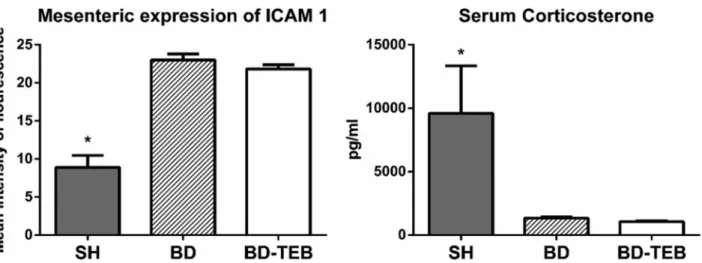
Expression of mesenteric intercellular adhesion molecule 1 and the serum corticosterone levels
The expression of ICAM-1 was investigated in the mesentery of rats that were not examined via intravital microscopy (Figure 3). The levels of ICAM-1 expression were similar between the BD and BD-TEB groups and exceeded those in the SH group. The serum levels of corticosterone were reduced in the BD and BD-TEB groups compared to the SH group three hours after BD induction (Figure 3).
Serum cytokine levels
Similar increases in the serum concentrations of TNF-a, IL-1b, IL-6, and IL-10 were observed in the BD, BD-TEB, and SH
groups 180 min after the surgical procedures. The reference values obtained in Naive rats were below the limit of detection of the assay. These results are summarized in Table 2.
’ DISCUSSION
In an attempt to attenuate the hemodynamic alterations triggered by BD, thoracic sympathetic blockade was induced in this study via epidural anesthesia with bupivacaine. We found that TEB effectively abolished the hypertensive event observed after BD induction but did not affect the following hypotensive episode. Furthermore, mesenteric hypoperfu-sion and inflammation, characterized by increased leukocyte migration and ICAM-1 expression, remained unchanged in the BD animals despite TEB. Moreover, the serum levels of cytokines and corticosterone did not change after the treatment of BD rats with TEB.
Hemodynamic instability is a major challenge in the treatment of BD donors because it leads to the impair-ment of organ perfusion and compromises the viability of these organs for transplantation. Although hypertension may occur during the phase of cerebral herniation, hypotension affects almost all patients after BD (18). Indeed, in animal models, the hypotensive event that accompanies BD is initiated by both rapid and slow augmentation of intracranial pressure (10,19–21).
Never-theless, the important question of how the modification of sympathetic activity after BD impacts microcirculation
Figure 3 -Expression of intercellular adhesion molecule (ICAM)-1 on mesenteric endothelial cells (p ANOVAo0.001) and the serum levels of corticosterone (p ANOVA=0.01) in sham-operated rats (SH), brain-dead non-treated rats (BD), and brain-dead rats under thoracic epidural blockade (BD-TEB) 180 min after the surgical procedures. The data are presented as the means±SEM. *po0.05 vs. the other groups.
Table 1 -The results of the evaluation of the rat mesenteric microcirculation via intravital microscopy.
SH BD BD-TEB pANOVA
Rollers 165 (12)* 44 (4) 31 (5) o0.001
Adherent 6.9 (0.6)* 4.5 (0.5) 3.2 (0.3) 0.002
Migrated 1.3 (0.2)** 2.5 (0.2) 1.8 (0.2) 0.01
WBC 11,771 (1,122) 10,450 (2,109) 8,667 (1,196) 0.3
Rollers: leukocytes/5 min; Adhered: leukocytes/100mm venule length; Migrated: leukocytes/5,000mm2; WBC: White blood cells/mm3. SH:
sham-operated rats; BD: brain-dead non-treated rats; BD-TEB: brain-dead rats under thoracic epidural blockade. These measurements were performed 180 min after the surgical procedures. The data are presented as the means±SEM. *po0.01 vs. the other groups. **po0.05 vs. the other groups.
Table 2 -The results of the evaluation of the serum cytokine levels via enzyme-linked immunosorbent assays.
TNF-a (pg/mL)
IL-1b (pg/mL)
IL-6 (pg/mL)
IL-10 (pg/mL)
SH 0.066±0.005 0.102±0.017 0.131±0.022 0.112±0.004 BD 0.054±0.004 0.074±0.004 0.130±0.043 0.089±0.007 BD-TEB 0.067±0.007 0.090±0.017 0.151±0.043 0.096±0.010
pANOVA 0.4 0.9 0.2 0.2
persistent hypoperfusion of the mesenteric microcirculation. Such hypoperfusion was previously demonstrated by our group in the mesentery (8) and by others in the pancreas and the liver (15,16). Additional real-time monitoring of the mesenteric microcirculation during the first 30 min after BD induction confirmed that mesenteric hypoperfusion occurs immediately after BD and concomitantly with the onset of the sustained hypotensive episode (unpublished data). This observation suggests that microcirculatory abnormalities can occur independently of an increase in arterial pressure, suggesting the involvement of other mechanisms triggered by BD in these microcirculatory abnormalities.
It has been demonstrated that BD leads to a decrease in organ perfusion and a progressive activation of endothelial cells and leukocytes (8,22–25). It is known that BD triggers a
series of inflammatory effects, including increased expression of adhesion molecules, leukocyte transmigration to the perivascular tissue in various organs, and increased serum levels of cytokines (2,3,26–28). The present data indicate that the inflammation in
the mesenteric microcirculation is not affected by TEB. The animals in both BD groups exhibited similar numbers of rolling, adherent, and migrating leukocytes, as well as comparably increased levels of ICAM-1 expression in endothelial cells; these characteristics were associated with a pronounced reduction in the serum corticosterone levels. It is known that BD impairs the endocrine system and decreases the release of hormones such as triiodothyronine, thyroxin, cortisol, and anti-diuretic hormone, suggesting an interruption along the hypothalamic-pituitary-adrenal axis (29,30). In this regard, it has been shown that in healthy subjects, endogenous glucocorticoids that are secreted in large amounts at the early stages of an inflammatory reaction regulate the functions of almost all cellular components of the microcirculation (31). Indeed, the SH rats exhibited increased levels of corticosterone in association with reduced ICAM-1 expression compared to the BD rats. Furthermore, the serum levels of cytokines were similar between the groups, suggesting that the release of cytokines triggered by BD-associated trauma (8,28) was not affected by TEB.
It has been well documented that abnormalities in micro-circulatory perfusion, including decreased vascular density, especially in the small vessels, and a large number of non-perfused and intermittently non-perfused small vessels, which occur in septic patients, are not affected by the global hemodynamic state or the use of adrenergic agents (32,33). The experimental findings are consistent with the clinical evidence that sepsis induces microvascular changes that may play an important role in the development of organ dysfunction (32,34,35). These results justify the attempts to revert these alterations.
that therapies aimed at maintaining the microcirculation may effectively compensate for the perfusion abnormalities after BD, thereby potentially improving the quality of donor organs for transplantation.
’ ACKNOWLEDGMENTS
This research was supported by Fundac¸ão de Amparo à Pesquisa do Estado
de São Paulo (FAPESP), Grant number 09/10759-9.
’ AUTHOR CONTRIBUTIONS
Simas R participated in the research design, performance of the research, the data analysis, and the writing of the paper. Ferreira SG, Menegat L, Zanoni FL, and Correia CJ participated in the performance of the research. Silva IA participated in the research design and the writing of the paper. Sannomiya P and Moreira LF participated in the research design, the data analysis, and the writing of the paper.
’ REFERENCES
1. Bittner HB, Kendall SW, Chen EP, Van Trigt P. Endocrine changes and metabolic responses in a validated canine brain death model. J Crit Care
1995;10(2):56–63, http://dx.doi.org/10.1016/0883-9441(95)90017-9.
2. Barklin A. Systemic inflammation in the brain-dead organ donor. Acta
Anaesthesiol Scand. 2009;53(4):425–35, http://dx.doi.org/10.1111/aas.
2009.53.issue-4.
3. Domínguez-Roldán JM, García-Alfaro C, Jimenéz-González PI, Hernán-dez-Hazañas F, Gascón Castillo ML, Egea Guerrero JJ. Brain death:
repercussion on the organs and tissues. Med Intensiva. 2009;33(9):434–41,
http://dx.doi.org/10.1016/j.medin.2009.03.008.
4. Audibert G, Charpentier C, Seguin-Devaux C, Charretier PA, Grégoire H, Devaux Y, et al. Improvement of donor myocardial function after treat-ment of autonomic storm during brain death. Transplantation. 2006;82
(8):1031–6, http://dx.doi.org/10.1097/01.tp.0000235825.97538.d5.
5. Shivalkar B, Van Loon J, Wieland W, Tjandra-Maga TB, Borgers M, Plets C, et al. Variable effects of explosive or gradual increase of intracranial pressure on myocardial structure and function. Circulation. 1993;87
(1):230–9, http://dx.doi.org/10.1161/01.CIR.87.1.230.
6. Wilhelm MJ, Pratschke J, Beato F, Taal M, Laskowski IA, Paz DM, et al. Activation of proinflammatory mediators in heart transplants from brain-dead donors: evidence from a model of chronic rat cardiac allograft
rejection. Transplant Proc. 2002;34(6):2359–60, http://dx.doi.org/
10.1016/S0041-1345(02)03283-9.
7. Floerchinger B, Oberhuber R, Tullius SG. Effects of brain death on organ
quality and transplant outcome. Transplant Rev (Orlando). 2012;26(2):54–9.
8. Simas R, Sannomiya P, Cruz JW, Correia Cde J, Zanoni FL, Kase M, et al. Paradoxical effects of brain death and associated trauma on rat mesenteric
microcirculation: an intravital microscopic study. Clinics. 2012;67(1):69–
75, http://dx.doi.org/10.6061/clinics.
9. Silva IA, Correia CJ, Simas R, Cruz JW, Ferreira SG, Zanoni FL, et al. Inhibition of autonomic storm by epidural anesthesia does not influence cardiac inflammatory response after brain death in rats. Transplant Proc.
2012;44(7):2213–8, http://dx.doi.org/10.1016/j.transproceed.2012.07.108.
10. Wauters S, Somers J, De Vleeschauwer S, Verbeken E, Verleden GM, van Loon J, et al. Evaluating lung injury at increasing time intervals in a
murine brain death model. J Surg Res. 2013;183(1):419–26, http://dx.doi.
11. Siaghy EM, Halejcio-Delophont P, Devaux Y, Richoux JP, Villemot JP, Burlet C, et al. Protective effects of labetalol on myocardial contractile
function in brain-dead pigs. Transplant Proc. 1998;30(6):2842–3, http://
dx.doi.org/10.1016/S0041-1345(98)00833-1.
12. Ojeda-Rivero R, Hernández-Fernández A, Dominguez-Roldán JM, Calderón E, Ruiz M, Lage E, et al. Experimental treatment with beta blockers of hemodynamic and myocardial changes in organ donors.
Transplant Proc. 2002;34(1):185–6, http://dx.doi.org/10.1016/S0041-1345
(01)02720-8.
13. Yeh T Jr, Wechsler AS, Graham L, Loesser KE, Sica DA, Wolfe L, et al. Central sympathetic blockade ameliorates brain death-induced cardiotoxicity and associated changes in myocardial gene expression. J Thorac Cardiovasc Surg.
2002;124(6):1087–98, http://dx.doi.org/10.1067/mtc.2002.124887.
14. Novitzky D, Wicomb WN, Cooper DK, Rose AG, Reichart B. Prevention of myocardial injury during brain death by total cardiac sympathectomy
in the Chacma baboon. Ann Thorac Surg. 1986;41(5):520–4, http://dx.doi.
org/10.1016/S0003-4975(10)63032-9.
15. Obermaier R, von Dobschuetz E, Keck T, Hopp HH, Drognitz O, Schareck W, et al. Brain death impairs pancreatic microcirculation. Am J Transplant.
2004;4(2):210–5, http://dx.doi.org/10.1046/j.1600-6143.2003.00317.x.
16. Yamagami K, Hutter J, Yamamoto Y, Schauer RJ, Enders G, Leiderer R, et al. Synergistic effects of brain death and liver steatosis on the hepatic
microcirculation. Transplantation. 2005;80(4):500–5, http://dx.doi.org/
10.1097/01.tp.0000167723.46580.78.
17. Simas R, Kogiso DH, Correia CJ, Silva LF, Silva IA, Cruz JW, et al. Influence of brain death and associated trauma on solid organ histological
characteristics. Acta Cir Bras. 2012;27(7):465–70, http://dx.doi.org/
10.1590/S0102-86502012000700006.
18. Dictus C, Vienenkoetter B, Esmaeilzadeh M, Unterberg, A., Ahmadi, R. Critical care management of potential organ donors: our current standard.
Clin Transplant. 2009;23 Suppl 21:2–9, http://dx.doi.org/10.1111/
ctr.2009.23.issue-s21.
19. Floerchinger B, Ge X, Lee YL, Jurisch A, Padera RF, Schmid C, et al. Graft-specific immune cells communicate inflammatory immune responses
after brain death. J Heart Lung Transplant. 2012;31(12):1293–300, http://
dx.doi.org/10.1016/j.healun.2012.09.005.
20. Hvas CL, Nielsen TK, Barklin A, Sørensen JC, Pedersen M, Andersen G, et al. Brain death induced by cerebral haemorrhage - a new porcine model
evaluated by CT angiography. Acta Anaesthesiol Scand. 2012;56(8):995–
1005, http://dx.doi.org/10.1111/aas.2012.56.issue-8.
21. Steen S, Sjöberg T, Liao Q, Bozovic G, Wohlfart B. Pharmacological nor-malization of circulation after acute brain death. Acta Anaesthesiol Scand.
2012;56(8):1006–12, http://dx.doi.org/10.1111/aas.2012.56.issue-8.
22. van Der Hoeven JA, Ter Horst GJ, Molema G, de Vos P, Girbes AR, Postema F, et al. Effects of brain death and hemodynamic status on function and immunologic activation of the potential donor liver in the
rat. Ann Surg. 2000;232(6):804–13,
http://dx.doi.org/10.1097/00000658-200012000-00009.
23. Itabashi K, Ito Y, Takahashi T, Ishii K, Sato K, Kakita A. Protective effects of urinary trypsin inhibitor (UTI) on hepatic microvasculature in
hypo-tensive brain-dead rats. Eur Surg Res. 2002;34(4):330–8, http://dx.doi.
org/10.1159/000063074.
24. van der Hoeven JA, Molema G, Ter Horst GJ, Freund RL, Wiersema J, van Schilfgaarde R, et al. Relationship between duration of brain death and hemodynamic (in)stability on progressive dysfunction and increased
immunologic activation of donor kidneys. Kidney Int. 2003;64(5):1874–82,
http://dx.doi.org/10.1046/j.1523-1755.2003.00272.x.
25. Zhou HC, Ding WG, Cui XG, Pan P, Zhang B, Li WZ. Carbon monoxide inhalation ameliorates conditions of lung grafts from rat brain death
donors. Chin Med J (Engl). 2008;121(15):1411–9.
26. Catania A, Lonati C, Sordi A, Gatti S. Detrimental consequences of brain
injury on peripheral cells. Brain Behav Immun 2009;23(7):877–84, http://
dx.doi.org/10.1016/j.bbi.2009.04.006.
27. Koudstaal LG,’t Hart NA, Ottens PJ, van den Berg A, Ploeg RJ, van Goor H,
et al. Brain death induces inflammation in the donor intestine.
Transplanta-tion. 2008;86(1):148–54, http://dx.doi.org/10.1097/TP.0b013e31817ba53a.
28. Schuurs TA, Morariu AM, Ottens PJ,’t Hart NA, Popma SH, Leuvenink
HG, et al. Time-dependent changes in donor brain death related
pro-cesses. Am J Transplant. 2006;6(12):2903–11, http://dx.doi.org/10.1111/
ajt.2006.6.issue-12.
29. Pratschke J, Wilhelm MJ, Kusaka M, Basker M, Cooper DK, Hancock WW, et al. Brain death and its influence on donor organ quality and outcome
after transplantation. Transplantation 1999;67(3):343–8, http://dx.doi.
org/10.1097/00007890-199902150-00001.
30. Wilhelm MJ, Pratschke J, Laskowski IA, Paz DM, Tilney NL. Brain death and its impact on the donor heart-lessons from animal models. J Heart
Lung Transplant. 2000;19(5):414–8,
http://dx.doi.org/10.1016/S1053-2498(00)00073-5.
31. Perretti M, Ahluwalia A. The microcirculation and inflammation: site of
action for glucocorticoids. Microcirculation. 2000;7(3):147–61, http://dx.
doi.org/10.1111/micc.2000.7.issue-3.
32. De Backer D, Creteur J, Preiser JC, Dubois MJ, Vincent JL. Microvascular blood flow is altered in patients with sepsis. Am J Respir Crit Care Med.
2002;166(1):98–104, http://dx.doi.org/10.1164/rccm.200109-016OC.
33. De Backer D, Donadello K, Taccone FS, Ospina-Tascon G, Salgado D, Vincent JL. Microcirculatory alterations: potential mechanisms and implications for therapy. Ann Intensive Care. 2011;1(1):27, http://dx.doi. org/10.1186/2110-5820-1-27.
34. Farquhar I, Martin CM, Lam C, Potter R, Ellis CG, Sibbald WJ. Decreased capillary density in?vivo in bowel mucosa of rats with normotensive
sepsis. J Surg Res. 1996;61(1):190–6, http://dx.doi.org/10.1006/
jsre.1996.0103.
35. Sakr Y, Dubois MJ, De Backer D, Creteur J, Vincent JL. Persistent micro-circulatory alterations are associated with organ failure and death in
patients with septic shock. Crit Care Med. 2004;32(9):1825–31, http://dx.