http://dx.doi.org/10.1590/1516-635x1703275-280
Metapneumovirus (aMPV)
Author(s)
Kohn LKI,II Foglio MAIII Rodrigues RAIII Sousa IM de OIII Martini MCII Padilla MAII Lima Neto DF deII Arns CWII
I Universidade de São Francisco, Rua Valdemar C. Silveira, 105, CEP 13045-510, Campinas, SP, Brazil
II Laboratory of Virology, Institute of Biology, CP 6109, University of Campinas – Unicamp, 13083-970, Campinas, SP, Brazil III Laboratory Research & Development of
Natural Products, Institute of Chemistry, CP 6154, University of Campinas – Unicamp, 13083-970, Campinas, SP, Brazil
IV Center for Chemical, Biological and Agricultural (CQBA) –University of Campinas – Unicamp, 13083-970, Campinas, SP, Brazil
Mail Address
Corresponding author e-mail address Arns, C.W.
Laboratory of Virology, Institute of Biology, CP 6109, University of Campinas – Unicamp, 13083-970, Rua Monteiro Lobato, Campinas, SP, Brazil Phone/Fax: +55 19 35216258 E-mail: clarns@gmail.com
Keywords
Antiviral, Avian metapneumovirus, Gaylussacia brasiliensis, Arrabidaea chica, Virola sebifera, Aspidosperma tomentosum.
Submitted: February/2014 Approved: December/2014
ABSTRACT
Avian metapneumovirus (aMPV) is a negative-sense single-stranded
RNA enveloped virus of the Metapneumovirus genus belonging to
the Paramyxoviridae family. This virus may cause significant economic losses to the poultry industry, despite vaccination, which is the main tool for controlling and preventing aMPV. The aim of this study was to evaluate the antiviral activity of extracts of four different native plants of the Brazilian Cerrado against aMPV. The antiviral activity against aMPV was determined by titration. This technique measures the ability of plant extract dilutions (25 to 2.5 μg mL-1) to inhibit the
cytopathic effect (CPE) of the virus, expressed as inhibition percentage (IP). The maximum nontoxic concentration (MNTC) of the extracts
used in antiviral assay was 25 μg mL-1 for Aspidosperma tomentosum
and Gaylussacia brasiliensis, and2.5 μg mL-1 for Arrabidaea chica and
Virola sebifera.Twelve different extracts derived from four plant species collected from the Brazilian Cerrado were screened for antiviral activity against aMPV. G. brasiliensis, A. chica, and V. sebifera extracts presented inhibition rates of 99% in the early viral replication stages, suggesting that these extracts act during the adsorption phase. On the other hand,
A. tomentosum inhibited 99% virus replication after the virus entered the cell. The biomonitored fractioning of extracts active against aMPV may be a tool to identify the active compounds of plant extracts and to determine their precise mode of action.
INTRODUCTION
Avian metapneumovirus (aMPV) is a negative-sense single-stranded
RNA enveloped virus of the Metapneumovirus genus belonging to
the Paramyxoviridae family. This family includes viruses responsible for causing respiratory diseases in turkeys and in humans. In chickens, the aMPV is involved in the etiology of a multi-factorial disease also known as the Swollen Head Syndrome (SHS). Another member of the
Paramyxoviridae family that is relevant to the poultry industry is the Newcastle disease virus. Avian metapneumovirus may cause significant economic losses to the poultry industry. Although vaccination is the main tool for the control and prevention of diseases caused by aMPV, pharmaceutical intervention is required under some circumstances. Furthermore, vaccinated subjects may acquire incomplete protection and/or there may be a reversion of the vaccine virus to pathogenicity (Catelli et al., 2006).
virus (e.g., herpes simplex virus, varicella zoster virus, and cytomegalovirus), hepatitis B virus, hepatitis C virus, or influenza virus. Most of the approved antiviral agents are nucleoside analogs, which inhibit viral DNA synthesis (herpes virus) or viral reverse transcription (HIV) (Mukhtar et al., 2008)
The use of different anti-microbial drugs derived from plants is currently limited to human diseases, with very few applications in veterinary medicine
(Pieroni et al., 2006). However, considering the
evidence accumulated to date, it is expected that their utilization will become increasingly important both in terms of limiting the environmental dissemination of pathogens and of animal health maintenance.
One of the most critical issues in the development of new antiviral drugs is the model to be used. The cell substrate requirements for virus replication limits the screening of new compounds. For this reason, few drugs have emerged as efficient antivirals (Abou-Karam & Shier, 1990 and Jassim & Naji, 2003). New technologies aimed at increasing the throughput of the antiviral screening process and the significant advances in viral molecular biology are opening new frontiers; however, no considerable success in therapeutics has been achieved yet.
A new antiviral screening technique has been used to optimize the discovery of new substances. The technique is widely used in the discovery of drugs against cancer in order to evaluate the pharmacological properties of the compounds. Additionally, experimental models should mimic the physiological properties of the processes in the human body, thus reducing the number of false positives (Smee et al., 2002 and Cos et al., 2006).
The screening of antiviral compounds requires the development of cell-culture systems that allow optimal viral replication and the selection of the most appropriate method to evaluate the action of these compounds against viruses (Smee et al., 2002).
The cytopathic effect assay is one of the most reliable and robust assays for the screening of large compound libraries, and it has been successfully adapted into a high throughput format for the identification of novel antiviral agents against a number of viruses. The influenza virus (Noah et al., 2007 and Severson
et al., 2008), the severe acute respiratory syndrome coronavirus (SARS-CoV) (Severson et al., 2007), the arenavirus (Bolken et al., 2006), and the bluetongue
virus (Li et al., 2009) were identified using this
technique.
This study aimed at evaluating the antiviral activity of four different Brazilian Cerrado plants against aMPV. In addition, aMPV could be a surrogate to evaluate
other members of the Paramyxoviridae family with
similar replication characteristics.
MATERIALS AND METHODS
Extraction of plant material
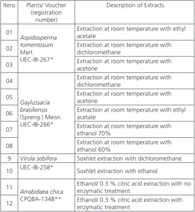
Plants were obtained from the Cerrado biome (the second largest biome in South America) and deposited at the University of Campinas, Campinas, SP, Brazil (Table 1).
Table 1 – Cerrado plant and description of each extracts used for aMPV screening.
Itens Plants/ Voucher (registration
number)
Description of Extracts
01
Aspidosperma tomentosum
Mart. UEC-IB-267*
Extraction at room temperature with ethyl acetate
02 Extraction at room temperature with dichloromethane
03 Extraction at room temperature with acetone
04
Gaylussacia brasiliensis
(Spreng.) Meisn. UEC-IB-266*
Extraction at room temperature with dichloromethane
05 Extraction at room temperature with acetone
06 Extraction at room temperature with ethyl acetate
07 Extraction at room temperature with ethanol 70%
08 Extraction at room temperature with ethanol 60%
9 Virola sebifera Soxhlet extraction with dichloromethane
10 UEC-IB-258* Soxhlet extraction with ethanol
11
Arrabidaea chica
CPQBA-1348**
Ethanol/ 0.3 % citric acid extraction with no enzymatic treatment
12 Ethanol/ 0.3 % citric acid extraction with enzymatic treatment
* UEC-IB-267; UEC-IB-266 and UEC-IB-258: Deposited at the Institute of Biology, State University of Campinas - Campinas/SP, Brazil.
** CPQBA-1348: Deposited at the Center for Chemical, Biological and Agricultural (CQBA) – State University of Campinas, Campinas/SP, Brazil
Extracts from the leaves of Aspidosperma
with 3.0g of citric acid, resulting in an enzyme crude extract (CE). A crude extract of A. chica (4) with no enzymatic treatment was also obtained (SE).
Virus and cell line
The CER cells (chicken embryo related cell) were
propagated as monolayer cultures using minimal
essential medium (MEM) with Earle’s salts and
supplemented with 10% fetal bovine serum (FBS).
Cells were serially diluted from 1:2 to 1:10, according to conventional procedures using 0.05% trypsin and 0.02% ethylenediaminetetraacetic acid (EDTA).
The aMPV strain SHS/669/03 was isolated by D’Arce
et al. in 2005 and belongs to aMPV subtype A. When the virus was incubated with the cells, the MEM medium was not supplemented with FBS.
Cell cytotoxic effect
Maximum nontoxic concentration (MNTC) was determined microscopically by the observation of morphological changes in the cells at 24, 48 and 72 hours of incubation. Cell suspensions were seeded at 100 μL/well in a 96-well culture plate at a density of 1 x 10⁵ cells/mL. The plates containing cells were
pre-incubated for 24 h at 37 °C to allow stabilizations prior to the addition of samples (100 μL) at four concentrations (0.25, 2.5, 25, and 250 μg.mL-1) (Kohn
et al., 2007).
Cytotoxicity after 72 h was measured using the sulforhodamine B (SRB) assay, performed as described by Kohn et al. (2007). Briefly, the cells were fixed using 50% Trichloroacetic Acid (TCA) at 4°C (50 μL per well, final concentration 10%) for 1h. The supernatant was discarded and the plates were washed five times with filtered water. The cells were stained for 30 min with 0.4% SRB in 1% acetic acid (50 μL per well) and subsequently washed four times with 1% acetic acid to remove any unbound dye. The plates were air-dried, and protein-bound dye was solubilized with 150 μL (100 mM) of Trizma buffer. The resulting optical density (OD) was read in a multi-well plate under a spectrophotometer at 540 nm.
Virus titration
The cells were seeded in 96-well culture plates at a density of 1 x 10⁵ cells/mL and then incubated at 37
ºC in a humidified atmosphere containing CO2 for 24 h. Serial dilutions of virus stocks were prepared and cells were infected accordingly. After an additional incubation period (1-2 days), the cytopathic effect was recorded. The 50% tissue-culture infective dose
(TCID50) per mL was calculated as previously described by Reed & Münch (1938).
Antiviral activity
The determination of the antiviral activity of evaluated plant extracts was based on cytopathic effect inhibition. All experiments were performed in triplicate. For the evaluation of inhibition, cells were seeded in 96-well culture plates. After 24h of incubation, the medium was replaced with 100 µL Dulbecco’s Modified Eagle’s Medium (DMEM), containing the plant extracts at MNTC, and 50 µL of logarithmic dilutions of the virus were added in quadruplicate; the plates were incubated for 3 days. Controls consisted of untreated infected (virus titer), treated non-infected (extract control), and untreated non-infected (cell control) cells. Viral titers was calculated as previously described by Reed and Münch (1938), and determined as 50% of the infective dose in tissue culture (TCID50/mL). Antiviral activity of each extract was determined as the logarithm reduction factor (log10) of the viral titer compared with untreated infected controls. Values were expressed as titer (TCID50/mL) and inhibition percentage (IP), as described in Koseki et al. (1990). The inhibition percentage was calculated according to the formula: (IP) = (1 – T/C) x 100, where T is the antilog of the extract-treated viral titers and C is the antilog of the control (without extract) viral titers. IP was considered positive if greater than or equal to 98%.
The antiviral activity was initially evaluated with a single dose at MNTC against different viral concentrations. The extract was considered positive if there was a 1.5 log decrease in the viral titer. In order to confirm antiviral activity, a concentration response curve with different extract concentrations in the
presence of 100 TCID50/mL was calculated using the
MTT1 assay to establish the half maximal effective
concentration (EC50). Briefly, an MTT solution at 5
mg.mL-1 in PBS was added to the 96-well culture
plates at 20 μL/well at each time point. Following 4h of incubation, 100μL of DMSO was added to each well and mixed thoroughly to dissolve the dark-blue formazan crystals from surviving cells. The resulting optical density (OD) was read in a multiwell plate in a spectrophotometer at 540 nm (Mosmann, 1983 and Scudiero et al., 1988).
Statistical analysis
The 50% cytotoxic (CC50) and 50% inhibition (IC50) concentrations were calculated from concentration-effect curves. The results were obtained from triplicate assays with at least five extract concentrations. The percentage of cytotoxicity was calculated as [(A – B)/A] x 100, where A and B are the OD 540 nm of untreated and of treated cells, respectively. The percentages of protection were calculated as [(A − B) × 100/(C − B)], where A, B and C indicate the absorbance of the extracts or fractions, virus and cell controls, respectively. Each obtained EC50 value was defined as the effective concentration that reduced the absorbance of infected cells to 50% when compared with cell and virus controls.
The CC50 and IC50 of each compound were obtained
from dose-effect curves (not shown). The CC50 and IC50 are the average of three assays with five concentrations within the inhibitory range of the compounds. The selective index was defined as CC50/IC50.
Potential stage of the viral infection cycle
Cells and viruses were incubated with the active plant extracts at different stages during the viral infection cycle in order to assess different modes of antiviral action (virus inactivation before infection or during the virus adsorption and replication phases). Cells were pretreated with the extracts prior to viral infection (virus inactivation); viruses were incubated with the extracts before infecting the cells (adsorption phase); the cells were infected with the virus and incubated with the plant extracts (replication phase). Each extract was used at its maximum noncytotoxic concentration.
Statistical analysis
The selectivity index (SI) was determined as the EC50 to IC50 ratio. All experiments were performed in triplicate, and three independent experiments were conducted. Data are presented as mean ± SD. The Student’s t-test was used to evaluate the difference between the test and control samples. Differences were considered statistically significant when p-value was < 0.05.
RESULTS AND DISCUSSION
Medicinal plants have been traditionally used for the treatment of different health conditions, including infectious diseases (Severson et al., 2008; Li et al., 2009). According to Cragg’s 2009 report, approximately 60% of the anti-tumor and anti-infective agents that are
commercially available or in late stages of clinical trials today are of natural product origin. Therefore, there is no doubt that traditional medicinal plants may serve as potential sources for the development of new antiviral agents in the future.
Developing new antiviral drugs is a difficult task due to the poor selective toxicity and fast selection of resistant viral variants that naturally arise given a selective pressure. The frequency of viral resistance to antiviral drugs is increasing and consequently, viral diseases remain difficult to treat.
The screening of plants as possible sources of antiviral agents has led to the discovery of potent inhibitors of
in-vitro viral replication, increasing the probability of identifying new bioactive plant compounds (Severson
et al., 2008; Li et al., 2009).
In-vitro assays usually rely on the virus ability to infect and replicate in specific cell lines in cell culture systems. Cell culture systems provide a rapid and reliable method to grow viruses at higher titers, to apply reverse genetics, and to test antiviral compounds.
A total of 12 extracts derived from four plant species collected from the Brazilian Cerrado were screened for antiviral activity against aMPV. Table 1 shows the plant names and extraction procedures. The results suggests that there is an increasing activity of the extract A. tomentosum acetone and G. brasiliensis
dichloromethane with SI higher than 1.5. However,
the most active extracts against AMPV were from A. chica and V. sebifera with an inhibition percentage of 99%, both extracted with acetone. The four extracts were tested to determine in which phase of the virus replication cycle they were most active.
The first stage of the antiviral assay is necessary to determine the maximum concentration (MNTC) of the extract that is not toxic to the cells. After the
MNTC of the extracts was determined, 25 µg mL-1
of Aspidosperma tomentosum and Gaylussacia brasiliensis, and 2.5 µg mL-1 of Arrabidaea chica
and Virola sebifera were applied. The concentration range used to assess the activity of the extracts did not induce significant toxicity to the host cells and the 50% cytotoxic concentration (CC50) determined for each extract were well tolerated by the CER cells. The
CC50 ranged from 25 to 0.25 µg mL-1 and none of
the extracts tested induced any visible changes in cell morphology and cell density.
extracts prior to infection and were added during or after the adsorption phase or during the intracellular replication period. Three different treatments were applied. The cells were infected with the virus, followed by addition of the extracts after 1 h in order to evaluate the viral replication phase, viruses were pretreated with the extract before infecting the cells (virus inactivation), while the extract were added to the cells before viral infection to evaluate any effects during viral adsorption. In all experiments, the extracts were used at their MNTC, and the results are shown in Figures 1 and 2.
Figure 1 – Antiviral activity, as determined by inhibition percentage (IP), using virus adsorption, replication and inactivation assays of Virola sebifera and Gaylussacia
brasiliensis extracts. Virus titer (TCID50) was determined three days after infection and
compared with untreated control cells. Virus and CER cells were pretreated with each extract at MNTC. Experiments were independently repeated and data presented here represent the average of three experiments.
Figure 2 – Antiviral activity, as determined by inhibition percentage (IP), using virus adsorption, replication and inactivation assays of Aspidosperma tomentosum and
Arrabidaea chica. Virus titer (TCID50) was determined three days after infection and
compared with untreated control cells. Virus and CER cells were pretreated with each extract at MNTC. Experiments were independently repeated and data presented here represent the average of three experiments.
The analyses of the results (Figures 1 and 2) suggest that the G. brasiliensis, A. chica, and V. sebifera extracts inhibit 99% of the virus during the early replication stage and that these extracts act during the viral adsorption phase. The fact that the extracts maintained their activity under these conditions suggests that they
establish stable bonds with virus receptors on the cell surface. However, this may also be due to other inhibitory mechanisms acting during the adsorption or penetration of the virus into the cell. Further studies should be conducted to determine the precise mode of action of these extracts. The 50% cytotoxic (CC50) and 50% inhibition (IC50) concentrations, as well as the selective index (SI) of the extracts active against aMPV were calculated and are listed in Table 2.
Table 2 – Inhibition percentage (IP), 50% cytotoxic (CC50) and 50% inhibition (IC50) concentrations, and selective index (SI) of the four extracts against aMPV in CER cell line.
Crude Extract IP CC50 IC50 SI
(%) µg.mL-1 ± SD
Aspidosperma
tomentosum 97 64.9 ± 0.05 45.86 ± 0.02 1.5
Gaylussacia brasiliensis 99 41.23 ±0.03 22.33 ± 0.12 1.8
Virola sebifera 99 2.7 ± 0.08 202 ± 0.05 0.1
Arrabidaea chica 99 117.1 ± 0.017 412.7 ± 0.018 0.3
CONCLUSION
Despite continuous advances made in antiviral therapy, viral diseases are still the leading cause of death globally. Being obligate intracellular parasites, the replication of viruses is dependent on the metabolic pathways of the host cell. Since viruses and hosts are intimately connected, the design of effective antiviral agents that attack viral enzymes or virus replication, without affecting the host cell, has proven to be difficult.
The results of the present investigation provide further evidence of the potential use of medicinal plants, which possibly represent a reservoir of pharmacologically active substances. The study showed that A. tomentosum, V. sebifera, G. brasiliensis, and
A. chica crude extracts are capable of inhibiting 99%
of aMPV in vitro. However, further studies on plant
fractions, aiming at isolating and identifying their active compounds and at determining their precise mode of action are required.
ACKNOWLEDGMENTS
This research was supported by Fundação de Amparo a Pesquisa do Estado de São Paulo (FAPESP) grant 2008/00700-4 to CWA. LKK thanks FAPESP for a post-doctoral scholarship (2008/00331-9), and Conselho Nacional de Desenvolvimento Científico e Tecnológico (CNPq) for the post-doctoral scholarship (503259/2011-1).
REFERENCES
Abou-Karan M, Shier WT. A simplified plaque reduction assay for antiviral agents from plants. Demonstration of frequent occurrence of antiviral activity in higher plants. Journal Natural Products 1990;53:340-44.
Bolken TC, Laquerre S, Zhang Y, Bailey TR, Pevear DC, Kickner SS, et al.
Identification and characterization of potent small molecule inhibitor of hemorrhagic fever New World arenaviruses. Antiviral Research 2006;69, 86–97.
Newman JD, Cragg GM. Natural product scaffolds as leads to drugs. Future Medicinal Chemistry 2009;1:1415-27.
Catelli M, Cecchinato CE, Savage RC, Jones CJ. Demonstration of loss of attenuation and extended field persistence of a live avian metapneumovirus vaccine. Vaccine 2006: 24:6476–6482.
Cos P, Vlietink AJ, Berghe DV, Maes L. Antiinfective potential of natural products: how to develop a stronger in vitro ‘proof-of-concept’. Journal of Ethnopharmacology 2006; 106:290-302.
De Clerq E. Antiviral drugs in current clinical use. Journal of Clinical Virology 2004;30:115-133.
Jassim SA, Naji MA. Novel antiviral agents: a medicinal plant perspective. Journal of Applied Microbiology 2003;95:412-27.
D’Arce RC, Coswig LT, Almeida RS, Trevisol IM, Monteiro MCB, Rossini LI,
et al. Subtyping of new Brazilian avian metapneumovirus isolates from
chikens and turkeys by reverse transcriptase-nested-polymerase chain reaction. Avian Pathology 2005;34(2):133-136.
Kohn LK, Pavam CH, Veronese D, Coelho F, De Carvalho JE, AlmeidaWP. Antiproliferative effect of Baylis-Hillman adducts and a new phthalide derivative on human tumor cell lines. European Journal of Medical Chemistry 2006;41:738-44.
Li Q, Maddox C, Rasmussen L, Hobrath JV, White LE. Assay development and high-throughput antiviral drug screening against Bluetongue virus. Antiviral Research 2009; 83:267–273.
Mosmann T. Rapid colorimetric assay for cellular growth and survival: application to proliferation and cytotoxicity assays. Journal Immunological Methods 1983;65:55-63.
Mukhtar M, Arshad M, Ahmad M, Pomerantz RJ, Wigdahl B, Parveen Z. Antiviral potentials of medicinal plants. Virus Research 2008;131:111-20.
Noah JW, Severson W, Noah DL, Rasmussen L, White EL, Jonsson CB. A cell-based luminescence assay is effective for high-throughput screening of potential influenza antivirals. Antiviral Research 2007;73:50–59.
Pieroni ME, Giusti C, de Pasquale C, Lenzarini E, Censorii MR, Gonzáles-Tejero C, Sánchez-Rojas CP, Ramiro-Gutiérrez JM, Skoula M, Johnson C, Sarpaki A, Della A, Paraskeva-Hadijchambi D, Hadjichambis A, Hmamouchi M, El-Jorhi S, El-Demerdash M, El-Zayat M, Al-Shahaby O, Houmani Z, Scherazed M. Circum-mediterranean cultural heritage and medicinal plant uses in traditional animal healthcare: a field survey in eight selected areas within the RUBIA project. Journal of Ethnobiological and Ethnomedical 2006; 2:16–28.
Reed LJ, Muench H. A simple method of estimating fifty percent endpoints. American. Journal of Public Hygiene 1938;27:493-497.
Scudiero DA, Shoemaker RH, Paull KD. Evaluation of a soluble tetrazolium/ formazan assay for cell growth and drug sensitivity in culture using human and other tumor cell lines. Cancer Research 1988;48:4827-4833.
Severson WE, McDowell M, Ananthan S, Chung DH, Rasmussen L, Sosa MI, White EL, Noah J, Jonsson CB. High-throughput screening of a 100,000-compound library for inhibitors of influenza A virus (H3N2). Journal of Biomolecular Screening 2008; 13:879–887.
Severson WE, Shindo N, Sosa M, Fletcher T, White EL, Ananthan S, Jonsson CB. Development and validation of a high-throughput screen for inhibitors of SARS CoV and its application in screening of a 100,000-compound library. Journal of Biomolecular Screening 2007;12:33–40.