A surveillance of enteropathogens in piglets from birth to
seven days of age in Brazil
1Eduardo C. Cruz Junior2, Felipe M. Salvarani2, Rodrigo O.S. Silva2, Marcos X. Silva2, Francisco C.F. Lobato2 and Roberto M.C. Guedes2*
ABSTRACT.- Cruz Junior E.C., Salvarani F.M., Silva R.O.S., Silva M.X., Lobato F.C.F. & Guedes R.M.C. 2013. A surveillance of enteropathogens in piglets from birth to seven days of age in Brazil. Pesquisa Veterinária Brasileira 33(8):963-969. Escola de Veterinária, Univer-sidade Federal de Minas Gerais, Av. Antônio Carlos 6627, Cx. Postal 567, Belo Horizonte, MG 30123-970, Brazil. E-mail: [email protected]
The purpose of the study was to evaluate the real importance of anaerobic enteropa-thogens and rotavirus in contrast to more common agents as cause of diarrhea in piglets
within the first week of life. Sixty 1- to 7-day-old piglets, 30 diarrheic and 30 non-diarrheic
(control), from 15 different herds were selected, euthanized and necropsied. Samples of the jejunum, ileum, colon, cecum and feces were collected from the piglets and analyzed to determine the presence of the following enteropathogens: enterotoxigenic Escherichia coli (ETEC), Clostridium perfringens types A and C, Clostridium difficile, rotavirus and Isospora suis. Among diarrheic piglets, 23.3% were positive for C. difficile, 70% for C. perfringens type A cpb2+, 14.3% for rotavirus and 10% for ETEC. Among non-diarrheic control piglets, 10% were positive for C. difficile, 76.7% for C. perfringens type A cpb2+, 0% for rotavirus, 3.3% for ETEC and 3.3% for I. suis. C. perfringens type C was not detected in any of the ani-mals. Histological lesions characteristic of C. difficile, E. coli and rotavirus were observed. However, no C. perfringens type A suggestive lesions were detected. There was a positive correlation between mesocolon edema and the presence of C. difficile toxins. Although C. perfringens type A cpb2+ was the most frequently detected enteropathogen, there was no association between its presence and diarrhea or macro or microscopic changes. C. difficile and Rotavirus were the most relevant pathogens involved with neonatal diarrhea in this
study, and histopathology associated with microbiological test proved to be the key to reach a final diagnosis.
INDEX TERMS: Piglet, swine, Clostridium perfringens, Clostridium difficile, enterotoxigenic Escheri-chia coli, rotavirus.
1 Received on February 5, 2013.
Accepted for publication on May 23, 2013.
2 Escola de Veterinária, Universidade Federal de Minas Gerais (UFMG),
Av. Antônio Carlos 6627, Cx. Postal 567, Belo Horizonte, MG 30123-970, Brazil. *Corresponding author: [email protected]
RESUMO.- [Levantamento dos enteropatógenos de lei-tões do nascimento a sete dias de idade no Brasil.] O objetivo do presente estudo foi avaliar a real importância de enteropatógenos anaeróbios e rotavirus em compara-ção à outros agentes mais comuns como causa de diarreia em leitões até cinco dias de idade. Leitões com 0 a 7 dias de vida, 30 diarreicos e 30 não diarreicos (controles) de 15 granjas diferentes foram eutanasiados e necropsiados. Amostras de jejuno, íleo, colon e ceco foram coletadas e
com lesões foi encontrada. C. difficile e Rotavirus foram os agentes mais relevantes associados à diarreia neonatal, e
ficou demonstrada a relevância de associação de histopato
-logia com testes de detecção microbiológica para se firmar
um diagnóstico.
TERMOS DE INDEXAÇÃO: Leitões, suínos, Clostridium perfringens, Clostridium difficile, Escherichia coli enterotoxigênica, rotavirus.
INTRODUCTION
Enteric disorders are the most common group of infectious
diseases in piglets during the first week of life. They are responsible for significant economic losses, mainly due to
reductions in weaning weight and increases in mortality rates and production costs. Thus, it is essential to obtain in-formation concerning the actual relevance of each
entero-pathogen to allow specific preventive measures and more efficient control methods to be used.
The most important pathogens associated with diar-rhea in piglets up to seven days of age are enterotoxigenic Escherichia coli (ETEC), Clostridium perfringens types A and C, Clostridium difficile, rotavirus and Isospora suis (Yaeger et al. 2002). However, some recent reports indicate that the frequency of these agents has been changing, with mi-croorganisms such as C. difficile becoming more prevalent (Yaeger 2007).
Additionally, the real importance of C. perfringens type
A as a primary agent of diarrhea in suckling piglets is not fully known. Information regarding the detection and pre -valence of intestinal pathogens in piglets is scarce. The purpose of our study was to evaluate the real importance of anaerobic enteropathogens and rotavirus in contrast to more common agents as cause of diarrhea in piglets within
the first week of life.
MATERIALS AND METHODS
Farms and animals
Stool samples were collected from 1- to 7-day-old piglets from 15 different farms with at least 500 sows in a three-site production system located in a densely swine-populated area of Minas Gerais, Brazil. On each farm, two diarrheic based on clini-cal signs and two apparently healthy piglets (n=60) were sedated with xylazine (Virbac®, Brazil; 0.3 to 5mg/kg IM), euthanized by electrocution and exsanguinated. Each piglet was selected from a different litter. Parity of dam wasn’t considered. Samples were col-lected for histology, bacteriology and molecular assays. Stool sam-ples were collected directly from the rectum and stored at 4°C for up to 48 hours. Samples of the jejunum, ileum, cecum and colon were fixed in 10% buffered formalin. All procedures performed were approved by the Ethics Committee on Animal Experiments of the Universidade Federal de Minas Gerais (CETEA-UFMG, pro-tocol no.30/09).
Enterotoxigenic Escherichia coli
Stool samples were plated on MacConkey agar (Biobrás®, Pro-dimol Biotechnology) followed by incubation at 37°C for 24 hours. Each isolated colony was subjected to biochemical testing as pre-viously described (Martins et al. 2000). All colonies identified as
E. coli by biochemical assay were subjected to DNA extraction by the phenol-chloroform method (Sambrook et al. 1989). DNA con -centrations were estimated in a spectrophotometer (Shimadzu®,
model UV160A). Samples were diluted to a final concentration of 10ng/μL prior to performing multiplex PCR for E. coli pathoge -nicity factor genes (Macêdo et al. 2007). Four strains, kindly pro -vided by the Veterinary Diagnostic Laboratory at the University of Minnesota, were used as positive controls for PCR: 2568 (STb, STaP, F18 and Stx2e), 2569 (STb, LT and K88), 2570 (987P and STaP) and 2571 (STaP, K99 and F41). Amplification products were visualized by electrophoresis in 6% polyacrylamide gels followed by silver staining (Macêdo et al. 2007).
Clostridium perfringens
For isolation of C. perfringens, 0.08 to 0.12g of feces was se-rially diluted from 10-1 to 10-6. Aliquots of about 50µL of each dilution were plated on sulfite polymyxin sulfadiazine agar (SPS, Difco Laboratories, Detroit, USA) and incubated anaerobically at 37°C for 24 hours. Following incubation, DNA was extracted from characteristic Clostridium sp. colonies (Baums et al. 2004) and used for PCR. Genes encoding beta-2 toxin (cpb2), enteroto-xin (cpe) and the major C. perfringens toxins (alpha, beta, epsilon and iota) were detected using multiplex PCR (Vieira et al. 2008). The products were visualized under UV light in a 2% agarose gel stained with ethidium bromide (Sigma-Aldrich, Saint Louis, USA).
Clostridium difficile
C. difficile A/B toxins were detected using an ELISA kit (Ridas -creen Clostridium difficile toxins A/B, R-Biopharm, Germany). All reactions were performed according to the manufacturer’s ins-tructions.
Rotavirus
Stool samples were diluted in Tris-HCl CaCl2 buffer as pre-viously described (Barbosa et al. 1998). RNA was extracted using the phenol-chloroform method (Theil et al., 1981; Sambrook et al., 1989). Each sample was then subjected to discontinuous polyacrylamide gel electrophoresis (PAGE; 7% running gel, 3.45% stacking gel; Sigma-Aldrich; according to Barbosa 1994) followed by silver staining (Herring et al. 1982).
Isospora suis
Coccidia oocysts (Eimeria spp. and I. suis) were detected using the flotation method with Sheather’s sugar solution followed by light microscopy (Hoffmann 1987).
Histopathology
Formalin-fixed samples of the jejunum, ileum, cecum and co -lon were subjected to dehydration, diaphanisation and paraffin inclusion. Five-micron sections of each part of intestine were pre-pared and stained with hematoxylin and eosin (Luna 1968), and histological evaluation was performed using light microscopy.
Statistical analysis
Fisher’s exact test (Data Analysis and Statistical Software - Stata, Tulsa, Oklahoma, EUA) was used to analyze the results. P values of <0.05 were considered significant.
RESULTS
Escherichia coli
Of the 60 piglets sampled, pathogens were not detected in nine of them, three diarrheic and six controls. All sam-ples from both groups showed lactose-positive colonies on
MacConkey agar. After biochemical testing, 22 isolates from
the diarrheic group and 24 isolates from the control group
Only four of the piglets, each from a different farm, were positive by PCR for the presence of virulence factor genes (Table 1). Three belonged to the diarrheic group and had the following virulence types: F41, 987P and 987P-LT. The one remaining piglet from the control group was positive for the 987P-LT gene. However, only one diarrheic piglet (987P-LT) had a large number of coccobacilli adhered to the enterocyte surface (Fig.1), which is a characteristic
histolo-gical finding of enterotoxigenic E. coli infection. No diffe-rence was observed between diarrheic and control groups regarding enterotoxigenic E. coli detection (p=0.741).
Clostridium perfringens
Suggestive colonies of C. perfringens were recovered from 22 diarrheic piglets and 26 control piglets. C. perfrin-gens type A was identified by PCR from all 15 farms investi -gated, demonstrating the high frequency of this agent, whi-ch is a normal part of the piglets’ microbiota. C. perfringens
type C was not found in any of the piglets. All farms had at least one piglet that was positive for C. perfringens type A with the beta-2 toxin gene (cpb2) (Table 1). In total, 44 strains (91.7%) of C. perfringens type A were positive for the cpb2 gene. Twenty-three of the strains (52.3%) came from non-diarrheic piglets.
Histopathological evaluation did not reveal any
altera-tions that could be directly linked to C. perfringens type A infection. No association was found between any type of histological lesion and the detection of C. perfringens type A cpb2+ (p=0.73). There was also no difference between the presence or absence of histopathological changes and the isolation of C. perfringens type A, even disregarding the presence of the cpb2 gene.
Clostridium difficile
Thirteen piglets from six farms had mesocolon edema (Table 2), a common macroscopic alteration associated with C. difficile infection. Of these 13 animals, eight (61.5%)
were from the diarrheic group and five (38.5%) were from
the control group. Mesocolon edema was associated with six farms; at one of the farms, all four of the sampled ani-mals presented with this macroscopic lesion.
Ten piglets (16.7%) from six different farms were posi-tive for C. difficile A/B toxins. Of these, seven were diarrheic piglets, and three were control piglets. Among the diarrheic piglets, two had mild neutrophilic colitis, one had moderate neutrophilic colitis and two had severe colitis (Fig.2b). The remaining two animals from the diarrheic group showed no histopathological alterations. All three of the control piglets that were positive for A/B toxins had neutrophilic colitis, two with severe and one with mild lesions.
Another six piglets, belonging to five different farms,
were negative for the detection of A/B toxins by ELISA but had histological lesions compatible with C. difficile infec-tion. Of the six, two belonged to the diarrheic group, one of which presented with mild colitis and the other with mo-Table 1. Frequency of enteric pathogens in piglets one to seven days old
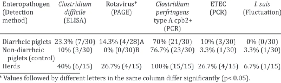
Enteropathogen Clostridium Rotavirus* Clostridium ETEC I. suis (Detection difficile (PAGE) perfringens (PCR) (Fluctuation) method) (ELISA) type A cpb2+
(PCR)
Diarrheic piglets 23.3% (7/30) 14.3% (4/28)A 70% (21/30) 10% (3/30) 0% (0/30) Non-diarrheic 10% (3/30) 0% (0/30)B 76.7% (23/30) 3.3% (1/30) 3.3% (1/30) piglets (control)
Herds 40% (6/15) 26.7% (4/15) 100% (15/15) 26.7% (4/15) 6.7% (1/15)
* Values followed by different letters in the same column differ significantly (p< 0.05).
Table 2. Mesocolon edema and histological changes in diarrheic and non--diarrheic piglets
Group Mesocolon edema Histopathological alterations*
Enteritis Typhocolitis
Diarrheic piglets 13.3% (4/30) mild 10% (3/30) mild 6.7% (2/30) mild 6.7% (2/30) moderate 6.7% (2/30) moderate 3.3% (1/30) moderate 6.7% (2/30) severe 13.3% (4/30) severe 10% (3/30) severe Non-diarrheic 6.7% (2/30) mild 20% (6/30) mild 6.7% (2/30) mild piglets (control) 6.7% (2/30) moderate 6.7% (2/30) moderate 3.3% (1/30) moderate 3.3% (1/30) severe 6.7% (2/30) severe 13.3% (4/30) severe * For piglets with more than one microscopic lesion, the most serious was considered. Fig.1. Piglet jejunum showing intense adhesion of coccobacilli in
derate colitis. In the control group, one piglet had mild coli-tis and the other three had severe colicoli-tis. Among all piglets with colitis, different degrees of reduction in the number of goblet cells were visualized, depending on the intensity of
the associated inflammation.
Rotavirus
Four samples were positive for rotavirus by PAGE analy-sis, all of which came from different farms and belonged to the diarrheic group (Table 1). Among them, three had multifocal neutrophilic enteritis ranging from moderate to severe, with enterocyte necrosis in the tip of the villi sug-gestive of recent rotavirus infection (Fig.3). The remaining piglet showed no histological lesions.
Isospora suis
Two piglets from the control group were positive for
fecal oocysts according to the flotation test (Table 1). Pa
-rasitization of the enterocytes, which is typical in piglets with coccidiosis, was not found during histopathological examination.
Coinfection
Fourteen piglets from eight different farms were po-sitive for more than one enteropathogen, nine from the
diarrheic group and five from the control group. Among
the diarrheic piglets, six were positive for C. perfringens type A cpb2+ and C. difficile toxins; one for C. perfringens cpb2+ type A and enterotoxigenic E. coli; and two for C. per-fringens type A cpb2+ and rotavirus. Among the five non --diarrheic piglets with coinfection, three were positive for C. perfringens type A cpb2+ and C. difficile A/B toxins; one for C. perfringens cpb2+ type A and enterotoxigenic E. coli; and one for C. perfringens type A cpb2+ and I. suis oocysts. On one farm, three of the four sampled animals were simul-taneously positive for C. difficile toxins and for other patho-gens. In addition, C. perfringens type A cpb2+ was involved in all coinfections.
DISCUSSION
Suggestive colonies of Escherichia coli according to plating
on MacConkey agar were obtained from all samples collec -ted. Without further detection of virulence factor genes by PCR, the role of this organism as a causal agent of diarrhea would have been over-estimated. Such misinterpretation
of results derived from non-routine identification metho -ds could explain the increase of antimicrobial-resistant strains of E. coli isolated from piglets; resistance may arise from the misuse of drugs given against pathogens not di-rectly associated with the disease being treated (Menin et al. 2008).
According to Francis (2002), newborn piglets with E. coli strains possessing virulence factors are commonly positive for LT, STB, K99 and 987P genes. Moreover, the same author reported that the presence of both toxin and
fimbriae are necessary for infection by enterotoxigenic E.
Fig.2. Colon sections of piglet with lesions suggestive of Clostridium difficile infection. (a) Severe goblet cell loss. HE, obj.4. (b) Severe necrotizing neutrophilic colitis with intense infiltration of neutrophils from the lamina propria to the intestinal lumen. HE, obj.40x.
coli and the development of diarrhea. In the present stu-dy, two out of four piglets positive for E. coli with
virulen-ce factors did not have either fimbriae or toxin genes. One of these four piglets expressed F41 fimbriae, but was po -sitive for Clostridium perfringens type A cpb2+. Thus, the role of each of these agents in causing diarrhea cannot be conclusively determined. The other piglet was positive for
a fimbrial encoding gene (987P) but negative for all other
pathogens studied. One possible explanation for the occur-rence of diarrhea in this piglet is the excessive
consump-tion of milk, a common cause of non-infectious diarrhea in
piglets (Jennings 1959). This could also potentially explain diarrhea occurring in other piglets testing negative for the studied pathogens.
A few decades ago, several studies reporting a high in-cidence of enterotoxigenic E. coli in piglets (Barcellos et al. 1980, Calderaro et al. 2001) resulted in ETEC becoming re-garded as the main cause of diarrhea in piglets up to seven days of age. However, recent studies have reported lower detection rates ranging from 3 to 20% (Yaeger et al. 2007). The low frequency (6%) of enterotoxigenic E. coli observed in our study supports these most recent studies and helps
confirm the reduced occurrence of colibacillosis in young
piglets. One factor that may be contributing to the decrea-sed frequency in ETEC-caudecrea-sed diarrhea is routine vaccina-tion against neonatal colibacillosis.
Although some authors associate C. perfringens type A
cpb2+ infection with the presence of inflammatory infiltra -tes in the lamina propria and with mild to moderate
ne-crosis of the enterocytes (Songer & Glock 1998, Hammer
& Walz 2008, Farzan et al. 2013), others report the absen-ce of pathological changes associated with this pathogen (Songer & Uzal 2005). In our study, we observed no his-tological alterations that could be attributed to this agent. Yaeger (2007) also found no microscopic lesions in piglets with diarrhea caused by C. perfringens type A cpb2+ and instead suggested that the agent was responsible for secre-tory diarrhea. However, any diagnosis of diarrhea due to this pathogen should be made cautiously, as C. perfringens type A is a normal part of the piglet intestinal microbiota. Detecting the presence of other possible pathogens or coin-fection is therefore essential. Another diagnostic
possibili-ty commonly cited in the literature is the quantification of
C. perfringens. In our study, quantification in SPS agar was performed, but no association was found between the pre-sence or abpre-sence of diarrhea and the number of organisms (data not shown), corroborating previous studies (Songer
& Glock 1998, Farzan et al. 2013) which reported that the quantification of C. perfringens colony-forming units in in-testinal or stool samples are not correlated with the pre-sence of diarrhea in piglets.
The high isolation rate of C. perfringens type A cpb2+ from control piglets (76.7%) was interesting because cpb2 strains are commonly associated with the occurrence of diarrhea or dysentery in pigs (Van Asten et al. 2010). The presence of cpb2+ in healthy animals; the absence of histo-logical lesions in C. perfringens-positive animals (diarrheic or non-diarrheic); and the absent correlation between the quantity of organism and clinical disease, all suggest that
the cpb2 gene is not an appropriate marker for determi -ning the pathogenicity of C. perfringens type A. This re-sults are similar to those recently reported by Farzan et al. (2013), which found no difference between the isolation of C. perfringens type A cpb2+ in diarrheic or non-diarrheic piglets. In addition, these authors also reported the absen-ce of correlation between the detection of the beta-2 toxin directly in the intestinal content in these two groups, su-ggesting that the isolation of C. perfringens type A cpb2+ or the detection of beta-2 toxin are not a useful approach
for making diagnosis of type A C. perfringens enteritis in pi-glets (Farzan et al. 2013).
Six of the thirteen piglets with mesocolon edema (46%) were positive for C. difficile A/B toxins by ELISA. This result supports Yaeger et al. (2007) who reported a low positive predictive value (54%) between the presence of mesocolon edema and the detection of C. difficile A/B toxins. However, all piglets with severe mesocolon edema were positive for
A/B toxins. Moreover, a significant association (p<0.004)
was found between the absence of macroscopic lesions and the non-detection of A/B toxins regardless of the presen-ce of diarrhea. Given these results, the presenpresen-ce of severe mesocolon edema should be considered an important in-dicator of C. difficile infection, while the complete absence of macroscopic alterations may indicate that the animal is negative for A/B toxin.
Of the 10 piglets positive for A/B toxins by ELISA, five
control piglets and three diarrheic piglets demonstrated
histological inflammatory lesions. These results also sup -port those of Yaeger et al. (2007) describing a higher positi-ve predictipositi-ve value (76%) for the occurrence of neutrophilic
inflammatory infiltrate in the lamina propria and detection of A/B toxins. In addition, a significant association was
found between the presence of microscopic alterations and
the detection of the A/B toxin (p<0.0001), confirming that the occurrence of inflammatory lesions is associated with
the toxin regardless of the presence or absence of diarrhea.
These findings suggest that in the absence of an assay for
the A/B toxin, histopathology could be used as a diagnostic tool to indicate possible involvement of C. difficile infection with diarrhea in piglets. In addition to these common
mi-croscopic inflammatory changes, Yaeger et al. (2007) also
observed a reduction in the number of goblet cells and in-creased mitotic activity in the crypts. We made similar
ob-servations, including a significant decrease in goblet cells
in the cecum and colon of piglets (Fig.2a).
Six piglets that were negative for A/B toxins had histo-logical lesions suggestive of C. difficile infection, which may be related to the low sensitivity of the ELISA used. Different studies evaluating various commercially available ELISA
kits using piglet stool samples indicated that they com
-monly lack sensitivity (Post et al. 2002, Songer & Anderson
2006, Keessen et al. 2011).
The association between macroscopic findings, histolo
-gical lesions and the presence of A/B toxins confirms that
complications related to C. difficile infection in piglets could occur without any clinical sign of diarrhea. Yaeger et al.
It should be emphasized that on a farm with a suspected C. difficile infection, sampling for diagnosis should include animals with clinical signs of diarrhea as well as some non--diarrheic piglets. Furthermore, an assortment of different
diagnostic tools should be used to confirm the diagnosis.
Among the four piglets testing positive for rotavirus by PAGE, only one presented with no histological lesions. This suggests that the piglet had only an initial infection because the onset of fecal shedding of rotavirus may precede the appearance of histological lesions (Linares et al. 2009). Our results demonstrate a 13.3% rotavirus- positive frequency
in diarrheic piglets, a significant association between pa
-thogen detection and the presence of diarrhea (p≤0.05).
Our rate in diarrheic piglets is higher than the 7.5% rate
reported by Lippke et al. (2011) and similar to the 13%
rate reported by Yaeger (2007). In contrast, Gregori et al. (2009) reported a rotavirus frequency of 29.9% (as de-termined by PAGE) in stool samples from 144 piglets with clinical diarrhea; unfortunately, the authors did not report the age of the sampled animals. According to Roehe et al. (1989), piglets are most commonly infected by rotavirus between 15 and 30 days of age. Early rotavirus infection
may therefore indicate a low colostrum intake or ineffec
-tive vaccination. Significantly, by 2011, the single commer -cially available rotavirus vaccine in Brazil included only the G5 and G4 serotypes of group A. However, Gregori et al. (2009) reports that the most common serotypes in Brazil
are (in order): G5, G10, the non-defined genotypes and G6. Moreover, studies on vaccination efficacy and the occurren -ce of cross-protection are still rare.
The low detection rate for I. suis observed in this study substantiates other recent reports (Karamon et al. 2007, Yaeger et al. 2007) suggesting that coccidiosis is rare in pi-glets between one and seven days of age. Previous studies indicate that infection with I. suis may be more frequent
du-ring the second and third weeks of life (Martineau & Cas -tillo 2000).
CONCLUSIONS
Clostridium perfringens type A cpb2+ and C. difficile were the most frequently detected enteropathogens in 1- to 7-day-old piglets, but an association between pathogen detection and histological alterations could only be obser-ved for C. difficile.
The more often detection of C. perfringens type A cpb2+ in health piglets proved the cpb2+ detection to be an
ine-fficient marker for determining the pathogenicity of C. per-fringens type A isolates.
Among these two anaerobic pathogens, C. difficile detec-tion results demonstrated the importance of testing diar-rheic piglets for this agent.
Rotavirus screening should always be performed for piglets with enteric disorders. The combination of histo-pathology with other detection methods provided the best results for accurate diagnosis.
Conflict of interest statement.- None of the authors has any financial or personal relationships that could inappropriately influence or bias the
content of the paper.
Acknowledgements.- This study was supported by funds from Capes, Fa-pemig and CNPq. RMC Guedes and FCF Lobato have a research fellowship
from CNPq. The authors would like to thank Dr. Blanko Kokotovic for the
discussions during the study design.
REFERENCES
Barbosa E.F. 1994. Vírus intestinais RNA de fita dupla em frangos de corte
no sudoeste catarinense: isolamento, caracterização e biologia molecu-lar. Tese de Doutorado em Medicina Veterinária, Universidade Federal de Minas Gerias, Belo Horizonte. 113p.
Barbosa E.F., Figueiredo H.C.P., Garcia A.M., Lobato Z.I.P. & Lage A.P. 1998. Rotavírus do grupo A em bezerros lactentes no estado de Minas Gerais. Ciência Rural 28:435-439.
Barcellos D.E.S.N., Guizzardi I.I. & Falavena L.C.B. 1980. Freqüência e cau-sa de diarréias bacterinanas em suínos nas zonas criatórias do Vale do Taquari e Missões, Rio Grande do Sul, Brasil. Bolm Inst. Pesq. Vet. Desi-dério Finamor 80:27-37.
Baums C.G., Schotte U., Amtsberg G. & Goethe R. 2004. Diagnostic multi-plex PCR for toxin genotyping of Clostridium perfringens isolates. Vet. Microbiol. 100:11-16.
Calderaro F.F., Baccaro M.R., Moreno A.M., Ferreira A.J.P., Jerez A.J. & Pena H.J.F. 2001. Frequência de agentes causadores de enterites em leitões lactentes provenientes de sistemas de produção de suínos do estado de São Paulo. Arqs Inst. Biológico, SãoPaulo, 68:29-34.
Farzan A., Kircanski J., DeLay J., Soltes G., Songer J.G., Friendship R. & Pres -cott J.F. 2013. An investigation into the association between cpb2-en-coding Clostridium perfringens type A and diarrhea in neonatal piglets. Can. J. Vet. Res. 77:45-53.
Francis D.H. 2002. Enterotoxigenic Escherichia coli infection in pigs and its diagnosis. J. Swine Health Prod. 10:171-175.
Gregori F., Rosales C.A.R., Brandão P.E., Soares R.M. & Jerez J.A. 2009. Di-versidade genotípica de rotavírus suínos no Estado de São Paulo. Pesq. Vet. Bras. 29:707-712.
Hammer J.M. & Walz M. 2008. Serological evaluation of a Clostridium per-fringens type A toxoid in a commercial swine herd. J. Swine Health Prod. 16:37-40.
Herring A.J., Inglis N.F., Ojeh C.K., Snodgrass D.R. & Menzies J.D. 1982. Ra-pid diagnosis of rotavirus infection by direct detection of viral nucleic acid in silver-stained polyacrylamide gels. J. Clinic. Microbiol. 16:473-477.
Hoffmann R.P. 1987. Diagnóstico de Parasitismo Veterinário. Editora Suli-na, Porto Alegre. 156p.
Jennings A.R. 1959. Gastro-enteritis in the pig. Vet. Rec. 71:766-771.
Karamon J., Ziomko I. & Cencek T. 2007. Prevalence of Isospora suis and Eimeria spp. in suckling piglets and sows in Poland. Vet. Parasitol. 147:171-175. DOI: 10.1016/j.vetpar.2007.03.029
Keessen E.C., Hopman N.E., Van Leengoed L.A., Van Asten A.J., Hermanus C., Kuijper E.J. & Lipman L.J. 2011. Evaluation of four different diag-nostic tests to detect Clostridium difficile in piglets. J. Clinic. Microbiol. 49:1816-21.
Linares R.C., Barry A.F., Alfieri A.F., Médici K.C., Feronato C., Grieder W. & Alfieri A.A. 2009. Frequency of group a rotavirus in piglet stool sam -ples from non-vaccinated Brazilian pig herds. Braz. Arch. Biol. Technol. 52:63-68. <http://dx.doi.org/10.1590/S1516-89132009000700009>
Lippke R.T., Borowski S.M., Marques S.M.T., Paesi S.O., Almeida L.L., More
-no A.M., Zlotowski P., Corbellini L.G. & Barcellos D.E.S.N. 2011. Matched
case-control study evaluating the frequency of the main agents associa-ted with neonatal diarrhea in piglets. Pesq.Vet. Bras. 31:505-510. Luna L.G. 1968. Manual of Histologic Staining Methods of the Armed
For-ces Institute of Pathology. McGraw-Hill Book Co., New York. 258p.
Macêdo N.R., Menezes C.P.L., Lage A.P., Ristow L.E., Reis A. & Guedes R.M.C. 2007. Detecção de cepas patogênicas pela PCR multiplex e avaliação da sensibilidade a antimicrobianos de Escherichia coli isoladas de leitões diarréicos. Arq. Bras. Med. Vet. Zootec. 59:1117-1123.
in-vestigations on field porcine coccidiosis: clinical, epidemiological and
parasitological paradigms? Parasitol. Res. 86:834-837.
Martins M.F., Nilce M.M.R., Ferreira A., Brocchi M., Yano T., Castro A.F.P. & Sil-veira W.D. 2000. Pathogenic characteristics of Escherichia coli strains isola-ted from newborn piglets with diarrhea in Brazil. Vet. Microbiol. 76:51-59.
Menin A., Reck C., Souza D., Klein C. & Vaz E. 2008. Agentes bacterianos en
-teropatogênicos em suínos de diferentes faixas etárias e perfil de resis -tência a antimicrobianos de cepas de Escherichia coli e Salmonella spp. Ciência Rural 38:1687-1693.
Post K.W., Jost B.H. & Songer J.G. 2002. Evaluation of a test for Clostridium
difficile toxins A and B for the diagnosis of neonatal swine enteritis. J. Vet. Diagn. Invest. 14:258-259.
Roehe P.M., Cunha A.C., Salvo E.O., Martins R.M. & Oliveira L.G. 1989. Rota-virus em suínos na região sul do Brasil. Pesq. Vet. Bras. 9:45-49.
Sambrook J., Fritsch E.F. & Maniatis T. 1989. Molecular Cloning: A laboratory
manual. 2nd ed. Cold Spring Harbor Laboratory Press, New York. 309p.
Songer J.G. & Anderson M.A. 2006. Clostridium difficile: an important pa-thogen of food animals. Anaerobe 12:1-4.
Songer J.G. & Uzal F.A. 2005. Clostridial enteric infections in pigs. J. Vet. Diagn. Investig. 17:528-536.
Songer J.G. & Glock R.D. 1998. Enteric infection of swine with Clostridium perfringens types A and C. J. Swine Health Prod. 6:223-225.
Theil K.W., McCloskey C.M., Saif L.J., Redman D.R., Bohl E.H., Hancock D.D.,
Kohler E.M. & Moorhead P.D. 1981. Rapid, simple method of preparing rotaviral double-stranded ribonucleic acid for analysis by polyacrylami-de gel electrophoresis. J. Clin. Microbiol. 14:273-280.
Van Asten A.J., Nikolaou G.N. & Gröne A. 2010. The occurrence of cpb2-to -xigenic Clostridium perfringens and the possible role of the beta2-toxin in enteric disease of domestic animals, wild animals and humans. Vet. Journal 183:135-140.
Vieira A.A.S., Guedes R.M.C., Salvarani F.M., Silva R.O.S., Assis R.A. & Lobato F.C.F. 2008. Genotipagem de Clostridium perfringens isolados de leitões diarréicos. Arqs Inst. Biológico, SãoPaulo, 75: 513-516.
Yaeger M.J., Funk N. & Hoffman L. 2002. A survey of agents associated with
neonatal diarrhea in Iowa swine including Clostridium difficile and por-cine reproductive and respiratory syndrome virus. J. Vet. Diagn. Invest. 14:281-287.
Yaeger M.J., Kinyon J.M. & Songer J.G. 2007. A prospective, case control stu-dy evaluating the association between Clostridium difficile toxins in the colon of neonatal swine and gross and microscopic lesions. J. Vet. Diagn. Invest.19:2-59.