Function
PAPER
Cite this:Food Funct., 2017,8, 2013
Received 21st February 2017, Accepted 17th April 2017
DOI: 10.1039/c7fo00297a
rsc.li/food-function
Non-edible parts of
Solanum stramoniifolium
Jacq.
–
a new potent source of bioactive extracts rich in
phenolic compounds for functional foods
Blanka Svobodova,
a,bLillian Barros,
*
a,cTomas Sopik,
bRicardo C. Calhelha,
aSandrina Heleno,
aMaria Jose Alves,
dSimone Walcott,
eVlastimil Kuban
band
Isabel C. F. R. Ferreira*
aExtracts prepared from leaves, roots, and stems ofSolanum stramoniifoliumJacq. (Solanaceae) in 80% ethanol have been tested for their in vitroantioxidant, anti-inflammatory, antimicrobial, and cytotoxic
activities with an aim tofind new sources of substances for functional foods and food additives. The root extract revealed the highest antioxidant activity in all assays exceeding the trolox capacity, and was the only extract that inhibited nitric oxide production in mouse macrophage cells, showing also the capacity
to suppress the growth of all tested human tumor cell lines (MCF-7, NCI-H460, HeLa and HepG2). The leaf extract showed the strongest antimicrobial activity inhibiting all tested clinical isolates. To the author’s best knowledge it was thefirst time that all individual parts of this plant were tested for biological activity
together with the phenolic compound characterization.
1.
Introduction
In recent years, food industry is interested in the application of naturally occurring phytochemical compounds with biologi-cal activity to food products to enhance their nutraceutibiologi-cal value, health benefits, safety and shelf-life.1 Moreover,
custo-mer demand for more natural and safer food additives and the growing number of chronic diseases motivate scientists to search for new substances that would meet such expectations.2
Plants from tropical regions, such as Trinidad and Tobago, grow in a highly competitive environment and therefore produce large amounts of secondary metabolites for their defense. These edible and medicinal plants, usually rich in polyphenols, are often a good source of new bioactive com-pounds.3 Solanum stramoniifolium Jacq. (coco-chat) is a hairy
fruited pea-eggplant of the Solanaceae family with distribution
in Asia, South America, Mesoamerica, and the Caribbean region. It is a perennial shrub, 1 to 2 meters high and about as broad; its stems, branches as well as leaves are sparsely prickly. Fruits are 1–2 cm in diameter, globose, hairy, orange or red when ripe.4The ripe fruits are consumed while leaves
and roots are used in traditional medicine to treat thrush, cold, venereal diseases, inflammation, asthma, arthritis, liver problems, malaria and cancer.5–8
In S. stramoniifolium plants originating from Thailand, fruits have been excessively tested, however other plant parts remain unexplored. The antioxidant activity (DPPH and ABTS tests, respectively) of water and methanol extracts was described as weak and explained by the low total phenolic content in the fruits.9,10Methanol and ethyl acetate extracts of
fruits inhibited Gram-negative bacteriaEscherichia coliin the disc diffusion test, however the same extracts showed no activity against Salmonella typhimurium, Shigella sonnei, Helicobacter pylori, Streptococcus pyogenase, Salmonella typhi, Staphylococcus aureus, Streptococcus viridians, and Enterococci sp.11 On the contrary, the water extract of seeds contained
small proteins (MW < 14.4 kDa) with significant antimicrobial activity against both Gram-positive and Gram-negative bacteria with Bacillus subtilis, Bacillus licheniformis and Pseudomonas aeruginosabeing the most sensitive in the disc diffusion test, and with no inhibition ofE. coliandKlebsiella pneumoniae.12
The bioactive compounds of this species are, nevertheless, unexplored. The ethanolic extract of roots revealed the presence of alkaloids, flavonoids, tannins, triterpenes and aMountain Research Centre (CIMO), ESA, Polytechnic Institute of Bragança, Campus
de Santa Apolonia, 1172, 5300-253 Bragança, Portugal. E-mail: [email protected], [email protected]; Fax: +351-273-325405, +351-273-325405; Tel: +351-273-303219, +351-273-303903
b
Department of Food Technology, Faculty of Technology, Tomas Bata University in Zlin, Vavreckova 275, 762 72 Zlin, Czech Republic
cLaboratory of Separation and Reaction Engineering (LSRE)
–Associate Laboratory
LSRE/LCM, Faculty of Engineering, University of Porto, Porto, Portugal
dEscola Superior de Saúde, Instituto Politécnico de Bragança, Av. D. Afonso V,
5300-121 Bragança, Portugal
e
Faculty of Science and Technology, University of West Indies, St Augustine Campus, Trinidad and Tobago
Published on 21 April 2017. Downloaded by Instituto Politecnico de Braganca on 28/01/2018 21:09:32.
saponins in a Brazilian study.13The only study on
phytochem-ical compounds of S. stramoniifolium from Trinidad and Tobago described the isolation of solamargine, a solasodine glycoalkaloid.14
According to the World Health Organization, chronic dis-orders such as cancer, diabetes and hypertension are becom-ing the major causes of mortality not only in Trinidad and Tobago, but also worldwide.15Therefore, it would be desirable
to search for new tropical plant sources rich in bioactive com-pounds that can be applied either as nutraceuticals or in func-tional foods to fight and prevent these diseases. The combi-nation of the health benefits, lately required by consumers, and the positive role in food safety and storage due to the strong antimicrobial and antioxidant activity of this plant may be of great interest to the modern food industry in develop-ment of new products.
To the author’s best knowledge, this is the first detailed study of individual parts, such as leaves, stems and roots of S. stramoniifolium reporting their inflammatory, anti-microbial, antioxidant, and cytotoxic activities associated with the phenolic compound profiles.
2.
Materials and methods
2.1. Reagents and standards
Acetonitrile 99.9% of HPLC grade was from Fisher Scientific (Lisbon, Portugal). The standards trolox (6-hydroxy-2,5,7,8-tetramethylchroman-2-carboxylic acid), β-carotene and ellipti-cine were purchased from Sigma-Aldrich (St Louis, MO, USA), as also acetic acid, phosphate buffered saline (PBS), sulfo-rhodamine B (SRB), and lipopolysaccharide (LPS). Phenolic compound standards were from Extrasynthèse (Genay, France). DPPH (2,2-diphenyl-1-picrylhydrazyl) was obtained from Alfa Aesar (Ward Hill, MA, USA). The Griess reagent system was purchased from Promega Corporation (Madison, WI, USA). The culture media Muller Hinton broth (MHB) and Tryptic Soy Broth (TSB) were obtained from Biomerieux (Marcy l’Etoile, France). The dye p-iodonitrotetrazolium chloride (INT) was purchased from Sigma-Aldrich (Spruce Street; St Louis, MO) and was used as a microbial growth indicator. All other
chemi-cals were of analytical purity and obtained from common sup-pliers. Water was treated via the purification system Milli-Q water (TGI Pure Water Systems, Greenville, SC, USA).
2.2. Plant material
Plant material was harvested during May 2015 in Santa Cruz area (Trinidad), after consultation with local healers. Table 1 presents the botanical name, local names, plant parts investi-gated and popular uses of the plant in natural medicine. The samples were authenticated by Dr Walcott at the National Herbarium, University of West Indies, St Augustine Campus, Trinidad and voucher specimen TRIN 40646 was deposited thereby.
2.3. Preparation of plant extracts
Leaves, stems and roots were air dried separately right after harvesting and ground to a fine powder by using an electric laboratory scale mill (Grindomix, Retsch, Germany). Each sample (1.5 g) was extracted twice with 30 mL of ethanol/water (80 : 20, v/v) for 1 hour at 150 rpm and room temperature. Subsequently, the supernatant was filtered through Whatman No. 4 filter paper. Ethanol was then evaporated under vacuum at 40 °C (Büchi R-210; Flawil, Switzerland) and the water residue was lyophilized (FreeZone 4.5 model 7750031, Labconco, Kansas City, MO, USA). The resulting fine powder (20 mesh) was mixed to yield homogenized crude extracts and stored in the dark at room temperature until tested. The meth-odology routinely used in our laboratory was modified accord-ing to ethnopharmaceutical requirements on solvents.16
2.4. Phenolic compounds’profile
A routine method used in our laboratory was followed.17Dry lyophilized extracts were re-dissolved in water/ethanol (80 : 20, v/v) using a sonic bath, filtered through a 0.22 µm nylon filter and subjected to HPLC analysis.
Chromatographic data were acquired using a Dionex Ultimate 3000 UPLC (Thermo Scientific, San Jose, CA, USA). This system consists of a diode array detector coupled to an electrospray ionization mass detector (LC-DAD-ESI/MSn), a quaternary pump, an auto-sampler (kept at 5 °C), a degasser
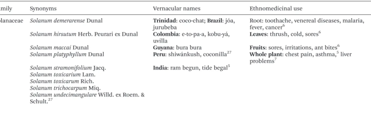
Table 1 Ethnomedicinal information onSolanum stramoniifoliumJacq
Family Synonyms Vernacular names Ethnomedicinal use
Solanaceae Solanum demerarenseDunal Trinidad: coco-chat;Brazil: jóa, jurubeba
Root: toothache, venereal diseases, malaria, fever, cancer6
Solanum hirsutumHerb. Peurari ex Dunal Colombia: e-to-pa-a, kobu-yá, uvilla
Leaves: thrush, cold, sores6
Solanum maccaiDunal Guyana: bura bura Fruits: sores, irritations, ant bites6
Solanum platyphyllumDunal Peru: shiwánkush, coconilla27 Whole plant: chest pain, asthma,5liver problems7
Solanum stramonifoliumJacq. India: ram begun, tide begal5
Solanum toxicariumLam.
Solanum toxicarumRich.
Solanum trichocarpumMiq.
Solanum undecimangulareWilld. ex Roem. & Schult.27
and an automated thermostated column section (kept at 35 °C). The Waters Spherisorb S3 ODS-2 C18 (3 μm, 4.6 ×
150 mm, Waters, Milford, MA, USA) column was used for chro-matographic separations. The solvents used were (A) 0.1% formic acid in water and (B) acetonitrile. The gradient elution applied was: 15% B (0–5 min), 15% B to 20% B (5–10 min), 20–25% B (10–20 min), 25–35% B (20–30 min), 35–50% B (30–40 min), the column was then re-equilibrated, using a flow rate of 0.5 mL min−1. Data were collected simultaneously with
a DAD (280 and 370 nm) and in a mass spectrometer. Negative mode was chosen for MS detection on a Linear Ion Trap LTQ XL mass spectrometer (ThermoFinnigan, San Jose, CA, USA). Sheath gas (nitrogen) was kept at 50 psi. Other parameter set-tings: source temperature: 325 °C, spray voltage: 5 kV, capillary voltage: −20 V, tube lens offset: −66 V, collision energy: 35 arbitrary units. The full scan captured the mass between m/z 100 and 1500. The Xcalibur® data system (ThermoFinnigan, San Jose, CA, USA) was used for data acquisition.
For identification of the phenolic compounds, retention times, UV-VIS and mass spectra were compared with available standards. Data from the literature were used to tentatively identify the remaining compounds. Calibration curves of avail-able phenolic standards were constructed based on the UV signal to perform quantitative analysis. Identified phenolic compounds with unavailable commercial standard were quan-tifiedviacalibration curves of the most similar standard avail-able. The results were expressed as mg g−1of dry extract.
2.5. Biological activity screening
Antibacterial activity. Clinical isolates from patients hospi-talized in the Local Health Unit of Bragança and Hospital Centre of Trás-os-Montes and Alto-Douro-Vila Real, Northeast of Portugal were used in the assay. Four Gram-positive bacteria
(Enterococcus faecalis isolated from urine; Listeria monocyto-genesisolated from cerebrospinal fluid; MSSA: methicillin-sen-sitiveStaphylococcus aureusisolated from wound exudate and MRSA: methicillin-resistantStaphylococcus aureus, isolated from expectoration), and six Gram-negative bacteria (Acinetobacter baumanniiandPseudomonas aeruginosaisolated from expectora-tion;Escherichia coli,Escherichia colispectrum extended produ-cer of β-lactamases (ESBL); Klebsiella pneumoniae, Klebsiella pneumoniaeESBL, all isolated from urine) were used to screen the antibacterial activity of the extracts. Microorganism identifi-cation and susceptibility tests were performed on the MicroScan panels (MicroScan®; Siemens Medical Solutions Diagnostics, West Sacramento, CA, USA) using the microdilution method. The interpretation criteria were based on Interpretive Breakpoints as indicated in the Clinical and Laboratory Standards Institute18 and in the European Committee on
Antimicrobial Susceptibility Testing.19
A microdilution method with rapidp-iodonitrotetrazolium chloride (INT) colorimetric assay according to Kuete et al.20
with some modifications was performed. The extract was diluted in appropriate media according to bacterial require-ments and successive dilutions were carried out in the wells (20 to 0.156 mg mL−1 of final concentration). Three negative
controls (MHB/TSB, the extract, and medium with antibiotic) and a positive control (MHB and each inoculum) were prepared. For the Gram-negative bacteria, negative control antibiotics, such as amikacin (K. pneumoniae ESBL and P. aeruginosa), tobramycin (A. baumannii), amoxicillin/ clavulanic acid (E. coli and K. pneumoniae) and gentamicin (E. coliESBL) were used. The concentration used was based on the MIC obtained (Table 2). For the Gram-positive bacteria, ampicillin (L. monocytogenes) and vancomycin (MSSA, MRSA andE. faecalis) were used (Table 3).
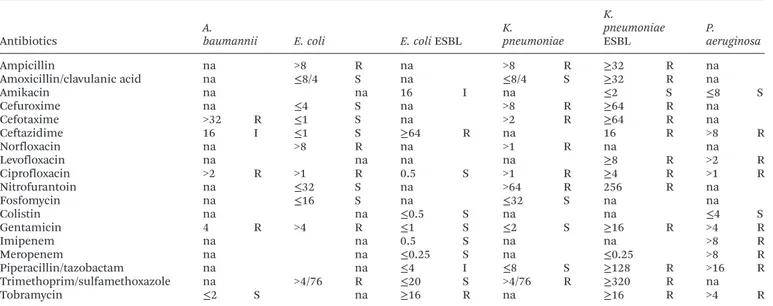
Table 2 Resistance profile of Gram-negative bacteria to different antibiotics; MIC values (µg ml−1)
Antibiotics
A.
baumannii E. coli E. coliESBL
K.
pneumoniae K.
pneumoniae
ESBL
P. aeruginosa
Ampicillin na >8 R na >8 R ≥32 R na
Amoxicillin/clavulanic acid na ≤8/4 S na ≤8/4 S ≥32 R na
Amikacin na na 16 I na ≤2 S ≤8 S
Cefuroxime na ≤4 S na >8 R ≥64 R na
Cefotaxime >32 R ≤1 S na >2 R ≥64 R na
Ceftazidime 16 I ≤1 S ≥64 R na 16 R >8 R
Norfloxacin na >8 R na >1 R na na
Levofloxacin na na na na ≥8 R >2 R
Ciprofloxacin >2 R >1 R 0.5 S >1 R ≥4 R >1 R
Nitrofurantoin na ≤32 S na >64 R 256 R na
Fosfomycin na ≤16 S na ≤32 S na na
Colistin na na ≤0.5 S na na ≤4 S
Gentamicin 4 R >4 R ≤1 S ≤2 S ≥16 R >4 R
Imipenem na na 0.5 S na na >8 R
Meropenem na na ≤0.25 S na ≤0.25 >8 R
Piperacillin/tazobactam na na ≤4 I ≤8 S ≥128 R >16 R
Trimethoprim/sulfamethoxazole na >4/76 R ≤20 S >4/76 R ≥320 R na
Tobramycin ≤2 S na ≥16 R na ≥16 R >4 R
S–susceptible; I–intermediate; R–resistant; classification according to the interpretative breakpoints suggested by Clinical and Laboratory Standards Institute (CLSI) and European Committee on Antimicrobial Susceptibility Testing (EUCAST); na–not applicable.
MIC was defined as the lowest extract concentration that prevented the color change (from yellow dye to dark pink), caused by viable microorganisms, and exhibited the complete inhibition of bacterial growth.
Antioxidant activity. Hydroethanolic extracts were re-dissolved in ethanol/water (80 : 20, v/v) to the final concentration of 20 mg mL−1 and further diluted to 0.156 mg mL−1 to be
subjected to the following assays. The antioxidant activity was evaluated by DPPH radical-scavenging activity, reducing power, inhibition ofβ-carotene bleaching in the presence of linoleic acid radicals and inhibition of lipid peroxidation using TBARS in brain homogenates.21The extract concentrations providing
50% of antioxidant activity or 0.5 of absorbance (EC50) were
calculated from the graphs of antioxidant activity percentages (DPPH,β-carotene bleaching and TBARS assays) or absorbance at 690 nm (reducing power assay) against extract concen-trations. Trolox was used as a positive control.
Anti-inflammatory activity.The method previously described by Correa et al.22 was performed in a concentration range
400–125μg mL−1. Dexamethasone (50μM) was used as a
posi-tive control. The mouse macrophage-like cell line RAW 264.7 stimulated with LPS was used in the assay. Nitric oxide (NO) production was studied with a Griess Reagent System kit. Results were expressed as EC50values (μg mL
−1) equal to the
sample concentration providing a 50% inhibition of NO production.
Cytotoxicity. Dry extracts (stock concentration 8 mg mL−1,
re-dissolved in water) were further diluted to different concen-trations to be subjected to in vitro antitumor activity and hepatotoxicity evaluation at final well concentrations (400–1.5μg mL−1). The cytotoxicity was determined using four
human tumour cell lines, HeLa (cervical carcinoma), HepG2 (hepatocellular carcinoma), MCF-7 (breast adenocarcinoma) and NCI-H460 (non-small cell lung cancer), following a pro-cedure already described by the authors.17The cell growth
inhi-bition was measured using sulforhodamine B assay, where the amount of pigmented cells is directly proportional to the total protein mass and therefore to the number of bounded cells.
For hepatotoxicity evaluation, a freshly harvested porcine liver, obtained from a local slaughter house, was used in order to obtain the cell culture, designated as PLP2. The growth inhi-bition was evaluated using the SRB assay, as previously described.23The results were expressed in GI
50values; sample
concentration that inhibited 50% of the net cell growth. Ellipticine was used as a positive control.
2.6. Statistical analysis
Three repetitions (or two repetitions in case of antimicrobial assay) of the samples were used and triplicates for each con-centration reading were carried out in all the assays. Results are expressed as mean values and standard deviations (SD). The results were analyzed using one-way analysis of variance (ANOVA) followed by Tukey’s HSD test with p = 0.05. When necessary, a Student’st-test was used to determine the signifi-cant difference among two different samples, with p = 0.05. Both statistical treatments were carried out using the SPSS v. 23.0 program.
3.
Results and discussion
3.1 Phenolic compounds’profile
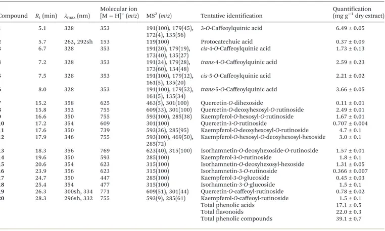
Tables 4 and 5 present chromatographic data and tentative determination of phenolic compounds in the hydroethanolic extracts of leaves, stems, and roots ofSolanum stramoniifolium Jacq. In leaves, 6 phenolic acid derivatives and 14 flavonoids (flavonol glycoside derivatives) were confirmed. Compounds2
and 6 were positively identified as protocatechuic acid and 5-O-caffeoylquinic acid (chlorogenic acid) after comparing the obtained LC-MS data with those of commercial standards. Compound5was tentatively assigned as the correspondingcis isomer of 5-O-caffeoylquinic acid based on its fragmentation pattern and lower levels compared with peak6. Furthermore, cis hydroxycinnamoyl derivatives would be expected to elute before the corresponding trans ones, as observed after UV irradiation (366 nm, 24 h) of hydroxycinnamic acids in our
Table 3 Resistance profile of Gram-positive bacteria to different antibiotics; MIC values (µg ml−1)
Antibiotics MRSA MSSA E. faecalis L. monocytogenes
Penicillin >8 R ≤0.12 S na na
Ampicillin na na ≤4 S ≤0.2 S
Oxacillin >0.25 R ≤0.25 S na na
Clindamycin na >0.5 R na na
Erythromycin na >2 R na na
Ceftaroline ≤1 S na na na
Gentamicin na ≤1 S na na
Ciprofloxacin na >1 R na na
Levofloxacin na >2 R na na
Nitrofurantoin na na ≤64 S na
Linezolid ≤4 S na na na
Trimethoprim/sulfamethoxazole na ≤2/38 S na ≤2/38 S
Vancomycin ≤2 S ≤2 S ≤2 S na
MSSA–methicillin-sensitiveStaphylococcus aureus; MRSA–methicillin-resistantStaphylococcus aureus; S –susceptible; I–intermediate; R–
resistant; classification according to the interpretative breakpoints suggested by Clinical and Laboratory Standards Institute (CLSI) and European Committee on Antimicrobial Susceptibility Testing (EUCAST); na–not applicable.
laboratory.24 cis and trans isomers of 4-O-ca
ffeoylquinic acid (compounds 3 and 4) and trans 3-O-caffeoylquinic acid (compound 1) were distinguished and identified by typical fragmentation patterns as described by Clifford et al.25,26 To
the best of our knowledge these compounds were described in Solanum stramoniifoliumJacq. for the first time.
The flavonol derivatives detected in the leaf extract were mainly glycosides of quercetin (λmaxaround 354 nm; MS2
frag-Table 4 Retention time (Rt), wavelengths of maximum absorption in the visible region (λmax), mass spectral data, and tentative identification of
phenolic compounds in the hydroethanolic extract ofSolanum stramoniifoliumleaves
Compound Rt(min) λmax(nm)
Molecular ion [M−H]−
(m/z) MS2(m/z) Tentative identification Quantification(mg g−1dry extract)
1 5.1 328 353 191(100), 179(45),
172(4), 135(56)
3-O-Caffeoylquinic acid 6.49 ± 0.05
2 5.7 262, 292sh 153 119(100) Protocatechuic acid 0.37 ± 0.09
3 6.7 328 353 191(20), 179(19),
173(40), 135(27)
cis-4-O-Caffeoylquinic acid 1.73 ± 0.13
4 7.2 328 353 191(24), 179(28),
173(60), 134(48)
trans-4-O-Caffeoylquinic acid 2.59 ± 0.23
5 7.5 328 353 191(100), 179(12),
161(5), 135(20)
cis-5-O-Caffeoylquinic acid 2.21 ± 0.02
6 8.0 328 353 191(100), 179(52),
161(5), 135(34)
trans-5-O-Caffeoylquinic acid 3.66 ± 0.05
7 15.2 358 625 463(5), 301(100) Quercetin-O-dihexoside 0.11 ± 0.01
8 15.8 352 755 609(33), 301(100) Quercetin-O-deoxyhexosyl-O-rutinoside 2.49 ± 0.01
9 16.6 350 755 593(100), 285(38) Kaempferol-O-hexosyl-O-rutinoside 1.67 ± 0.01
10 17.2 354 609 301(100) Quercetin-3-O-rutinoside 0.707 ± 0.004
11 17.6 350 739 593(36), 285(95) Kaempferol-O-deoxyhexosyl-O-rutinoside 4.7 ± 0.1
12 17.9 346 755 593(100), 469(50),
285(72)
Kaempferol-O-hexosyl-O-deoxyhexosyl-hexoside 3.0 ± 0.1
13 18.3 356 769 623(40), 315(100) Isorhamnetin-O-deoxyhexoside-O-rutinoside 1.57 ± 0.01
14 19.6 350 593 285(100) Kaempferol-3-O-rutinoside 1.8 ± 0.1
15 20.6 354 623 315(100) Isorhamnetin-O-deoxyhexosyl-hexoside 1.31 ± 0.05
16 23.9 356 623 315(100) Isorhamnetin-3-O-rutinoside 0.366 ± 0.007
17 24.7 350 447 285(100) Kaempferol-3-O-glucoside 0.45 ± 0.03
18 25.4 354 477 315(100) Isorhamnetin-3-O-glucoside 1.5 ± 0.1
19 26.3 300sh, 334 771 609(51), 301(44) Quercetin-O-caffeoyl-rutinoside 0.78 ± 0.02
20 28.3 296sh, 332 755 593(9), 285(61) Kaempferol-O-caffeoyl-rutinoside 1.5 ± 0.1
Total phenolic acids 17.1 ± 0.5
Total flavonoids 22.0 ± 0.3
Total phenolic compounds 39.1 ± 0.7
Table 5 Retention time (Rt), wavelengths of maximum absorption in the visible region (λmax), mass spectral data, and tentative identification of phenolic compounds in the hydroethanolic extract ofSolanum stramoniifoliumroots and stems
Compound Rt(min) λmax(nm)
Molecular ion [M−H]−
(m/z) MS2(m/z) Tentative identification
Quantification (mg g−1
dry extract)
Student’s
t-test
Roots Stems
5 7.3 328 353 191(100), 179(12),
161(5), 135(20)
cis-5-O-Caffeoylquinic acid 2.62 ± 0.22 1.26 ± 0.01 <0.001
6 7.9 328 353 191(100), 179(52),
161(5), 135(34)
trans-5-O-Caffeoylquinic acid 5.03 ± 0.14 3.42 ± 0.02 <0.001
21 17.4 236, 296,
320sh
472 350(40), 308(31) Bis(dihydrocaffeoyl) spermidine isomer 1
1.86 ± 0.16 0.43 ± 0.01 <0.001
22 20.3 226, 294,
322sh
799 637(100), 515(6),
472(10), 350(3), 308(3)
Tris(dihydrocaffeoyl) spermidine hexoside
0.63 ± 0.10 1.17 ± 0.01 <0.001
23 24.3 284 637 515(23), 472(47),
350(15), 308(8)
Tris(dihydrocaffeoyl) spermidine
9.51 ± 0.08 1.06 ± 0.02 <0.001
24 29.4 226, 284,
316sh
472 350(32), 308(38) Bis(dihydrocaffeoyl) spermidine isomer 2
0.78 ± 0.02 0.46 ± 0.05 <0.001
25 31.1 226, 292,
320sh
472 350(30), 308(48) Bis(dihydrocaffeoyl) spermidine isomer 3
0.55 ± 0.09 1.08 ± 0.05 <0.001
Total phenolic compounds and derivatives
20.98 ± 0.81 8.89 ± 0.01 <0.001
mentm/z301), isorhamnetin (λmaxaround 356 nm; MS2 frag-mentm/z317), and kaempferol (λmaxaround 348 nm, MS2 frag-mentm/z285).
Quercetin-3-O-rutinoside (rutin; compound 10), kaemp-ferol-3-O-rutinoside (nicotiflorin; compound 14), isorhamne-tin-3-O-rutinoside (narcissin; compound 16), kaempferol-3-O-glucoside (astragalin; compound 17) and isorhamnetin-3-O-glucoside (compound18) were positively identified upon com-parison of their retention times, UV-Vis characteristics and mass spectra with available commercial standards.
Compound7presented a pseudomolecular ion [M−H]−
at m/z625, releasing a MS2fragment atm/z301 ([M−H−162−
162]−
, loss of two hexosyl moieties), which led to its tentative identification as quercetin-O-dihexoside. Compounds 8, 11, and13provided the same fragmentation losses of deoxyhexose (146 u) and deoxyhexosyl-hexose (308 u), indicating the location of each residue on different positions of the aglycons of quercetin, kaempferol, and isorhamnetin ([M−H]−
atm/z755, 739, and 769, respectively). Similarly, MS2fragments of peaks9
and12revealed the alternative loss of hexosyl (m/zat 593;−162 u) and deoxyhexosyl-hexose (m/zat 285;−308 u) residues. The positive identification of present rutinosides, including querce-tin-3-O-rutinoside, in the samples may suggest a rutinoside identity for the deoxyhexosyl-hexose residues in peaks8,9,11
and13. However, in the case of peak12, the information about the identity of the sugar moieties and location onto the aglycon could not be confirmed, therefore the compound was tentatively identified as kaempferol-O-hexosyl-O-deoxyhexosyl-hexoside. Compound15([M−H]−
atm/z623) presented the same pseudo-molecular ion as compound16, but showed an earlier retention time. The observation of just a single MS2fragment (m/zat 315; −308 u), could indicate that the two sugar units were linked together and the compound was tentatively assigned as isorham-netin-O-deoxyhexosyl-hexoside.
Compounds19([M− H]−
atm/z771) and20([M−H]−
at m/z755) could correspond to compounds including an acyla-tion with a phenolic acid. The observaacyla-tion in their fragmenta-tion of a product ion atm/z609 and 593, respectively, from the losses of caffeoyl residue (162 u), could also be coherent with that identity, as well as the late elution, since the presence of the hydroxycinnamoyl residue implies a decrease in polarity. Therefore, these molecules were tentatively assigned to querce-tin-O-caffeoyl-rutinoside and kaempferol-O-caffeoyl-rutinoside.
The root and stem extracts gave a similar phenolic profile, obtaining different quantities of seven identified compounds. Compounds 5 and 6 were identified as 5-O-caffeoylquinic isomers cis- and trans- as described above. The root extract gave higher amounts of these substances than the stem extract. Compounds21,24, and25([M−H]−
atm/z472) were thought to represent polyamine derivatives, namely three isomers of N,N′-bis(dihydrocaffeoyl)spermidine as described in the literature by Parr et al.27 Similarly, and taking into
account the findings reported by Gancelet al.28compound23
([M−H]−
atm/z637) lead toN,N′,N″ -tris(dihydrocaffeoyl)sper-midine and its hexoside, compound22; [M−H]−
atm/z799, which gives a MS2 fragment at m/z 637 [M − H − 162]−
.
Nevertheless, a complete identification of the position of dihydrocaffeoyl groups on the spermidine skeleton was not possible. Compound 23 was the most abundant compound present in both parts of this species.
Flavonoids were the most abundant group of phenolic com-pounds identified in the present study. Nevertheless, polyamine derivatives (spermidines) were dominant in the root and stem extracts. To date, no record exists on spermidine derivatives in S. stramoniifolium, however, their presence was frequently described in other representatives of Solanum genus, such as potato (S. tuberosum) or naranjilla fruit (S. quitoense).28,29
3.2. Biological activity
The increasing number of bacterial strains resistant to severe available antibiotics remains a huge problem and is a driving force for the search of new compounds with antimicrobial activity.30Furthermore, the food industry calls for natural anti-microbial additives that would be efficient and safe for human consumption at the same time. Various natural peptides, poly-saccharides, terpenes, and phenolic compounds have been applied as food preservatives with no toxicity, such as thymol, carvacrol, chitosan, and nisin.31
The crude extracts of leaves, stems, and roots of S. stramoniifoliumwere tested for antimicrobial activity against selected clinical isolates representing both Gram-positive and Gram-negative bacteria: Acinetobacter baumannii, Klebsiella pneumoniae, Pseudomonas aeruginosa and Staphylococcus aureus, all known to exhibit multi-resistance to antibiotics and labeled as the ESKAPE pathogens (Enterococcus faecium, Staphylococcus aureus,Klebsiella pneumoniae,Acinetobacter bau-mannii,Pseudomonas aeruginosa, andEnterobacterspecies).32It is established that the Gram-negative bacteria possess stronger resistance due to their protective outer membrane rich in lipo-polysaccharides,33which is missing in Gram-positive bacteria.
In Table 6, the results obtained from a broth microdilution method with INT colorimetric evaluation are displayed. As can
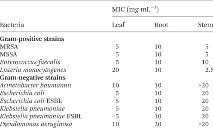
Table 6 Antibacterial activity ofSolanum stramoniifolium
hydroethano-lic extracts (MIC; mg mL−1)
Bacteria
MIC (mg mL−1 )
Leaf Root Stem
Gram-positive strains
MRSA 5 10 5
MSSA 5 10 5
Enterococcus faecalis 5 10 10
Listeria monocytogenes 20 10 2.5
Gram-negative strains
Acinetobacter baumannii 10 10 >20
Escherichia coli 5 10 20
Escherichia coliESBL 5 10 20
Klebsiella pneumoniae 5 10 20
Klebsiella pneumoniaeESBL 5 10 20
Pseudomonas aeruginosa 10 20 >20
ESBL = spectrum extended producer ofβ-lactamases. MIC = minimal inhibition concentration. MRSA = methicillin-resistantStaphylococcus aureus. MSSA = methicillin-sensitiveStaphylococcus aureus.
be seen, all three extracts exhibited antimicrobial activity to all the assayed bacteria, and MICs ranged from 2.5 to 20 mg mL−1. In two cases, the MIC was above the maximal tested
concentration (stem extract against A. baumannii and P. aeruginosa). In general, the Gram-positive bacteria were more sensitive to the extracts than Gram-negative bacteria, as expected. However, the root extract presented non-selective inhibition providing the same MIC values for 9 of 10 bacterial strains (10 mg mL−1). On the other hand, the stem extract was
significantly more active against Gram-positive bacteria. Listeria monocytogeneswas the most susceptible organism pro-viding the lowest MICs in stem extract (2.5 mg mL−1).
P. aeruginosa was the least inhibited organism in the assay. Overall, the leaf extract was the most effective inhibitor with MICs of 5 mg mL−1 obtained for 7 clinical isolates. Notably,
the bacteria with special characteristics, such as methicillin-resistant MRSA or β-lactamase producing E. coli and K. pneumoniae, did not present higher MICs than their more sensitive analogues. The water extract of seeds from S. stramoniifolium(Thailand) showed significant multispectral inhibition (S. aureus, P. aeruginosa, Bacillus. subtilis, Bacillus licheniformis,Xanthomonassp.,Salmonella typhi), however inhi-bition ofE. coli andK. pneumoniae were not observed in the disc diffusion test.12
From the phenolic compounds identified in the plant parts, nicotiflorin, rutin, and chlorogenic acid were previously related with antimicrobial activity in the Solanum genus34and
therefore can contribute to the inhibitory potential of this species.
The results of antioxidant, anti-inflammatory and cytotoxic activity are included in Table 7, due to their possible relation-ship previously described in the literature.35,36 Polyphenol
extracts have been used in the food industry as they often exert
multiple biological activities in protection against spoilage and oxidationviasynergism of the compounds they contain.31
The antioxidant activity was evaluated using four in vitro assays covering various mechanisms, such as hydrogen atom transfer (HAT) and single electron transfer (SET), to fully unfold the antioxidant capacity of the studied samples.37
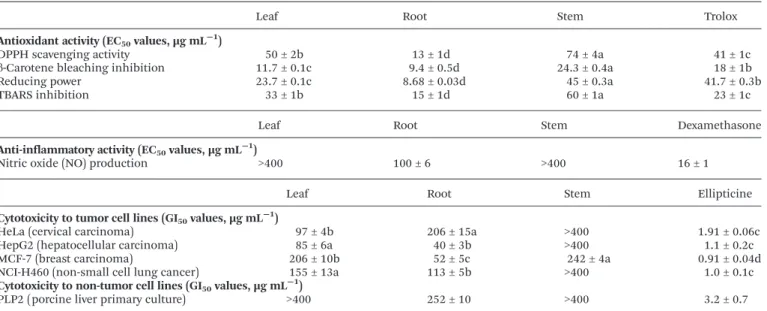
As it can be observed in Table 7, all plant part extracts showed significant antioxidant potential in the four assays (DPPH; reducing power, β-carotene bleaching inhibition and TBARS). The root extract stands out when compared to the other plant parts. It was significantly more effective than trolox standard in all antioxidant assays, providing lower EC50values
in each of the tested assays. Regarding DPPH scavenging capacity assay, the plant parts were declining as follows: root > leaf > stem with the corresponding EC50values of 13 ± 1; 50 ±
2 and 74 ± 4 µg mL−1, respectively. In reducing power assay,
two extracts provided better results than the standard trolox (EC50 = 41.7 ± 0.3 µg mL
−1), namely root and leaf (EC 50 of
8.68 ± 0.03 and 23.7 ± 0.1 µg mL−1, respectively). The order of
activity in reducing power was: root > leaf > stem, as observed in DPPH assay as well. Moreover, the same two extracts proved to be better β-carotene bleaching inhibitors than trolox, as only the stem extract gave a higher EC50value than this
stan-dard (23.4 ± 0.4 versus18 ± 1 µg mL−1). In the TBARS
inhi-bition test, only the root extract exceeded trolox capacity, however the results were still quite promising (root > leaf > stem; EC50values corresponding to 15 ± 1; 33 ± 1 and 60 ±
1 µg mL−1, respectively). Previously, Wetwitayaklung and
Phaechamud10observed low scavenging activity for the
metha-nol fruit extract ofS. stramoniifoliumin TEAC assay using the ABTS+• radical (IC
50 = 1133.08 µg compared to 10.14 µg for
trolox) and correlated it to the low presence of total phenolic compounds (1.55 g gallic acid equivalents per 100 g extract).
Table 7 Biological activity of hydroethanolic extracts from different parts ofSolanum stramoniifoliumJacq
Leaf Root Stem Trolox
Antioxidant activity (EC50values, µg mL−1)
DPPH scavenging activity 50 ± 2b 13 ± 1d 74 ± 4a 41 ± 1c
β-Carotene bleaching inhibition 11.7 ± 0.1c 9.4 ± 0.5d 24.3 ± 0.4a 18 ± 1b
Reducing power 23.7 ± 0.1c 8.68 ± 0.03d 45 ± 0.3a 41.7 ± 0.3b
TBARS inhibition 33 ± 1b 15 ± 1d 60 ± 1a 23 ± 1c
Leaf Root Stem Dexamethasone
Anti-inflammatory activity (EC50values, µg mL−1)
Nitric oxide (NO) production >400 100 ± 6 >400 16 ± 1
Leaf Root Stem Ellipticine
Cytotoxicity to tumor cell lines (GI50values, µg mL−1)
HeLa (cervical carcinoma) 97 ± 4b 206 ± 15a >400 1.91 ± 0.06c
HepG2 (hepatocellular carcinoma) 85 ± 6a 40 ± 3b >400 1.1 ± 0.2c
MCF-7 (breast carcinoma) 206 ± 10b 52 ± 5c 242 ± 4a 0.91 ± 0.04d
NCI-H460 (non-small cell lung cancer) 155 ± 13a 113 ± 5b >400 1.0 ± 0.1c
Cytotoxicity to non-tumor cell lines (GI50values, µg mL−1)
PLP2 (porcine liver primary culture) >400 252 ± 10 >400 3.2 ± 0.7
Trolox, dexamethasone and ellipticine, respectively, were used as positive controls in the assays. All values are means ± SD (n= 9) and in each row different letters represent significant differences (p< 0.05).
Lipid peroxidation products (e.g.malondialdehyde), as well as free radicals, may damage important cell macromolecules, such as DNA, proteins, and lipids and contribute to the devel-opment of pathological processes, including aging, cancer, atherosclerosis, coronary heart disease or neurodegenerative problems.38 Despite the effectiveness of endogenous
anti-oxidant systems, an exogenous source of antianti-oxidants is necessary in the case of excessive presence of oxidative species. Therefore, prevention or limitation of oxidative stress might be achieved by dietary antioxidants, such as phenolic-rich plant extracts.
From the tested plant parts, only the root revealed activity in the NO production (EC50= 100 ± 6 µg mL
−1) as stated in
Table 7. Leaf and stem did not show any activity within the maximal concentration tested (400 µg mL−1), which is
surpris-ing accordsurpris-ing to the traditional choice of leaves for external inflammation. It can be suggested that other than NO pro-duction-related mechanisms are involved and different assays shall be evaluated in future to study this activity.
More than 60% of agents used in cancer therapy are from natural sources, especially tropical plants.39 The Solanum
genus is a good source for anticancer substances, such as sola-nine or solamargine.40,41 The antitumor potential was
evalu-ated against four human tumor cell lines represented by MCF-7 (breast carcinoma), NCI-H460 (non-small cell lung cancer), HeLa (cervical carcinoma) and HepG2 (hepatocellular carcinoma), and porcine liver primary culture PLP2 was selected for cytotoxicity assessment against non-tumor cells. Observing the results presented in Table 7, it can be concluded that leaf and root are the most promising plant parts with anti-tumor compounds as they inhibited all anti-tumor cell lines used in the study. The highest inhibition was found for HepG2, yielding the lowest GI50(40 ± 3 µg mL
−1 for root and 85 ± 6
µg mL−1for leaf extract). The stem extract was efficient only in
MCF-7 cell line inhibition (GI50= 242 ± 4 µg mL
−1). The most
sensitive cell line was MCF-7, which was inhibited by all three extracts in the following order root > leaf > stem. Interestingly, the root extract provided lower GI50 for HepG2, MCF-7 and
NCI-H460 than leaf, but was less effective against HeLa cell line. Compared to ellipticine, the extracts revealed medium activity. Nevertheless, ellipticine has a very strong inhibiting power on all presented tumor cell lines, but also exhibits high hepatotoxicity to non-tumor PLP2 cell line. In our case, only root showed mild hepatotoxicity towards PLP2 (GI50 = 252 ±
10 µg mL−1), however it did not exceed active concentrations
against the tumor cell lines (40 ± 3 µg mL−1 in HepG2; 52 ±
5 µg mL−1in MCF-7; 113 ± 5 µg mL−1in NCI-H460; and 206 ±
15 µg mL−1in HeLa).
Consequently, although the leaf and root extracts of S. stramoniifoliumcould be useful in the development of new anticancer products, the leaf is the most promising part, since it did not present unspecific toxicity, as suggested by results obtained with the PLP2 assay.
Due to the possible synergetic effect of present compounds, the plant crude extracts can often be a more powerful anti-oxidant tool than individual substances. Moreover, the natural
matrices in the form of crude extracts possess usually very low toxicity compared to individual chemicals and therefore are currently experiencing a renaissance in both the phytopharma-cological and food industry.31
4.
Conclusions
This study highlights the potential of different parts of Solanum stramoniifoliumJacq. as a rich source of biologically active compounds suitable for applications in the food indus-try, for example in the development of novel functional foods and nutraceutical formulations. Ethanol/water extracts from leaves, stems, and roots demonstrated to have a strong biologi-cal activity. The root extract gave the highest antioxidant poten-tial exceeding trolox standard values. It also significantly inhibited the growth of MCF-7 and HepG2 tumor cell lines. The leaf extract showed the best results in the antimicrobial assay inhibiting all the clinical bacterial isolates. Furthermore, it did not possess any cytotoxicity, unlike the root extract, and therefore might be a better candidate for the food industry. The phenolic compounds in the extracts revealed the content of compounds known for their biological activities, such as caffeoylquinic acid derivatives, flavonoids and polyamines. The presence of these compounds could be correlated with the high biological activity shown by these extracts. Several com-pounds were determined for the first time in this plant.
Con
fl
ict of interest
No conflict of interest.
Acknowledgements
The authors thank the Foundation for Science and Technology (FCT, Portugal) and FEDER under Programme PT2020 for financial support to CIMO (UID/AGR/00690/2013) and L. Barros (SFRH/BPD/107855/2015), S. Heleno (SFRH/BPD/ 101413/2014) and R. C. Calhelha (SFRH/BPD/BPD/68344/2010) grants. To POCI-01-0145-FEDER-006984 (LA LSRE-LCM), funded by FEDER, through POCI-COMPETE2020 and FCT. This study was financially supported by Internal Grant Agency of Tomas Bata University in Zlin, project no. IGA/FT/2016/003.
References
1 M. Carocho and I. C. F. R. Ferreira, Food Chem. Toxicol., 2013,51, 15–25.
2 O. Paredes-Lopez, M. L. Cervantes-Ceja, M. Vigna-Perez and T. Hernandez-Perez,Plant Foods Hum. Nutr., 2010,65, 299– 308.
3 D. A. Herms and W. J. Mattson,Q. Rev. Biol., 1992,67, 283– 335.
4 M. D. Whalen, D. E. Costich and C. B. Heiser, Gentes Herbarum, 1981,12, 41–129.
5 H. B. Das, K. Majumdar, B. K. Datta and D. Ray,Nat. Prod. Radiance, 2009,8, 172–180.
6 R. A. DeFilipps, S. L. Maina and J. Crepin,Medicinal Plants of the Guianas (Guyana, Surinam, French Guiana), Department of Botany, National Museum of Natural History, Smithsonian Institution, Washington, D. C., 2004, 491 p.
7 C. T. Pedrollo, V. F. Kinupp, G. Shepard Jr. and M. Heinrich,J. Ethnopharmacol., 2016,186, 111–124. 8 Y. Estevez, D. Castillo, M. T. Pisango, J. Arevalo, R. Rojas,
J. Alban, E. Deharo, G. Bourdy and M. Sauvain, J. Ethnopharmacol., 2007,114, 254–259.
9 W. Samee, M. Engkalohakul, N. Nebbua, P. Direkrojanavuti, C. Sornchaithawatwong and N. Kamkaen, Thai Pharm. Health Sci. J., 2006,1, 196–203.
10 P. Wetwitayaklung and T. Phaechamud,Res. J. Pharm., Biol. Chem. Sci., 2011,2, 146–154.
11 A. Sakunpak and P. Panichayupakaranant, Food Chem., 2012,130, 826–831.
12 R. Sarnthima and S. Khammuang,Int. J. Agric. Biol., 2012,
14, 111–115.
13 I. C. S. Aires, R. A. Lima and A. G. S. Braga, in 64o
Congresso Nacional de Botânica, Belo Horizonte, Brazil, 2013.
14 R. Pingal, Thesis, University of West Indies, 2008.
15 PAHO/WHO, 2013. http://www.paho.org/hq/index.php? option=com_content&view=article&id=9135%3A2013-cancer- mortality-declining-some-countries-americas-new-paho-who-report&catid=740%3Apress-releases&Itemid=1926〈=en (accessed May 2016).
16 H. Chandoura, J. C. M. Barreira, L. Barros, C. Santos-Buelga, I. C. F. R. Ferreira and L. Achour,Ind. Crops Prod., 2015,65, 383–389.
17 L. Barros, E. Pereire, R. C. Calhelha, M. Duenas, A. M. Carvalho, C. Santos-Buelga and I. C. F. R. Ferreira, J. Funct. Foods, 2013,5, 1732–1740.
18 CLSI, Performance Standards for Antimicrobial Susceptibility Testing, 18th informational supplement. CLSI document M100-S18, Clinical and Laboratory Standards Institute, Wayne, PA, USA, 2008.
19 EUCAST, European Society of Clinical Microbiology and Infectious Diseases, 2013. http://www.eucast.org/ ast_of_bacteria/previous_versions_of_documents/ (accessed March 2016).
20 V. Kuete, P. Y. Ango, G. W. Fotso, G. D. W. F. Kapche, J. P. Dzoyem, A. G. Wouking, B. T. Ngadjui and B. M. Abegaz, BMC Complementary Altern. Med., 2011,11, 42–46.
21 J. Pinela, L. Barros, M. Dueñas, A. M. Carvalho, C. Santos-Buelga and I. C. F. R. Ferreira, Food Chem., 2012, 135, 1028–1035.
22 R. C. G. Correa, A. Henrique Pereira de Souza, R. C. Calhelha, L. Barros, J. Glamoclija, M. Sokovic, R. M. Peralta, A. Bracht and I. C. F. R. Ferreira,Food Funct., 2015,6, 2155–2164.
23 R. M. V. Abreu, I. C. F. R. Ferreira, R. C. Calhelha, R. T. Lima, M. Helena Vasconcelos, F. Adega, R. Chaves and M.-J. R. P. Queiroz,Eur. J. Med. Chem., 2011,46, 5800–5806. 24 L. Barros, M. Dueñas, A. M. Carvalho, I. C. F. R. Ferreira
and C. Santos-Buelga,Food Chem. Toxicol., 2012,50, 1576– 1582.
25 M. N. Clifford, K. L. Johnston, S. Knight and N. Kuhnert, J. Agric. Food Chem., 2003,51, 2900–2911.
26 M. N. Clifford, J. Kirkpatrick, N. Kuhnert, H. Roozendaal and P. R. Salgado,Food Chem., 2008,106, 379–385.
27 A. J. Parr, F. A. Mellon, I. J. Colquhoun and H. V. Davies, J. Agric. Food Chem., 2005,53, 5461–5466.
28 A. L. Gancel, P. Alter, C. Dhuique Mayer, J. Ruales and F. Vaillant,J. Agric. Food Chem., 2008,56, 11890–11899. 29 C. E. Narváez-Cuenca, J. P. Vincken and H. Gruppen,Food
Chem., 2012,130, 730–738.
30 G. Normanno, G. La Salandra, A. Dambrosio, N. C. Quaglia, M. Corrente, A. Parisi, G. Santagada, A. Firinu, E. Crisetti and G. V. Celano, Int. J. Food Microbiol., 2007,115, 290–296.
31 M. Carocho, M. F. Barreiro, P. Morales and I. C. F. R. Ferreira, Compr. Rev. Food Sci. Food Saf., 2014,13, 377–399.
32 J. N. Pendleton, S. P. Gorman and B. F. Gilmore,Expert Rev. Anti-Infect. Ther., 2013,11, 297–308.
33 D. I. Andersson and D. Hughes,Nat. Rev. Microbiol., 2010,
8, 260–271.
34 A. Kröner, N. Marnet, D. Andrivon and F. Val,Plant Physiol. Biochem., 2012,57, 23–31.
35 A. Winczura, D. Zdzalik and B. Tudek, Free Radical Res., 2012,46, 442–459.
36 A. M. Pisoschi and A. Pop,Eur. J. Med. Chem., 2015,97, 55– 74.
37 R. L. Prior, X. L. Wu and K. Schaich,J. Agric. Food Chem., 2005,53, 4290–4302.
38 B. N. Ames, M. K. Shigenaga and T. M. Hagen,Proc. Natl. Acad. Sci. U. S. A., 1993,90, 7915–7922.
39 E. Elisabetsky and G. C. de Souza,Farmacogn. da planta ao Medicam., 2004,2, 87–99.
40 M. Amir and S. Kumar,J. Sci. Ind. Res., 2004,63, 116–124. 41 K. W. Kuo, S. H. Hsu, Y. P. Li, W. L. Lin, L. F. Liu,
L. C. Chang, C. C. Lin, C. N. Lin and H. M. Sheu,Biochem. Pharmacol., 2000,60, 1865–1873.