UNIVERSIDADE TÉCNICA DE LISBOA
Faculdade de Medicina Veterinária
DIAGNOSTIC VALUE OF MRI IN DOGS WITH INFLAMMATORY NASAL DISEASE
ANA RITA RAMALHO FURTADO
CONSTITUÍÇÃO DO JÚRI ORIENTADOR
Doutora Maria da Conceição da Cunha e Vasconcelos Peleteiro
Professor Michael E. Herrtage
Doutor Fernando Manuel d’Almeida Bernardo CO-ORIENTADOR
Doutor António José Almeida Ferreira Doutor António José Almeida Ferreira 2010 LISBOA
i
Acknowledgments
This Master project was carried out in the Department of Diagnostic Imaging, at the Queen’s Veterinary School Hospital, Cambridge Veterinary School, University of Cambridge from 21th of September 2009 to the 30th of April 2010.
I am sincerely grateful to my supervisor, Professor Michael Herrtage, for accepting me to this prestige institution, but mainly for the encouragement, guidance and support from the initial to the stages of the project. I consider myself very fortunate for being able to work with a very considerate and encouraging Professor like him.
This thesis would not have been possible without the help of Abby Caine, whose guidance, knowledge and patience enabled me to develop an understanding of the subject. Her enlightening suggestions and encouragements made me feel I was not isolated in my research. I owe my deepest gratitude to Fernando Constantino Casas for providing me all the histopathology data needed, without his help, it would have been impossible to reach all the aims of this project.
It was a great pleasure to learn from respectful and international known Professors and researchers that are truly inspirational in many areas of veterinary medicine such as diagnostic imaging, internal medicine, soft tissue surgery, orthopaedics, neurology and oncology. I hope one day I could cooperate in other scientific project(s) with these expertise Professors.
I am also indebted with my co-supervisor Professor António José Almeida Ferreira for the continued encouragement, support and help during my Master’s degree.
I would like to thank my mother, Leonor Ramalho, for helping me with the statistical and formatting aspects, without her help, time and patience this project would not have been possible, in such a short period time.
I would like to show my gratitude to my lovely family, in especially my father João Furtado and my grandparents Amélia Salavessa, Manuel Ramalho, Maria de S.João Silva e Armando Furtado, for the support and endless efforts during all my academic life.
I would like to thank all my UK friends, including the Link House people, Ana Luisa Silva, Pedro Saavedra, Pedro Correa de Sampaio, Mark Barber, Ana Barbosa and Miguel Solano for all their friendly words of encouragement during my stay in the Cambridge.
I would like to extent my thanks to all my friends in Portugal, especially to Mafalda Pires Gonçalves, Ana Maria Azevedo, Rafaela Araújo, Inês Moutinho, Margarida Costa, Madalena Centeno, Carolina Silva, Inês Salvado, João Vieira Lopes, Vera Pereira, Luís Lagoa, Carlos Baptista, Patrícia Paredes, Alexandra Couto, Adriana Mendes, Tiago Mota, Sara Alfacinha, João Mauritti, Vânia Sá, Joana Leitão, Bernardo Garcia, Inês Lopes, Mariana Horta, Duarte Bettencourt, Lourenço Simas, Ana van Uden and Tomás Rodrigues. These individuals always
ii
helped me to keep my life in context. Graduate school is not the most important thing in life, but good friends, good times and happiness are.
iii
Congress Presentation
Part of the results of this research will be presented in the Southern European Veterinary Conference (Barcelona from 30 September to 3 October, 2010) in both abstract and poster formats.
v
DIAGNOSTIC VALUE OF MRI IN DOGS WITH INFLAMMATORY NASAL DISEASE
This study determines the value of low-field magnetic resonance imaging in differentiating nasal aspergillosis from chronic rhinitis in dogs. The Queen’s Veterinary School Hospital magnetic resonance imaging database (2002-2009) was searched for dogs that had undergone MRI of the nasal cavity. Forty-one cases were included of which twenty five were classified as Rhinitis and sixteen as Aspergillosis. On MRI, destruction of the turbinates was classified as mild, moderate or severe. The cribriform plate and vomer/nasal septum destruction were classified as present or absent as well as the involvement of the frontal sinus. Images were examined to assess the signal intensity of mucus and turbinates and classified as hypointense, hyperintense and isointense on the brightest area on the same slice. On T1W images, the intensity was compared with muscle and on T2W images with periorbital fat and brain.
Turbinate destruction was statistically associated (p=0.005) with aspergillosis. Most of the Rhinitis cases (48%) had no turbinate destruction but a significant number had mild destruction (40%). Six Aspergillosis cases (37.5%) had moderate turbinate destruction and only three cases (18.8%) had severe destruction. Cribriform plate and vomer/nasal septum destruction, although not statistically associated with any pathology, were more frequent in Aspergillosis. There was no statistical association with Rhinitis or Aspergillosis with respect to frontal sinus involvement. On T1W scans, Aspergillosis was associated with turbinate hyperintensity, whilst Rhinitis was associated with turbinate hypointensity (p=0.007). On T2W scans, the turbinate intensity was not statistical significant with Rhinitis or Aspergillosis, however the majority (60%) of Rhinitis cases exhibited hypointense turbinates, whereas the majority (56.3%) of Aspergillosis cases had isointense turbinates. On T1W, mucus intensity was not statistical associated with Rhinitis or Aspergillosis, but it was noticed that mucus hyperintensity was the most significant feature in both groups. On T2W scans, there was no statistical significance when comparing with fat and brain, with mucus, nonetheless it was noted that the majority of cases showed hyperintense mucus. It was concluded that T1W scans provided more information since the turbinate hyperintensity seen was associated with Aspergillosis while hypointensity was with Rhinitis. On T2W images relevant differences were seen but these were not associated with specific pathology.
vii
VALOR DIAGNÓSTICO DA RESSONÂNCIA MAGNÉTICA NUCLEAR EM DOENÇAS INFLAMATÓRIAS NASAIS NO CÃO
Este estudo pretende determinar o valor da ressonância magnética nuclear (RMN) de baixo campo no diagnóstico diferencial entre aspergilose e rinite crónica em cães. Foram pesquisados registos de cães que realizaram RMN da cavidade nasal no Queen’s Veterinary School Hospital em Cambridge. A população era constituída por 41 casos dos quais 25 foram classificados como Rinite e 16 como Aspergilose. Nas imagens de RMN, a destruição das conchas nasais foi classificada como ligeira, moderada e grave. A destruição da placa cribiforme e do vómer/septo nasal e o envolvimento do seio frontal foram classificadas como presente ou ausente. Analisaram-se as imagens para avaliar a intensidade do muco e das conchas nasais, que foram classificados como hipointensas, hiperintensas ou isointensas na zona mais brilhante do mesmo corte. Nas imagens de T1W, a intensidade foi comparada com o músculo e nas imagens em T2W com a gordura periorbital e cérebro. Verificou-se que a destruição das conchas nasais estava estatisticamente associada com a Aspergilose (p=0.005). A maior parte dos casos de Rinite (48%) não apresentavam destruição das conchas nasais, no entanto um número considerável tinha uma destruição ligeira (40%). Seis casos (37.5%) de Aspergilose tinham uma destruição moderada das conchas nasais e só três casos (18.8%) tinham destruição grave. Apesar de a destruição da placa cribriforme e do vómer/septo nasal se ter observado com mais frequência nos casos de Aspergilose, não se demonstrou qualquer associação estatisticamente significativa. Também não se observou associação estatisticamente significativa entre qualquer das duas patologias e o envolvimento do seio frontal. Em T1W, a Aspergilose estava estatisticamente associada com a hiperintensidade das conchas nasais, enquanto a Rinite estava associada com a hipointensidade das mesmas (p=0.007). Em T2W, não se observou nenhuma associação com significado estatístico entre a intensidade das conchas nasais e qualquer das duas patologias, contudo a maioria dos casos de Rinite (60%) apresentava conchas nasais hipointensas, enquanto a maioria dos casos de Aspergilose (56.3%) tinha conchas nasais isointensas. A intensidade do muco em T1W, não estava estatisticamente associada a nenhuma das patologias, de facto a hiperintensidade do muco foi a característica mais frequentemente encontrada em ambos os grupos. Quando foi comparada a intensidade do muco em T2W com a gordura e o cérebro, também não foram encontradas quaisquer associações estatisticamente significativas, sendo de realçar que a maioria dos casos apresentava muco hiperintenso. Em conclusão, pode-se afirmar que as imagens em T1W têm mais valor diagnostico que as de T2W, uma vez que se descreveram associações estaticamente significativas nesta sequência.
ix
Table of Contents
1
Externship Report ... 1
2
Literature review ... 5
2.1 Introduction ... 5
2.2 Respiratory system general considerations ... 5
2.3 Anatomy of the nasal passages of the dog ... 8
2.4 Importance of clinical history in dogs in nasal disease ... 9
2.5 Clinical signs of nasal disease in the dog ... 10
2.5.1 Nasal discharge ... 10
2.5.2 Epistaxis ... 11
2.5.3 Stertor/reverse-sneezing and stridor ... 12
2.5.4 Sneezing ... 13
2.5.5 Other reported signs ... 13
2.6 Physical Examination ... 13
2.7 Differential Diagnoses ... 14
2.8 Imaging studies ... 15
2.8.1 Radiography ... 16
2.8.2 Computed tomography ... 18
2.8.3 Magnetic resonance imaging ... 19
2.9 Other ancillary diagnostic tests ... 29
2.9.1 Rhinoscopy ... 29 2.9.2 Nasal biopsy ... 31 2.9.3 Histopathology ... 32 2.9.4 Nasal cultures ... 33 2.9.5 Serology ... 34 2.9.6 Leukogram ... 35 2.10 Respiratory diseases ... 36 2.10.1 Lymphoplasmacytic Rhinitis ... 37 2.10.2 Allergic rhinitis ... 40 2.10.3 Mycotic Rhinitis ... 41 2.11 Systemic aspergillosis ... 45 2.12 Treatment ... 47 2.12.1 Anti-fungal treatment ... 47
2.12.2 Current treatment for canine nasal aspergillosis ... 51
2.12.3 Surgical considerations ... 53
x
2.14 Conclusions from the literature review and research aims ... 55
3
Materials and Methods ... 57
3.1 Case Selection criteria ... 57
3.2 Diagnostic Criteria ... 57
3.3 Procedures ... 57
3.3.1 Clinical signs and Leukogram ... 58
3.3.2 Radiology ... 58
3.3.3 Magnetic Resonance Imaging (MRI) ... 59
3.3.4 Rhinoscopy and Nasal Biopsies ... 60
3.3.5 Histopathology ... 62
3.3.6 Bacteriology and Mycology ... 62
3.3.7 Final Diagnosis ... 63 3.3.8 Statistics analysis ... 63
4
Results ... 65
4.1 Patient Characteristics ... 65 4.2 Clinical Signs ... 65 4.3 Leukogram ... 684.4 Ancillary diagnostic tests ... 69
4.4.1 Radiography ... 69
4.4.2 Magnetic Resonance Imaging (MRI) ... 70
4.4.3 Rhinoscopy ... 80 4.4.4 Histopathology ... 82 4.4.5 Bacteriology ... 88 4.4.6 Mycology ... 88 4.5 Statistic summary ... 89 4.6 Final diagnosis ... 90 4.7 Treatment ... 90 4.8 Follow-up ... 91
5
Discussion ... 93
6
Conclusions ... 107
7
Bibliography ... 111
xi
Figures
Figure 1 - Saggital section of the skull, medial view ... 9
Figure 2 – Transverse section of the nasal cavity. ... 9
Figure 3 - Positioning of the head. LL and DV projections. ... 16
Figure 4 – Positioning of the head. Open mouth RV-DCd and intra-oral DV projections. ... 16
Figure 5 – T1W (left) and T2W (right) sequences of the same dog ... 22
Figure 6 – Transverse sections through the nasal cavity, tongue and lower jaw ... 25
Figure 7 – Dorsal sections through the nasal cavity and cranium ... 25
Figure 8 - Saggital section through the nasal cavity and cranium ... 26
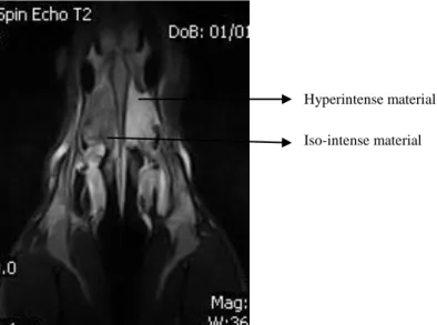
Figure 9 – Hyperintense and iso-intense material in the nasal cavity on a T2W image ... 27
Figure 10 – Nasal discharge on a dog with aspergillosis (19/04/2010) ... 42
Figure 11 – Horizontal beam frontal sinus skyline view x-ray at QVSH, Radiology Department (10/11/2009) ... 59
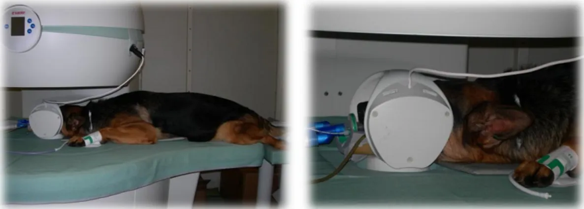
Figure 12 - Nasal MRI scan on a German Shepherd dog at the Veterinary MRI Unit (10/11/2009) ... 59
Figure 13 - Rhinoscopy on a German Shepherd dog at the QVSH, Surgery Department (10/11/2009)61 Figure 14 - Biopsy taken during rhinoscopy (10/11/2009) ... 62
Figure 15 – How to reach a final diagnosis in inflammatory nasal diseases ... 64
Figure 16 – Radiography descriptive results ... 69
Figure 17 – Dorsal section. Destruction of the septum/vomer on T1W. ... 71
Figure 18 – Transverse section. Frontal sinus involvement seen on T1W. ... 72
Figure 19 – Turbinate destruction seen on transverse and saggital sections. ... 73
Figure 20 – Dorsal sections on T2W sequence. ... 77
Figure 21 – Mucus Hyperintensity on T1W ... 78
Figure 22 – Mucus hyperintensity on T2W when compared with fat and brain, on a dog with aspergillosis ... 79
Figure 23 – Aspergillosis treatment with clotrimazole infusion (left) followed by clotrimazole cream deposition (right). ... 91
Figure 24 –Aspergillosis treatment with enilconazol flush through frontal sinus tubes ... 92
Tables
Table 1 – Anatomic limits of upper respiratory tract and defining clinical signs adapted from (Ford, 2005) ... 13Table 2 – Diagnostic criteria for nasal aspergillosis ... 15
Table 3 – Relaxation sequences formation... 21
Table 4 - The magnetic resonance criteria evaluated ... 60
Table 5 - Classification * Epistaxis Cross-tabulation ... 66
Table 6 - Classification * Nasal Depigmentation Cross-tabulation ... 67
Table 7 - Classification * X-ray Cross-tabulation ... 70
Table 8 – Classification * Turbinate Destruction Cross-tabulation ... 72
Table 9 -Turbinate Destruction * Final Inflammation Grade Cross-tabulation ... 73
Table 10 - Rhinoscopy * Turbinate Destruction (2) on MRI cross-tabulation ... 74
Table 11 - Turbinate Destruction (2) * Final Inflammation Grade Cross-tabulation ... 75
Table 12 - Classification * Turbinate Intensity on T1W Cross-tabulation ... 76
Table 13 - Epistaxis * Turbinate Intensity on T1W cross-tabulation ... 76
Table 14 – Classification * Frontal sinus involvement Cross-tabulation ... 80
xii
Table 16 - Rhinoscopy * MRI diagnosis Cross-tabulation ... 82
Table 17 - Classification * Fungi Cross-tabulation ... 87
Table 18 - Classification * Serology Cross-tabulation ... 89
Charts
Chart 1-Population skull Size ... 65Chart 2 - Clinical Signs ... 66
Chart 3 - Leukogram results ... 68
Chart 4 – Most prevalent abnormalities found in the leukogram ... 68
Chart 5 – Distribution of Turbinate Destruction (2) according to Rhinitis and Aspergillosis ... 74
Chart 6 - Turbinate Intensity on T2W ... 77
Chart 7 – Mucus intensity on T2W ... 79
Chart 8 – Rhinoscopy results distribution according to the Classification ... 81
Chart 9 – Cell type frequency distribution according to Rhinitis and Aspergillosis ... 84
Chart 10 – Distribution of inflammation grade according to Rhinitis and Aspergillosis ... 84
Chart 11 – Distribution of the different type cell hyperplasia according to Rhinitis and Aspergillosis 86 Chart 12 – Distribution of oedema according to Rhinitis and Aspergillosis ... 86
Chart 13 - Bacteriology results ... 88
xiii
Abbreviations and symbols
AGDD – Agar-Gel Double ImmunoDiffusion AR- Allergic Rhinitis
BID – bis in die
CD – Cluster of Differentiation CNS- Central Nervous System CSF – Cerebrospinal Fluid CT – Computed tomography
ELISA – Enzyme-Linked Immuno Sorbent Assay FeLV – Feline Leukaemia Virus
FIP – Feline Infectious Peritonitis
FLAIR – Fluid Attenuation Inversion Recovery IDST – Intradermal skin test
IFN - Interferon Ig – Immunoglobulin Il - Interleukin Kg – Kilograms
LPR – Lymphoplasmacytic rhinitis
MHC – Major Histocompatibility Complex Ml - Milliliters
MR – Magnetic Resonance
MRI- Magnetic Resonance Imaging
NSAID- Non Steroidal Anti-Inflammatory Drug PO – Per os
QVSH – Queen’s Veterinary School Hospital RF – Radiofrequency
SE – Spin Echo SID – Semel in die SP – Species
SPSS - Statistical Package for the Social Sciences STIR – Short Time Inversion Recovery
TE – Echo time
TGF – Transforming growth factor TR – Repetition Time
1
1 Externship Report
The externship was done at the Queen’s Veterinary School Hospital (QVSH), Department of Veterinary Medicine of the University of Cambridge, between the 21th of September 2009 and the 30th of April 2010 (41 weeks).
The QVSH is a teaching and referral hospital with a national and international reputation as a centre for clinical excellence. It offers referral and advice in the areas of small animal studies (orthopaedics, soft tissue surgery, internal medicine, ophthalmology, oncology, neurology and anaesthesia and intensive care), farm animal studies, equine studies and diagnostic imaging (radiography, ultrasound, MRI and scintigraphy).
Diagnostic imaging department
The diagnostic imaging department offers the following activities:
Three dedicated radiography suites for imaging small and large animals
A digital radiography system
Digital image intensification for functional studies and interventional radiology, e.g. balloon valvuloplasty and pacemaker implantation
High resolution ultrasound machines for imaging small and large animals including echocardiography
Scintigraphy and nuclear medicine used mainly for equine patients, but also occasionally for small animals
A dedicated magnetic resonance imaging for small animals as well as magnetic imaging for standing horses. Indications for small animal MRI include: neurological and spinal disease, tumour assessment for surgical or radiotherapy planning, evaluation of nasal disease, obscure lameness and discharging sinuses.
Thirty one of the 41 weeks spent at the QVSH, were at the diagnostic imaging department. The clinical staff present during the externship was: Michael Herrtage, Barbara Posh, Valentina Piola, Abby Caine, Nicholas Rousset and Julie Sales.
The following activities were done:
Assisted at several seminars, including orthopaedics, hip and elbow dysplasia, skull, abdominal and thoracic imaging.
Participated in morning rounds, in which x-rays and MRI scans from the day before were evaluated and discussed by board-certified specialists.
2
Was able to see several contrast studies on x-rays and under fluoroscopy.
Trained ultrasonographic techniques, learnt how to make differentials based on ultrasonographic results, and assisted several ultrasonographic guided biopsies and fine-needle aspirates.
Several pathologies were seen on MRI like: Chiari-
like malformation and Syringomyelia in the Cavalier King Charles Spaniel, spinal disc herniation and brain tumours.
Other activities assisted included: echocardiography, joints and tendons ultrasonography and myelography.
All the x-rays and MRI scans from 2000 were available for consult during all the stay at the hospital
Small animal hospital
During the externship I was able to rotate through different areas of small animal medicine. I spent 2 weeks on neurology, 2 weeks on internal medicine, 1 week on oncology, 1 week in soft tissue surgery and 1 week in orthopaedic surgery. Besides this I did 20 night-shift (12 hrs), in which I was able to follow and study medications for intensive and critical care, as well as study pain management in small animals.
Since the hospital is open 24 hours, I was also able to help the interns during emergencies such as intoxications, bites, road-traffic accidents and seizures. The following activities were done, during the weeks at the small animal hospital:
Neurology
During the two weeks in neurology, I assisted morning rounds, consults, imaging, surgeries and treatment of dogs with neurologic deficits. The neurology clinical staff, with whom I was able to work, included Nick Jeffery, An Vanhaesebrouck, John Parker and Tom Harcourt-Brown.
During consults I was able to learn how to do a neurologic exam and what further exams we should do to an animal with neurologic deficits. The main pathologies seen were: Myastenia Gravis, Lafora disease, idiopathic epilepsy, chronic seizures, atlanto-axial subluxation, Cavalier King Charles falling syndrome, spinal disc herniation, chiari malformation, brain tumours, hydrocephalus and epilepsy/narcolepsy in horses.
The surgeries seen included ventral slot, dorsal laminectomy and stabilisation of an atlanto-axial subluxation by a transarticular screw fixation.
3
Internal Medicine
During the 2 weeks in the internal medicine department I was able to do consults, admit patients at the hospital, write referral letters and participate on morning and afternoon rounds. I studied pathophysiology and treatment of cases, such as diabetes mellitus, hypoadrenocorticism, hyperadrenocorticism, aspergillosis, renal insufficiency (chronic and acute), pancreatitis, leptospirosis, hypertiroidism, cardiomyopathies, rhinitis, urolithiasis and immune-mediated diseases. I was also able to see 2 rhinoscopy procedures and 1 pace-maker implantation.
I had the opportunity to work with Michael Herrtage, Penny Watson, Barbara Skelly, Mark Reading, Ben Harris and Allison Collings.
Oncology
The oncology department facilities include orthovoltage and megavoltage radiotherapy and radiation treatment planning assisted by MRI, chemo- and photodynamic therapy for animals with very superficial carcinomas. The clinical staff included: Jane Dobson, Malcolm Brearley and Frances Taylor
During the week in the oncology department, I was able to see consults (first or check-ups) and assist and help during radiotherapy and chemotherapy sessions. Some of the pathologies seen wereepitheliotropic cutaneous lymphoma, mast cell tumour, nasal tumours, multicentric lymphoma, osteosarcoma, haemagiosarcoma, glioma, transitional cell carcinoma, histiocytic sarcoma and anal sac gland carcinoma.
Soft Tissue Surgery
On soft tissue surgery, I participated on several surgeries such as spays and castrations, epulis removal, dental surgery on a rabbit, removal of a pancreatic limb due to an insulinoma, removal of an infiltrative lipoma and wound closures. Besides morning and afternoon rounds, I learn how to do dressings and how to do pain scores in surgical patients. During the week, I worked with Jane Ladlow, Jackie Demetriou and Graham Hayes.
Orthopaedics
During the week that I spent in orthopaedics, I participated in morning and afternoon rounds, did consults and referral letters and evaluated post-operative x-rays. I was also able to participate on surgeries, such as total hip replacement, TPLO, distal femoral osteotomy and fracture reduction with locking plates. The clinical staff with whom I worked was Sorrel Langley-Hobbs and Ian Nicholson.
4
The student The supervisor
5
2 Literature review
2.1 Introduction
Respiratory system diseases are considered to be common and important causes of morbidity and mortality in animals and humans, mainly because the respiratory tract is in direct contact with the physical environment and is exposed to airborne microorganisms such as viruses, bacteria, fungi and parasites. The nose is the first part of the respiratory tract and is more likely to be subjected to these insults. Clinicians of all the veterinary fields are daily contacted to diagnose and treat respiratory tract disorders.
Aspergillosis and lymphoplasmacytic rhinitis are the most common inflammatory nasal diseases, and because of that their correct diagnose is essential. With the increased availability of non-invasive diagnostic techniques, such as magnetic resonance, it has started to be used more often to evaluate animals with nasal disease.
The purpose of this chapter is: i) to provide background information about the most common nasal inflammatory diseases in dogs, ii) to review the salient literature about nasal imaging on dogs, iii) to provide recent knowledge about other ancillary diagnostic methods and treatment and lastly iv) to draw conclusions from the literature review, to set the research aims of the present study.
2.2 Respiratory system general considerations
Respiration is essential for all the animals, including humans, since it enables gas exchange between the external environment and a circulatory system. Animals have adapted differently in order to perform such exchanges, for instance, in amphibians besides lungs, the skin plays a vital role in gas exchange, birds have a unique organ called air sacs which besides gas exchanging also helps balancing during the flight, reptiles do not possess a diaphragm, instead intercostal muscles are the responsible for breathing movements. Insects possessed a series of external openings called spiracles, and the gas exchanges are performed by simple diffusion in a tracheal system and fishes use the oxygen in water, in order to survive, in most of them the gas exchanges are performed through the gills.
For mammals, the respiratory system is essential thus it allows gas exchange through all the parts of the body and it will be discussed in more detail in the following chapters.
Based on anatomical features the respiratory system can be divided into upper respiratory tract (nasal passages, pharynx and larynx) and lower respiratory tract (trachea, bronchi, bronchiole, and lungs). For this study, the author decided to adopt a classification based on physiology and function, which can be divided into conducting, transitional and gas exchange
6
systems (López, 2007). The conducting system includes the nasal cavity, paranasal sinuses, pharynx, larynx, trachea and extrapulmonary and intrapulmonary bronchi, all of them lined by a pseudostratified ciliated columnar cells plus a variable proportion of secretory goblet and serous cells (López, 2007).
The nasal cavity is the facial portion of the respiratory passage way, and it’s composed of bony and cartilaginous parts and extends from the nostril to the choanae (Haagen & Herrtage, 2010) and apart from olfaction it has the important function of modifying the incoming air before presented to the lower respiratory passages (Dyce, Sack, & Wensing, 2010). Dilation of the nostrils changes the pattern of flow of inspired air, so to sample environmental odours, the nostrils are dilated, and forced inspiration occurs, allowing the air to flow around the ethmoidal conchae, where the olfactory receptors are situated (Haagen & Herrtage, 2010). The canine nasal cavity lacks organized mucosal lymphoid aggregates but does have diffuse lymphoid populations. IgA plasma cells predominate over IgM and IgG plasma cells within the nasal mucosa, and are particularly distributed around glandular tissue (Day, 2009).
Mast cells are also prominent within in the nasal mucosa (beneath mucosal epithelium) and are present in greater number than in the lower respiratory tract. Similarly, there are significant numbers of antigen presenting cells such as CD1 (dendritic cell marker) and MHC. T cells are, also more prominent within the nasal mucosa of adult dogs compared with puppies (Day, 2009). All these observations are consistent with the greater exposure of the nasal mucosa to inhaled antigens.
The paranasal sinuses are diverticula of the nasal cavity that excavate the skull bones, largely after birth (Dyce, et al., 2010). Although the function of the paranasal sinus is still obscure, in the literature they are considered to offer thermal and mechanical protection to the rostral portions of the brain, orbits and nasal cavities (Dyce, et al., 2010; Haagen & Herrtage, 2010). The larynx forms a cartilaginous connection between the pharynx and the tracheobronchia tree, and was originally developed as a device to protect the lower respiratory passages against inundation, and although this remains as its primary role, phonation is also a role to be considered (Dyce, et al., 2010). Trachea and bronchi (tracheobronchial tree) form a continuous system of tubes conducting the air between the larynx and bronchioles (Dyce, et al., 2010).
The transitional system of the respiratory tract is constitute by the bronchioles (primary, secondary and tertiary), which serve as a transition zone between the ciliated conducting system and the gas exchange system. In this system, goblet cells are replaced by neuroendocrine and Clara cells, which play an important role in detoxification of xenobiotics (López, 2007).
7
Alveolar ducts and alveoli form the gas exchange system; the last ones are superficially lined by two types of cells: type I pneumocytes and type II pneumocytes (López, 2007).
The portals of entry of pathogens, allergens and toxic substances can be divided into three major groups: aerogenous, haematogenous and direct extension. The aerogenous way is the most common route in transmission of most infections in domestic animals, through it pathogens, toxic gases and foreign particles such as food can gain access to the respiratory system via inspired air (López, 2007). The haematogenous way is commonly seen in septicaemias, sepsis, protozoal infections and viruses that target endothelial cells (López, 2007) and the direct extension is less common but occurs when the pathogens reach the pleura and lungs through penetrating injuries (López, 2007).
Mechanisms of defence are closely related to the mechanisms of mucosa cleaning, so they will be approached together as a single mechanism. The final goal of this mechanism is to keep the lungs sterile, preventing insoluble and soluble particles, allergens and bacteria reaching the lower respiratory tract.
This mechanism can be divided into two phases: the deposition and the clearance (López, 2007). Deposition is the process by which particles are trapped in the pseudostratified mucosa in the nose, which consists of ciliated cells, intermediate cells, basal cells and goblet cells, with the tall ciliated cells with microvilli the most predominant type (Haagen & Herrtage, 2010). Clearance is the process by which those particles are destroyed, neutralized, or removed from the mucosal surfaces (López, 2007), being the principal mechanisms involved the sneezing reflex (stimulation of the sensory receptors in the nasal mucosa) (Haagen & Herrtage, 2010), the coughing reflex, phagocytosis and the propulsive movement of mucociliary cells, that transports the mucus blanket with insoluble particles towards the pharyngeal end of the oesophagus, the soluble particles are caught in the periciliary area and are absorbed (Haagen & Herrtage, 2010).
In addition to this, other cells contribute to the defence mechanism like: M cells (microfold cells), which are modified epithelial cells covering the bronchial associated lymphoid tissue (BALT) and antigen-presenting cells (APC’s) like macrophages and dendritic cells (López, 2007).
Immunoglobulin (Ig) A, IgG and IgM, produced by mucosal plasma cells, plays an important role in local immunity of the conducting system, especially in preventing attachment of pathogens to the mucociliary blanket (López, 2007).
Alveoli lack ciliated and mucus-producing cells but instead its defence is provided by the pulmonary alveolar macrophages, which attach bacteria and any other particle that reaches the alveolar region (López, 2007). These particles are then phagocytised and discriminated
8
between “self” and “foreign” antigens, by the Fc receptors on the surface of macrophages like: complement receptors (C3b, C3a, C5a), tumour necrosis factor (TNF) and CD40 receptors (López, 2007).
2.3 Anatomy of the nasal passages of the dog
The domestic dog (Canis familiaris) has a big variation in body size and skull size, which comes in more shapes than any other mammal (Evans, 1993). Generally, the canine skull is classified according to its shape: dolichocepahic if it is long and narrow, like Border Collies; brachycephalic if it is short and wide, like Boxer, and mesaticephalic if it has medium proportions like Labrador Retriever (Evans, 1993).
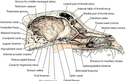
The nose in the broad sense comprises the external nose, the paired nasal cavities, and the paranasal sinuses (Dyce, et al., 2010). Within the nasal cavity are three sets of nasal conchae (dorsal, ventral and ethmoidal), which are cartilaginous or slightly ossified scrolls covered with nasal mucosa (Harcourt-Brown, 2006a). The dorsal nasal concha is the first endoturbinate and the longest. It arises from the dorsal part of the cribriform plate caudally as well as from the medial part of the roof plate (Evans, 1993), and is formed by a single curled scroll of bone (Harcourt-Brown, 2006a). The second endoturbinate, is the ventral nasal concha, it arises from its basal lamina near the middle of the lateral lamina (Evans, 1993), and it is divided by a series of tightly folded scrolls (Harcourt-Brown, 2006a). The ethmoidal conchae occupy the caudal part of the nasal cavity and consist of a number of bony scrolls that are into two groups: ectoturbinates and endoturbinates (Harcourt-Brown, 2006a), that viewed from the medial side have the same general form as the second endoturbinates (Evans, 1993). The ethmoidal conchae are attached to the cribriform plate and extend dorsally into the frontal sinus (Harcourt-Brown, 2006a).
The frontal sinus, is divided into rostral, medial and lateral compartments, and is localized dorsocaudal to the nasal meati (dorsal, middle and ventral) (Harcourt-Brown, 2006a).
The cribriform plate is part of the ethmoid bone and articulates ventrally with the presphenoid to form the spheno-ethmoid suture and with the vomer bone to form the vomero-ethmoid suture (Evans, 1993).
9
Figure 1 - Saggital section of the skull, medial view
Roman numerals I, II, III, IV indicate endoturbinates in the nasal cavity; Arabic numerals 1, 2, 3 are ectoturbinates located in the frontal sinus (Adapted from Evans & Lahunta, 2010).
Figure 2 – Transverse section of the nasal cavity.
(Adapted from Evans & Lahunta, 2010)
2.4 Importance of clinical history in dogs in nasal disease
The medical history of a dog with a disease of the nasal cavity and paranasal sinuses is very important, since nasal disease signs like depigmentation, inflammation, crust formation, dehydration and hyperkeratosis are usually noted by the owner (Haar, 2006).
The reported presence or absence of epistaxis, sneezing, stertor, coughing, gagging, facial deformity and pain, mouth breathing, systemic signs of illness and in rare instances central nervous system give much useful information (Hawkins, 2009; Sullivan, 1987).
The susceptibility of the breed, sex and age, should also be considered by the clinician. The literature shows that dolicocephalic and mesaticephalic breeds are prone to intranasal disease but brachycephalic breeds are less affected (Knotek, Fichtel, Kohout, & Benák, 2001;
10
Mathews & Sharp, 2006; J. Saunders, et al., 2004; Sharp, Sullivan, & Harvey, 1992), maybe because the latter is mainly a mouth-breathing breed and has less nasal tissue (Sullivan, 1987). The same study shows that the knowledge of the age range of the most frequent diseases (nasal neoplasia, aspergillosis and chronic hyperplastic rhinitis) can be helpful (Sullivan, 1987). The majority of nasal tumours occur in older dogs, while aspergillosis and chronic hyperplasic rhinitis are found in younger dogs (Sullivan, 1987; Tasker, et al., 1999). Male dogs appeared to be at greater risk than female dogs (J. Saunders, et al., 2004). It is also important to determine the duration of signs, whether clinical signs are static or evolving, if the patient has ever exhibited the presenting signs before, if treatment was initiated in the past and the success or failure of such treatment (Padrid, 2007).
Additional questions should be asked about the animal´s general condition (Haagen & Herrtage, 2010) like exercise intolerance, food habits, drinking and other changes in habits. Questions about the environment and geographic origin can also be useful. It has been described in the literature that questions concerning the habits of the owners can sometimes bring some important information like if there is any smoker in the house, changed carpeting recently, or any house significant renovation in the last 6 months (Padrid, 2007). Travel history should also be assessed, to determine if the pet has an increased risk of diseases such as mycotic infection or heartworm (Padrid, 2007). It is also reported that aspergillosis affects more frequently farm dogs, with no specific access to heavy concentrations of fungal spores (Sullivan, 1987).
2.5 Clinical signs of nasal disease in the dog
Clinical sings related to the upper respiratory tract, are among the most common presenting complaints encountered in small animal practice and are frequent reasons for referral to specialty practices and veterinary teaching hospitals (Ford, 2005). The following paragraphs are a brief enumeration and explanation of the most common signs of nasal disease.
2.5.1 Nasal discharge
It is most commonly associated with disease localized within the nose, nasal cavity and paranasal sinuses, although it may also develop with disorders of the lower respiratory tract (Ford, 2005), such as bacterial pneumonia and infectious tracheobronchitis, or systemic disorders such as coagulopathies and systemic hypertension. It is reported that discharge occurring only when sneezing indicates a less productive mucosal disease than does continuous discharge (Haagen & Herrtage, 2010).
11
Based on the nature of the nasal discharge, rhinitis can be classified as serous, which may be seen initially with a variety of nasal diseases (Kuehn, 2009) but can also be normal (Hawkins, 2009), catarrhal which is a slightly more severe sign, and its characterized by an increased of mucus production due to hyperplasia of goblet cells (López, 2007). Purulent or mucopurulent implies inflammation (Hawkins, 2009) and secondary bacterial invasion (Kuehn, 2009) and a neutrophilic exudates occurs (López, 2007), fibrinous when there is an increase of vascular permeability, resulting in exudation of plasma fibrinogen, which coagulates into fibrin and granulomatous when infiltration of numerous active macrophages and lymphocytes occurs, and it is characteristic of chronic allergic inflammation and nasal fungal infection (López, 2007).
The profuse, purulent nature of the discharge in aspergillosis helps to differentiate this condition from nasal neoplasia in which the discharge tends to be more serous and intermittent (Sharp, et al., 1992).
2.5.2 Epistaxis
A previous study (Bissett, Drobatz, McKnight, & Degernes, 2007) describes that owners and veterinarians see epistaxis episodes as an emergency. Epidemiologically it was seen in the same study, that epistaxis was more likely to occur in old dogs (≥ 6 years old), male, and large (≥ 26 Kg.) breeds.
It has been commonly suggested in the veterinary literature that epistaxis is most often a result of local disorders, unilateral, chronic and associated with other nasal tract signs (Bissett, et al., 2007; Dhupa & Littman, 1992). However, a recent study (Mylonakis, et al., 2008), reported systemic disorders to be the most common cause of epistaxis.
When epistaxis is a result of a systemic disorder is said to be bilateral and acute, although it was shown that unilateral epistaxis was not pathognomonic for a local disorder (Bissett, et al., 2007).
Epistaxis can be associated with mucopurulent exudates from any aetiology but when prolonged is usually associated with trauma, local aggressive diseases like neoplasia or mycotic infection, systemic hypertension or bleeding disorders (Hawkins, 2009).
One of the most well-known causes of epistaxis, in dogs, is trauma (Bissett, et al., 2007; Dhupa & Littman, 1992; Gough, 2007), due to the fractures of the maxillae or displaced nasal bones fragments that contribute to the occlusion of the airway (Chandler, Thompson, Sutton, & Price, 1991). However, until the present date, there were no published studies that related trauma with epistaxis in dogs.
12
Local disease processes have been reported to be a common cause of epistaxis in dogs. Previous studies showed that nasal and paranasal neoplasia (Bissett, et al., 2007; Strasser & Hawkins, 2005) like adenocarcinoma, lymphoma and squamous cell carcinoma (Gough, 2007) and mycotic rhinitis (Bissett, et al., 2007; Mathews, et al., 1998; Strasser & Hawkins, 2005) due to Aspergillus spp. (Gough, 2007; Tasker, et al., 1999) have long been regarded as the most important causes.
Other causes of local diseases included idiopathic rhinitis (Bissett, et al., 2007; Strasser & Hawkins, 2005; Windsor, Johnson, Herrgesell, & Cock, 2004), parasitic rhinitis (Strasser & Hawkins, 2005), nasal foreign body (Bissett, et al., 2007), periapical abscesses (Bissett, et al., 2007; Gough, 2007), and arteriovenous malformation. (Bissett, et al., 2007; Hawkins, 2009) Systemic disorders previously reported to cause epistaxis in dogs include hereditary/congenital diseases like immune-mediated thrombocytopenia (Mylonakis, et al., 2008; Tilley & Smith, 2007) and von Willebrand disease (Hawkins, 2009; Tilley & Smith, 2007), and various systemic infections like canine distemper virus (Gough, 2007), leishmaniasis, monocytic ehrlichiosis (Mylonakis, et al., 2008) and babesiosis (Tilley & Smith, 2007). Other less common causes reported are rodenticide toxicity and systemic arterial hypertension (Hawkins, 2009; Mylonakis, et al., 2008).
2.5.3 Stertor/reverse-sneezing and stridor
The second most common clinical sign associated with upper respiratory disease in dogs is stertor, which can be intermittent, yet persistent or continuous snorting, also called stertorous breathing (Ford, 2005), caused by air passing through a narrowed nasopharynx, pharynx or trachea and meeting resistance because of partial obstruction of these regions (Tilley & Smith, 2007). Reverse-sneezing is characterized by paroxistical stertous, which is believed to be a patient’s attempt to displace matter trapped in the nasopharynx and move it into the oropharynx to be swallowed (Ford, 2005).
Stridor is a common sign for obstructive nasal disease (Haagen, 2009) and it is characterized by a higher-pitched sounds that result when rigid tissues are vibrated by the passage of air, usually is a result of nasal or laryngeal partial or complete obstruction (Haagen & Herrtage, 2010; Tilley & Smith, 2007).
These signs are commonly seen in brachycephalic breeds, and the most frequent causes are between others foreign bodies (Gough, 2007), brachycephalic obstructive syndrome (Stanley, 2007), neoplasia most frequently due to squamous cell carcinoma and adenocarcinoma (Haagen, 2009), stenosis, secretions in the airway lumen and upper airway infection and haemorrhage (Tilley & Smith, 2007).
13 2.5.4 Sneezing
A sneeze is an explosive release of air from the lungs through the nasal cavity and mouth and is a protective reflex to expel irritants from the nasal cavity (Hawkins, 2009). It can due to any mucosal irritation or inflammation (Tilley & Smith, 2007), but usually respiratory disorders cause acute-onset and persistent (Hawkins, 2009).
The most common causes of sneezing are excess of nasal secretions, foreign bodies, neoplasia, parasites (Tilley & Smith, 2007), infections (viral, fungal or bacterial) and inflammatory nasal disease like lymphoplasmacytic rhinitis (Gough, 2007).
2.5.5 Other reported signs
Other reported signs, included nasal depigmentation and nasal pain, which become more obvious when the animal begins to react adversely to the owner´s customary petting (Haagen & Herrtage, 2010). Dyspnoea expresses difficulty in breathing or respiratory distress (Tilley & Smith, 2007). The major causes related to upper airway diseases are obstruction (Haagen & Herrtage, 2010; Tilley & Smith, 2007) due to stenotic nares, infection, inflammation, neoplasia or trauma (Tilley & Smith, 2007).
Coughing is a sudden forceful expiration of air through the glottis, preceded by an exaggerated inspiratory effort and usually accompanied by an audible sound (Tilley & Smith, 2007). It can be caused by lower and upper respiratory tract diseases which include rhinitis, sinusitis, foreign body and neoplasia (Tilley & Smith, 2007).
2.6 Physical Examination
After clinical history has been taken, a thorough clinical examination of the respiratory tract should be followed. Firstly, it is advised to examine the oral cavity, with emphasis on the maxilla, hard palate (trauma and congenital cleft palate on puppies) and canine teeth, which despite appearing normal can occult periodontal disease (Ford, 2005). Soft palate elongation may be also easily appreciated during visual inspection of the posterior pharynx in many patients without sedation (Padrid, 2007).
Table 1 – Anatomic limits of upper respiratory tract and defining clinical signs adapted from (Ford,
2005)
Anatomic limits Clinical Signs
Nose, Nasal Cavity and
Paranasal Sinuses Sneezing and/or Nasal Discharge Nasopharynx, choanae and soft palate Stertor and Reverse-Sneezing
14
Palpation of the nasal architecture may reveal structural abnormalities like humps, nasal pain and nasal asymmetry suggestive of bone distortion from neoplasia or mycotic infection (Padrid, 2007).
Unfortunately, even when reported by the owners, the nature of the nasal discharge and side(s) involved can be difficult to assess since in many dogs the discharge is intermittent and therefore absent at the time of the examination (Sullivan, 1987). Checking the patency of the nasal airway is also very important and be easily accessed by using a thread or wisp of cotton wool and suspending it in front of each nostril. In this way an undisclosed bilateral lesion may be detected (Haagen & Herrtage, 2010; Sullivan, 1987).
2.7 Differential Diagnoses
Nasal diseases usually requires ancillary diagnostic techniques in order to reach a final diagnose, however through the history, signs and physical examination, the clinician needs to have in mind some differentials in order to proceed with the investigation.
Nasal disease in dogs can be a result of several conditions like neoplasia, inflammation, infection (primarily fungal), trauma, foreign body or, less commonly, parasitic infestation (Miles, Dhaliwal, Moore, & Reed, 2008). Determine the underlying cause in dogs with nasal disease can be challenging and frustrating, and often necessitating multiple diagnostic procedures at substantial cost to the client (Miles, et al., 2008).
Several studies report neoplasia as the most common disease in middle-aged to older dogs of mesaticephalic and dolicocephalic conformations, with chronic nasal signs (Harcourt-Brown, 2006a; Petite & Dennis, 2006; Sullivan, 1987; Tasker, et al., 1999) being the most common tumour the adenocarcinoma followed by chondrosarcoma (Tasker, et al., 1999).
Two studies reported chronic hyperplastic rhinitis as the second most common disease (Sullivan, 1987; Tasker, et al., 1999), although one reported aspergillosis being the second most frequent nasal disease (Harcourt-Brown, 2006b).
The diagnosis of aspergillosis can be challenging, and the clinician should consider that a correct diagnosis should be confirmed by at least two or three independent methods (Table 2) in order to avoid false positive or negative results (Sharp, et al., 1992).
15
Table 2 – Diagnostic criteria for nasal aspergillosis
Other diseases less commonly reported in the literature as frequent (Harcourt-Brown, 2006b; Sullivan, 1987; Tasker, et al., 1999), were foreign bodies, which should always be considered when a patient presents with a sudden onset of violent and persistent sneezing (Chandler, et al., 1991), dental problems that result in bone necrosis and subsequently communication between oral and nasal cavity (Ford, 2005), oronasal fistula characterised by the presence of food material in the nasal cavity, which has gained entrance via a defect in either the hard or soft palate (Chandler, et al., 1991), destructive rhinitis, polyp and undiagnosed diseases. Diagnostic tests are obviously required to confirm the presence of these respiratory diseases, however respiratory medicine is considered to be underdeveloped subspecialty in veterinary medicine, and most of the commonly available tests are used to point the clinician in the right direction, and to rule out the presence of other potentially confounding disorders (Padrid, 2007).
2.8 Imaging studies
Nasal imaging is a decisive component of the diagnostic assessment of animals with signs of nasal disease, allowing the study of the bone and soft tissue structures that are not visible by physical examination or rhinoscopy (Hawkins, 2009).
However, it must be taken into consideration that imaging studies should always be performed prior to the rhinoscopy and biopsy procedures (J. Saunders, et al., 2004). The two major reasons for that are: the results of nasal imaging can help directing biopsy instruments to the most abnormal regions and the resultant haemorrhage from rhinoscopy and biopsy may obscure subtle soft tissue lesions and induce imaging abnormalities (Hawkins, 2009; J. Saunders, et al., 2004).
16 2.8.1 Radiography
The canine head consists of 50 bones and numerous soft-tissue structures, which create challenges when making and interpreting survey radiographs of the nasal cavity and paranasal sinuses (Pownder, Rose, & Crawford, 2006).
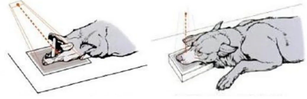
Radiographs should be made under general anaesthesia to achieve perfect positioning (Pownder, et al., 2006). The standard radiographic examination for nasal diseases consists of a latero-lateral (LL) and dorso-ventral (DV) projection of the skull (Haagen, 2005) (Figure 3), however the value of the DV is limited by the superimposition of the mandible over much of the nasal chamber (Dennis, Kirberger, Wrighley, & Barr, 2001; Haagen & Herrtage, 2010).
Figure 3 - Positioning of the head. LL and DV projections.
(Adapted from Waibl, Mayrhofer, Matis, Brumberg, & Köstlin, 2005)
In examination of the nose and nasal sinuses, special projections are required (Figure 4), such as the open mouth rostroventral-dorsocaudal (RV-DCd) and intra-oral DV projections (Haagen, 2005), providing the last one minimal superimposition of structures over the area of interest (Haagen & Herrtage, 2010) and the rostrocaudal (skyline) projection, which is useful for the evaluation of frontal sinuses (Haagen & Herrtage, 2010).
Figure 4 – Positioning of the head. Open mouth RV-DCd and intra-oral DV projections.
Adapted from (Waibl, et al., 2005)
Radiographic evaluation of a diseased nasal cavity is difficult because of the superimposition of bony structures and the complexity of the nasal turbinates (J. Saunders, et al., 2004). There are nasal structures that are never visible on radiographs like nasal septum and caudal
17
recesses, others are only sometimes visible like orbital lamina of the maxillary recesses. The cribriform plate, the naso-orbital wall and the vomer are well visualised, but changes like destruction need to be severe to be seen(Petite & Dennis, 2006; J. Saunders & Bree, 2003; J. Saunders, et al., 2004). This proves that conventional radiography has a low diagnostic value for evaluation of cerebral involvement and aggressive nasal disease, which is a very important prognostic factor (Petite & Dennis, 2006).
The radiographs may provide all the information that is needed or they may serve as a primary examination for prioritizing the differential diagnosis (Haagen, 2005). In fact, if an appropriate technique is used, they are considered useful for identifying the extent and severity of the disease, localizing sites for biopsy within the nasal cavity (Hawkins, 2009) and showing opacifications of the nasal cavity and frontal sinuses (except when lesions are bilateral and subtle) (J. Saunders, et al., 2004).
Nasal radiographs should be evaluated for increased fluid density, loss of turbinates, lysis of facial bones, radiolucency at the tips of tooth roots and the presence of radiodense material (Hawkins, 2009).
In the past, it was believed that radiography was not reliable for diagnosis of chronic nasal disease (Gibbs, Lane, & Denny, 1979). However, a study (Russo, Lamb, & Jakovljevic, 2000) identified some of the features that are most often found in radiographs of dogs with inflammatory nasal disease, and showed that dogs with rhinitis often get lucent foci in the nasal cavity (82%), usually the frontal sinus is not involved, focal or multifocal loss of turbinate detail could be present and localized soft tissue opacities could be seen. The majority of these lesions were frequently found in the rostral part of the nasal cavity (Russo, et al., 2000).
A more recent study showed that radiography like CT, provides a diagnosis mainly on the basis of the cavitated appearance of the nasal cavity and the presence of hyperostotic bone (J. Saunders, et al., 2004). However, it is particularly important to notice that in some cases, lesions identified on CT and MRI that could be significant with regard to prognosis and treatment are not visible on radiographs (Petite & Dennis, 2006).
Russo et al., showed that radiography had a high degree of accuracy for distinguishing between nasal neoplasia and aspergillosis, and a more recent study demonstrated that it has a sensitivity of 72-84% for aspergillosis (J. Saunders & Bree, 2003). Nevertheless, trapped fluid can be misinterpreted as solid tissue on some radiographs, and tumour size may be overestimated in these cases (Petite & Dennis, 2006).
In fact, a very high sensitivity was obtained in dogs with generalized lesions, whereas, the sensitivity was lower in dogs with localized lesions (J. Saunders & Bree, 2003; Jimmy
18
Saunders, et al., 2002), frontal sinus lesions or bilateral and subtle lesions (Gibbs, et al., 1979; J. Saunders, et al., 2004).
2.8.2 Computed tomography
CT is a reliable, noninvasive technique for use in the diagnosis of nasal disease in dogs (J. Saunders, Bree, Gielen, & Rooster, 2003), and has been used in dogs with a possible diagnosis of fungal rhinitis, nasal tumours, nonspecific rhinitis, and foreign-body rhinitis (Drees, Forrest, & Chappell, 2009; J. Saunders & Bree, 2003; J. Saunders, et al., 2003; Jimmy Saunders, et al., 2002).
Nasal CT is a powerful tool and greatly enhances the ability to establish an accurate and definitive diagnosis of chronic nasal disease in the dog, since cross sectional imaging provides good assessment of the extent of nasal disease, identifies areas of the nose to examine via rhinoscopy (Lefebvre, Kuehn, & Wortinger, 2005), and permits the evaluation of the character of lesions in the nasal cavities (Rycke, Saunders, Gielen, Bree, & Simoens, 2003; J. Saunders, et al., 2003).
CT uses the same basic physical principles as diagnostic x-ray, except it depicts the shades of gray in cross-section (Gavin & Bagley, 2009). The interpretation of CT scans is also very similar in principle to radiographic interpretation, with mineralised and bony material appearing radiopaque, fluid and soft tissue producing intermediate grey shaded and fat being more radiolucent. However, unlike radiography, differentiation between soft tissue and fluid is possible and internal soft tissue architecture is visible (Dennis, 2003).
It was found to be more sensitive than radiography for diagnosis of nasal aspergillosis in dogs, because the axial images permits accurate visualization of all the structures that are not visible on radiography (two-dimensional projections) as well as abnormalities like cribriform plate lysis (dorsal reconstructions), bilaterality and retrobulbar involvement (J. Saunders & Bree, 2003; Jimmy Saunders, et al., 2002).
Furthermore, CT was found to be more sensitive than radiography for diagnosis of nasal aspergillosis in the dog because of a better demonstration of mucosal thickening, frontal sinus lesions and a cavitated–like process (J. Saunders & Bree, 2003). The most common CT findings in dogs with nasal aspergillosis included moderate to severe turbinate destruction with a variable amount of abnormal soft tissue in the nasal passages, non-specific thickening of the mucosa along the bones of the frontal sinus, maxillary recess and nasal cavity; and thickened reactive bone (Jimmy Saunders, et al., 2002).
The diagnosis of rhinitis has also been studied for the past few years; non-specific rhinitis was seen as a non-destructive process affecting both entire nasal cavities of the dog with a
19
minimal to moderate amount of fluid in the frontal sinus (J. Saunders, et al., 2003). CT also enables the detection of highly attenuating foreign bodies, such as metal or glass. When not visible, the foreign body rhinitis is characterized with features similar to a localized nasal aspergillosis, which can make the differentiation between them difficult (J. Saunders, et al., 2003).
In conclusion, nasal computed tomography is a powerful tool and greatly enhances the ability to establish an accurate definitive diagnosis of nasal disease, since it helps differentiating idiopathic inflammatory disease from fungal rhinitis and neoplasic diseases from non-neoplasic diseases (Kuehn, 2006a).
2.8.3 Magnetic resonance imaging
2.8.3.1 Basic Physics
MRI was utilized in veterinary medicine primarily as a research tool in the 1980´s and early 1990´s (Gavin & Bagley, 2009), and over the past years, has been replacing computed tomography (CT) as a method of diagnostic imaging (Dennis, 1998).
Magnetic resonance imaging allows investigators to make multiplanar images without repositioning the dog (Rycke, et al., 2003). The three common planes are transverse (axial), dorsal (coronal) and the sagittal plane. The images are made from different slices within these planes, which are formed from the three magnetic gradients used (Gavin & Bagley, 2009). The transverse plane allows better evaluation of the nasal turbinates, which is essential for the diagnosis of diseases that affect the nasal region. Therefore, this is the reference plane for the assessment of nasal pathologic changes (Rycke, et al., 2003).
The dorsal plane is most appropriate for use in determining the integrity of the cribriform plate in dogs with nasal tumours or nasal aspergillosis. Images through the dorsal plane also provide a more general view of the entire nasal cavities allowing easier characterization of disease processes (Rycke, et al., 2003).
The sagittal plane is of more use for spinal studies, because in small animals the whole spine can easily be included, and abnormalities like disc prolapsed, cord compression and nerve root impingement can easily be recognised (Elliot & Skerritt, 2010).
The current clinical applications for MRI rely on visualization of the resonance of the hydrogen atom nucleus (Gavin & Bagley, 2009), because a single hydrogen atom produces a relatively large magnetic moment and resonates very well, and in addition to this is very abundant within the body (Elliot & Skerritt, 2010). The size of the magnetic field is
20
dependent on the speed of movement (magnetic movement) and the size of the charge, since the hydrogen nucleus has a small electric charge it spins very fast (Gavin & Bagley, 2009). However, in reality, it is not only hydrogen that can resonate, any atom with an odd mass number such as carbon (13), sodium (23) and phosphorous (31) would be suitable, since they also possess the ability to resonate and produce images (Elliot & Skerritt, 2010).
The hydrogen proton, as all other protons, carries a positive electrical charge and spins permanently on its own axis, which creates a corresponding magnetic field (magnetic moment) around the proton that possesses properties of size and direction (Dennis, 1998; Elliot & Skerritt, 2010; Gavin & Bagley, 2009). The body has many billions of microscopic magnetic moments that are completely randomly orientated, so they cancel each other out in a manner that their macroscopic magnetic field is zero (Elliot & Skerritt, 2010).
When an animal is placed within a large external magnetic field (scanner), the randomly spinning protons will come into alignment with the external field, although some of them will align against the field (anti-parallel state), largely cancelling each other out (Gavin & Bagley, 2009).
If protons are then bombarded with a series of RF pulses at a similar frequency to the rotational movement, they resonate (Dennis, 1998). This is due to the fact that the RF pulse imparts sufficient energy to allow more protons to adopt the anti-parallel state and it brings all the hydrogen spins into phase with each other, this means that the spins are no longer cancelling each other out, but instead each microscopic magnetic field is in unison with its neighbours (Elliot & Skerritt, 2010). After each brief RF pulse, the protons try to realign with the magnetic field, but are quickly unbalanced again by the next RF pulse, this produces cross-sectional images of tissues using a combination of magnetic fields and RF signals (Dennis, 1998).
This process continues for several minutes during which time the protons themselves emit a detectable RF signal which is related in strength and frequency to their chemical environment and position within the tissues (Dennis, 1998).
When RF transmission is turned off, three things happen simultaneously but independently of each other. Firstly, the absorbed RF energy is retransmitted as the useable MR signal, which depends on how much hydrogen there is in a particular tissue (proton density). Secondly, the spins that were in phase begins to slow down relative to others (dephase), this event is referred to as T2, transverse or spin spin relaxation. The other event, occurs when the extra excited protons that were using the RF energy to adopt the anti-parallel state begin to return to their usual state, this is called T1, longitudinal recovery or spin lattice relaxation (Elliot & Skerritt, 2010; Gavin & Bagley, 2009).
21
The emitted RF signal is detected by an aerial and rapidly converted by a computer into a series of cross-sectional images which can be viewed within a few seconds of the end of the study, the images are 'maps' of the locations of hydrogen nuclei (protons) within the tissues, based on their different chemical environments (Dennis, 1998) as well as their T1 and T2 relaxation (Elliot & Skerritt, 2010).
Thus, MRI is able to make high-quality images, not only because of the energy of the spinning protons, but also due to the abundance of hydrogen protons present in the body (Gavin & Bagley, 2009). Tissues containing a high concentration of hydrogen protons such as fat and CSF will generate more signal than others, containing little or no hydrogen protons, like cortical bone and lung (Elliot & Skerritt, 2010).
The spin echo sequences, uses an additional 180º RF pulse, which is divided into two 90º pulses. The first one will bring spins back into phase whilst the second will continue to push spins out of phase but in the opposite direction, this makes fast moving spins proceeding in the same direction but behind slow moving ones. Once the RF pulse has been turned off, they gradually catch up, and are said to rephrase or refocus, the corresponding regrowth of signal is detected in the receiver coil, referred to as an echo (Elliot & Skerritt, 2010).
The refocusing process forms the basis of spin echo (SE) pulse sequences and manipulation of the timing of these 90º and 180º RF pulses determines image contrast (Elliot & Skerritt, 2010). However a single echo is not sufficient to give an image, so this process is repeated hundreds of times (Elliot & Skerritt, 2010).
The two relaxation processes, T1 and T2, occurred simultaneously but are completely independent of each other, so by choosing the right echo time and repetition time (time between one 90º RF pulse and the next) the sequence can be optimized in favor of the desired contrast. Thus images are said to be T1/T2 weighted or T1W/T2W (Elliot & Skerritt, 2010). The proton density weighted minimizes both T1 and T2 effects, so any contrast in this image is down to absolute numbers of hydrogen atoms rather than either of the relaxation processes (Elliot & Skerritt, 2010).
Table 3 – Relaxation sequences formation
TR TE Short Long
Short T1W
Optimize both T1 and T2
(very poor quality images) Long Proton density weighted
22
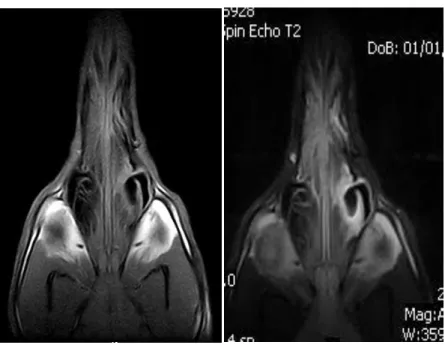
Since fluid contains a lot of hydrogen protons, it will required long TR values to allow a full T1 relaxation, so it appears bright or hyperintense on T2-weighted and PDW images and dark or hypointense on T1-weighted images (Dennis, 2003; Gavin & Bagley, 2009). On the contrary, fat is hyperintense on PDW and T2 and T1-weighted images (Gavin & Bagley, 2009).
Figure 5 – T1W (left) and T2W (right) sequences of the same dog
Pulse sequences, describe the sequence and timing of RF pulses and gradient applications required to produce an image (Elliot & Skerritt, 2010). The STIR sequence allows for a T2 -weighted type of image with uniform loss of the fat signal, which would normally be bright on conventional SE sequences (Elliot & Skerritt, 2010; Gavin & Bagley, 2009). It is used to demonstrate lesions in parts of the body such as the abdomen or orbits, which contain lots of fat (Elliot & Skerritt, 2010).
The FLAIR sequence is similar to the previous one, but it coincides with null point of water, so they show low signal intensity in areas of free fluid (Elliot & Skerritt, 2010; Gavin & Bagley, 2009), bound fluid, on the other hand, has a quicker relaxation time because it is able to impart energy to the molecules with it is bound (Elliot & Skerritt, 2010). In this sequence, oedema, tumour, necrosis or other pathology will retain a high signal, whilst areas of free fluid, such as CSF will appear dark (Elliot & Skerritt, 2010). This is particularly useful in brain studies, since it demonstrates periventricular lesions (Elliot & Skerritt, 2010) and gets rid of the usually hyperintese fluid signal from the vertebral spinal fluid (Gavin & Bagley, 2009).
Contrast is the mechanism in image quality, which allows differentiation of the various tissues according to their signal intensity (Elliot & Skerritt, 2010). Paramagnetic contrast media such