U
NIVERSIDADE DE
L
ISBOA
FACULDADE DE FARMÁCIA
DEPARTAMENTO DE MICROBIOLOGIA E IMUNOLOGIA
E
NGINEERING NEW STRATEGIES TO IMPROVE
PHARMACOKINETICS OF THERAPEUTIC PROTEINS
C
ÁTIAS
OFIA DEC
ARVALHOC
ANTANTEDoutoramento em Farmácia
Especialidade em Biotecnologia Farmacêutica
U
NIVERSIDADE DE
L
ISBOA
FACULDADE DE FARMÁCIA
E
NGINEERING NEW STRATEGIES TO IMPROVE
PHARMACOKINETICS OF THERAPEUTIC PROTEINS
C
ÁTIAS
OFIA DEC
ARVALHOC
ANTANTETese orientada pelo Professor Doutor João Gonçalves e Doutor Frederico
Aires-da-Silva, especialmente elaborada para a obtenção do grau de doutor em
Farmácia (Biotecnologia Farmacêutica)
Lisboa
Cátia Sofia de Carvalho Cantante was financially supported by a PhD fellowship (SFRH/BD/48598/2008) from Fundação para a Ciência e a Tecnologia (FCT), Lisbon, Portugal.
De acordo com o disposto no ponto 1 do artigo nº 45 do Regulamento de Estudos Pós-Graduados da Universidade de Lisboa, deliberação nº 4624/2012, publicado em Diário da República – Série nº 65 – 30 Março de 2012, a autora desta dissertação declara que participou na concepção e execução do trabalho experimental, interpretação dos resultados obtidos e redação dos manuscritos.
The research work described in the present Thesis was performed from April 2009 until May 2014, under the supervision of Professor João Gonçalves (PhD) and Frederico Aires-da-Silva (PhD), mainly at the URIA/CPM of the Faculdade de Farmácia da Universidade de Lisboa, Lisbon, Portugal. This project was also performed in collaboration with the Pharmacokinetics, and Metabolism and Genetics group from iMed.UL, Faculdade de Farmácia, ITN (Instituto Superior Técnico), Faculdade de Medicina Veterinária, IMM (Faculdade de Medicina) da Universidade de Lisboa, Lisbon, Portugal; with CQFB/REQUIMTE, Faculdade de Ciências e Tecnologia, Universidade Nova de Lisboa, Caparica, Portugal; and with TechnoAntibodies (TechnoPhage S.A.), Lisbon, Portugal.
The results described in this thesis were included in manuscripts already published or in preparation, in peer-reviewed journals, and in posters in international and national scientific meetings:
Morais, M., Cantante, C., Gano, L., Santos, I., Lourenço, S., Santos, C., Fontes, C., Aires da Silva, F., Gonçalves, J., Correia, J.D. (2014) Biodistribution of a 67Ga-labeled anti-TNF VHH single-domain antibody containing a bacterial albumin-binding domain (Zag). Nucl. Med.
Biol. pii: S0969-8051(14)00012-22
Cantante, C., Lourenço, S., Morais, M., Leandro, J., Gano, L., Silva, N., Leandro, P., Fontes, C., Correia, J.D.G., Aires-da-Silva, F., Gonçalves, J. (2014) Albumin-binding domain (Zag) from Streptococcus zooepidemicus as a strategy to improve half-life of therapeutic proteins. Submitted to Journal of Biological Chemistry
Cantante, C., Lourenço, S., Oliveira, S., Leandro, J., Morais, M., Gano, L., Silva, N., Leandro, P., Fontes, C., Correia, J.D.G., Aires-da-Silva, F., Gonçalves, J. (2014) Albumin-binding domain (ProtH) from Streptococcus pyogenes, a new approach to increase half-life of therapeutic proteins. Submitted to Journal of Biological Chemistry
Cantante C., Lourenço S., Leandro J., Morais M., Oliveira S., Gano L., Fontes C., Correia J., Leandro P., Silva F., Gonçalves J. “Albumin-binding domains Zag and ProtH improve pharmacokinetics and affect biodistribution of an anti-TNF VHH”. 5th PEGS Europe, Protein & Antibody Engineering Summit, Lisbon, Portugal, 4-8th November, 2013
Correia J., Morais M., Gano L., Santos I., Cantante C., Lourenço S., Santos C., Fontes C., Silva F., Gonçalves J. “Pharmacokinetic properties of a 67Ga-labeledanti-TNF VHH single domain antibody containing a bacterial albumin-binding domain (Zag)”. 2nd International Workshop on Innovative Personalized Radioimmunotherapy, Pharmacy Faculty, Nantes, France, 9-12th July, 2013
Cantante C., Lourenço S., Leandro J., Morais M., Gano L., Fontes C., Correia J., Leandro P., Silva F., Gonçalves J. “Albumin-binding domain from Streptococcus pyogenes protein H increases half-life and affect blood clearance of anti-TNF VHH”. 5th Postgraduate iMed.UL students meeting, Faculty of Pharmacy-University of Lisbon, Lisbon, Portugal, 18th July, 2013
Cantante C.,Lourenço S., Leandro J., Morais M., Gano L., Fontes C., Correia J.,Leandro P., Silva F., Gonçalves J. “Albumin-binding domain from Streptococcus pyogenes protein H increases half-life and affect blood clearance of anti-TNF VHH”. 4th PEGS Europe, Protein & Antibody Engineering Summit, Vienna, Austria, 6-8th November, 2012
Cantante C.,Lourenço S., Morais M., Gano L., Santos C., Fontes C., Correia J.,Silva F., Gonçalves J. “Albumin-binding domain (Zag) from Streptococcus zooepidemicus increases half-life and affect blood clearance of anti-TNF VHH”. 8th PEGS Summit Boston, Protein & Antibody Engineering Summit, Boston, USA, 30th April- 4th May, 2012
vii
ABSTRACT
Recombinant antibody formats and therapeutic proteins have found increasing applications as therapeutics, e.g. for the treatment of cancer or inflammatory diseases. While whole antibodies have an exceptionally long half-life, small antibody formats and small molecular weight therapeutic proteins often suffer from rapid elimination from circulation. Usually, this involves frequent administration and repeated infusions with the drug application intravenous or subcutaneously, using a slow adsorption into the blood stream. These limitations of small size protein drugs have led to the development and implementation of several half-life extension strategies to prolong circulation of these molecules and thus improve their administration, pharmacokinetic and pharmacodynamic properties. Over the recent years, plasma protein binding has been shown to be an effective approach to improve pharmacokinetic properties of short half-life molecules. Within this context, in order to improve the half-life of therapeutic proteins we have investigated new strategies based on protein fusions with two albumin-binding domains (ABDs) from streptococcal cell surface proteins. These two ABDs were the Zag and protein H (ProtH) derived from Streptococcus
zooepidemicus and Streptococcus pyogenes, respectively. To validate our ABDs half-life
strategy and as a proof-of-concept, the Zag and ProtH domains were fused with an anti-TNFα VHH single-domain antibody. Our results showed that Zag and ProtH ABDs alone and fused to the anti-TNFα VHH could be stably produced in vitro, present nanomolar binding to human, rat and mouse albumins, quantified by ELISA, and for VHH-Zag, using surface plasmon resonance (SPR). VHH, VHH-Zag and VHH-ProtH proteins presented high thermal properties, and human and mouse serum stabilities as well. Moreover, the fusion of the ABDs with the anti-TNFα VHH did not affect the efficacy of the therapeutic molecule against TNFα. Furthermore and importantly, fusion with Zag and ProtH domains strongly increases the half-life and stability of the anti-TNFα VHH. Comparing with the VHH (terminal half-life of 47 min), VHH-Zag (terminal half-life of ~31 h) and VHH-ProtH (terminal half-life of ~21 h) presented prolonged circulation times of 39-fold and 26-fold, respectively. Therefore, after injection, the residence in mouse serum presented higher time extensions until 72 h for VHH-Zag and 48 h for VHH-ProtH (versus 2 h exhibited by VHH). The biodistribution profile of
viii
VHH-Zag was determined with technetium 99 m (99mTc) and gallium 67 (67 Ga)-radiolabelling. The prolonged circulation time with both radionuclides lead to a reduction of the 99mTc-VHH-Zag in kidneys and an increase of the presence of the fusion anti-TNFα VHH in blood and other organs, e.g. liver, intestine, muscle and lungs. The biodistribution profile of VHH-ProtH was evaluated with 99mTc, presenting a similar organ distribution that the observed for the nanobody alone. Accordingly, 99mTc-VHH-ProtH is retained in kidneys and showed a trend to accumulate within highly perfused organs like and intestine.
Though, Zag and ProtH unmodified ABDs preserved the albumin-binding ability and thermal stability of the fusions with the recombinant antibody, these domains present very different amino acid sequences and unit organization. Whereas, Zag ABD presents one domain with 52 aa, ProtH ABD possess three C repeats (C1C2C3) that were tested in ELISA assays to evaluate the binding of each unit alone (C1, C2 and C3) or several combinations of these repeats (C1C2, C2C3 and C1C2C3), to human serum albumin. The results obtained showed that C1 repeat is a key unit for the albumin-binding, presenting the highest HSA binding with similar values that those observed with the three repeats. The predicted in silico of the three-dimensional structure of ProtH showed an α-helical conformation. Nuclear magnetic resonance (NMR) assignments show that Zag domain presents a α-helix three-dimensional structure. Circular dichroism (CD) measurements confirmed the α-helical conformation of these motifs and also indicate that the conformation of both ABDs is maintained in the presence of HSA.
In summary, the results presented on this Thesis strongly demonstrate that Zag and ProtH ABDs are promising strategies to increase the half-life of therapeutic proteins. Both ABDs will be certainly an alternative to the recent most used PEGylation or N-glycosilation approaches to improve therapeutic applications of recombinant proteins. Furthermore, Zag and ProtH can be engineered and be used on therapeutic applications as protein scaffolds.
Keywords: Albumin-binding domain (ABD), biodistribution, half-life, pharmacokinetics, streptococcal protein H, streptococcal protein ZAG, therapeutic proteins.
ix
RESUMO
Nos últimos anos, os formatos de anticorpos recombinantes e as proteínas terapêuticas têm sido desenvolvidos e utilizados para diversas aplicações terapêuticas, como por exemplo, para o tratamento de cancro e doenças inflamatórias, entre outras. Enquanto que os anticorpos no seu formato natural, ou imunoglobulinas, possuem um tempo de semi-vida excepcionalmente longo, os pequenos derivados de anticorpos e as proteínas terapêuticas são, geralmente, rapidamente eliminadas da circulação sanguínea. Para aplicações terapêuticas esta característica faz com que, frequentemente, seja necessário recorrer-se a uma administração frequente e a infusões repetidas com uma aplicação intravenosa ou subcutânea, utilizando uma adsorção lenta destes medicamentos biológicos na corrente sanguínea. Estas limitações, inúmeras vezes relacionadas com o reduzido tamanho destas moléculas, têm resultado no desenvolvimento e implementação de diversas estratégias de extenção do tempo de semi-vida, para prolongar a sua presença na circulação sanguínea. Desta forma, é possível melhorar o seu modo de administração e as suas propriedades farmacocinéticas e farmacodinâmicas, sem afectar a sua eficácia terapêutica.
Actualmente, algumas das estratégias mais utilizadas pela indústria farmacêutica para aumentar o tempo de circulação deste tipo de moléculas terapêuticas, como a PEGilação e a glicosilação, apresentam várias desvantagens. Entre as mesmas encontram-se os elevados custos na produção, o facto de o processo de conjugação nem sempre produzir moléculas com iguais propriedades terapêuticas e de não serem métodos que permitam uma reprodutividade das características da molécula terapêutica, na sua produção. Para evitar estes problemas, existe uma necessidade de desenvolvimento de metodologias alternativas para o mesmo fim.
Deste modo, neste projecto científico desenvolvemos novas estratégias para melhorar o tempo de semi-vida de proteínas terapêuticas, utilizando dois domínios de ligação à albumina, provenientes de proteínas da superfície celular de estreptococos (proteína ZAG e a proteína H). A proteína ZAG é uma proteína da superfície membranar de Streptococcus
zooepidemicus, que apresenta um domínío de ligação à albumina (Zag) constituído por 52
aminoácidos. Esta proteína também possuí uma região de ligação às imunoglobulinas. Assim como a proteína ZAG, a proteína H, proveniente da superfície celular de Streptococcos
x
pyogenes, também apresenta um domínio de ligação à albumina (ProtH), constituído por três
unidades denominadas repetições C (C1C2C3) e uma região de ligação às imunoglobulinas. Pensa-se que estas proteínas possam ser factores de virulência bacterianos e que possam funcionar como mecanismo de escape das bactérias à vigilância do sistema imunitário, camuflando estes microorganismos com proteínas plasmáticas do hospedeiro.
Uma vez que a albumina é das proteínas plasmáticas mais abundantes e apresenta um tempo de semi-vida muito elevado (~19 dias em humanos), o objectivo deste trabalho científico foi utilizar a capacidade natural de ligação à albumina destes domínios para, em fusão com um proteína terapêutica, melhorar o desempenho farmacocinético destas moléculas, aumentando assim o seu tempo de semi-vida em circulação. Deste modo, os domínios de ligação à albumina (ABDs, do inglês albumin-binding domains) Zag e ProtH foram testados utilizando, como prova-do-conceito, um anticorpo recombinante (VHH, designado também por nanobody) desenvolvido pela empresa de biotecnologia belga Ablynx contra o TNFα (factor de necrose tumoral alfa), para o tratamento da artrite reumatóide. Este
nanobody foi desenvolvido para bloquear a interacção entre o TNFα e o seu receptor,
prevenindo assim o desencadeamento da resposta inflamatória responsável pela formação das junções artritícas, nos doentes com artrite reumatóide. Este tipo de moléculas têm sido sido extensivamente estudadas ao longo dos últimos anos para o tratamento da artrite reumatóide e encontram-se em ensaios clínicos de fase II, com o nome de ATN-103 (actualmente Ozoralizumab).
Os resultados obtidos mostraram que ambos os ABDs sozinhos ou em fusão com o VHH podem ser estavelmente produzidos em bactérias, preservando as suas propriedades naturais de ligação à albumina, com uma ligação às albuminas humana, de rato e de ratinho na ordem do nanomolar e apresentando também elevadas estabilidades térmica e no soro humano e de ratinho. Adicionalmente, a fusão destes domínios com o VHH não afectou a ligação ao TNFα nem a eficácia terapêutica do nanobody. A fusão dos domínios Zag e ProtH com a molécula terapêutica aumentaram eficazmente o tempo de semi-vida da mesma. Comparando com o anticorpo recombinante não-modificado (tempo de semi-vida terminal de 47 min), o VHH-Zag (tempo de semi-vida terminal de ~31 h) e o VHH-ProtH (tempo de semi-vida terminal de ~21 h) apresentaram aumentos de 39 e de 26 vezes, respectivamente, no tempo de circulação plasmática em murganhos. Após injecção, observou-se a presença no sangue de ratinho até às 72 h para o VHH-Zag e até às 48 h para o VHH-ProtH, comparando com o curto tempo apresentado para o VHH de apenas 2 h.
xi
O perfil de biodistribuição do VHH-Zag foi avaliado utilizando a marcação com os radioisótopos tecnécio 99 m (99mTc) e gálio 67 (67Ga). Nos estudos feitos com os dois radioisótopos, verificou-se que a extensão do tempo de circulação do VHH-Zag também influenciou o perfil de biodistribuição, conduzindo a uma redução da presença do anticorpo nos rins e um aumento da quantidade de proteína no sangue e nos orgãos mais irrigados, como o fígado, intestino, músculos e pulmões. O perfil de biodistribuição do VHH-ProtH foi determinado, usando apenas a marcação com o radioisótopo 99mTc. Deste modo, o 99m Tc-VHH-ProtH mostrou um perfil de biodistribuição diferente do 99mTc-VHH-Zag, mas semelhante ao do 99mTc-VHH, ou seja, com uma retenção mais acentuada nos rins e uma menor presença no sangue. O 99mTc-VHH-ProtH também mostrou uma tendência para se acumular em orgão altamente perfundidos, como o intestino. Contudo, embora o 99m Tc-VHH-Zag e o 99mTc-VHH-ProtH apresentem um perfil de biodistribuição diferente, ambos revelaram um elevado tempo de semi-vida, comparando com o nanobody não-modificado.
Deste modo, o nosso objectivo de desenvolver estes domínios como estratégias para aumentar o tempo de semi-vida de proteínas foi alcançado. Após se determinar que os ABDs Zag e ProtH em fusão com o anticorpo mantinham a sua capacidade de ligação à albumina, fomos investigar se estes domínios não-modificados mantinham as mesmas propriedades. Para atingir este objectivo, as proteínas Zag e ProtH foram clonadas num vector de expressão bacteriano, expressas em E. coli e, posteriormente purificadas. Seguidamente, verificamos que os domínios de ligação à albumina Zag e ProtH não-modificados preservam as propriedades de ligação às albuminas humana, de rato e de ratinho observadas anteriormente, assim como também mantêm a estabilidade térmica observada quando se encontram em fusão com o VHH.
Estas proteínas possuem uma sequência de aminoácidos e organização de domínios muito diferentes. Enquanto que o domínio Zag é composto por uma única unidade de 52 aminoácidos, o domínio ProtH é constituído por três unidades, denominadas de sequências repetidas C ou repetições C (C1C2C3). Estas unidades apresentam, entre si uma elevada homologia entre as suas sequências aminoacídicas. Para avaliar a ligação que cada uma destas unidades à albumina, várias combinações destes domínios (C1, C2, C3, C1C2, C2C3, C1C2C3) foram expressas em bactéria e purificadas. De todas as combinações, a unidade C1 tem uma elevada ligação à albumina humana semelhante à ligação observada com os três domínios juntos, mostrando assim que esta unidade apresenta um papel muito importante para a ligação à albumina de todo o domínio. De todas as repetições C, a C1 também é a que apresenta uma maior estabilidade térmica. A previsão in silico da estrutura tridimensional do
xii
ABD ProtH revelou uma possível conformação de alfa-hélix. Os resultados de resonância magnética nuclear (RMN) mostraram que o ABD Zag possui uma estrutura tridimensional de tri-α-hélix. Os dados de dicroismo circular confirmam os resultados referidos anteriormente e indicam que, em ambos os domínios, a ligação à albumina não altera a conformação de nenhuma destas proteínas, mantendo a sua conformação helical.
Como perspectivas futuras, seria interessante determinar a estrutura tridimensional do domínio ProtH e investigar por RMN ou cristalografia de raio-X as interacções entre a albumina e estas proteínas. Assim será possível determinar o local de ligação à albumina de cada um destes domínios.
Em conclusão, estes resultados demonstram que os domínios de ligação à albumina Zag e ProtH podem ser utilizados como estratégias promissoras, para o aumento do tempo de semi-vida de proteínas terapêuticas, como alteranativa às metodologias mais utilizadas actualmente pela indústria farmacêutica. Outra potencial aplicação para estas proteínas será a seu desenvolvimento como scaffolds terapêuticos para o tratamento de inúmeras doenças, como por exemplo, a artrite reumatóide.
Palavras-chave: Biodistribuição, domínio de ligação à albumina (ABD), farmacocinética, proteína H de estreptococos, proteínas terapêuticas, proteína ZAG de estreptococos, tempo de semi-vida.
xiii
ACKNOWLEDGMENTS
(AGRADECIMENTOS)
Em primeiro lugar, gostaria de agradecer ao Professor Doutor João Gonçalves, orientador desta tese. Agradeço-lhe por me ter proposto começar a trabalhar neste projecto há mais de 6 anos atrás e por me ter incutido o entusiasmo e curiosidade para desenvolver este trabalho e ter permitido que fosse muitos vezes além do que era possível fazer no laboratório. Muito obrigada por ter acreditado nas minha capacidades, por me ter acompanhado neste caminho nem sempre fácil, por ter possibilitado a realização deste trabalho e pela amizade, ajuda e apoio ao longo destes anos.
Quero agradecer também ao Doutor Frederico Aires-da-Silva (TechnoPhage e FMV-UL), co-orientador desta tese. Obrigada também por me teres proposto este projecto e me teres acolhido nos primeiros anos deste trabalho e me teres ensinado muito sobre anticorpos, engenharia de proteínas, farmacocinética entre outras coisas. Agradeço-te também pela tua constante disponibilidade e por sempre me teres acompanhado, apoiado e ajudado ao longo destes anos. Muito obrigada pela tua amizade e por teres sempre uma palavra de força nas piores alturas. E por sempre incentivares o pensamento criativo e o ir mais além do que o que está acessível. Obrigada pela grande ajuda na correcção da tese e dos artigos.
Obrigada a todos os meus colegas de laboratório que me acompanharam neste precurso. Muitos deles já não estão no laboratório, mas além de colegas muitos tornaram-se amigos. À Catarina Santos (dos anticorpos) que sempre me acompanhou até agora e me ajudou na parte das proteínas, ensaios com HIV e colaborou comigo em muitos projectos que nos foram propondo. À Soraia que também me ajudou em vários ensaios com anticorpos e também colaborou comigo em partes do trabalho. À Ana por me ter dado uma ajuda enorme com a parte da produção de proteínas e clonagens difíceis, que começou no laboratório do Professor Doutor Carlos Fontes (FMV-UL) e continou na nossa companhia. Ao Vasco pela ajuda com os protocolos, até horas muito tardias, de expressão e purifcação de proteínas para NMR. À Catarina (ACS), ao Luís, ao Pedro, à Lídia, ao André, e à Catarina Milho (do laboratório da Professora Doutora Madalena Pimentel) obrigada pela ajuda e companheirismo sempre que
xiv
precisava de alguma coisa, nem que que fosse para discutir ideias ou fazer uma pausa estratégica no fim do dia e pelo vossa amizade. À Sofia Romano e ao Galber pela ajuda, companhia e amizade. À Rita e Acil, por muitas vezes discutirem comigo problemas com os anticorpos, me fazerem companhia fora de horas e me fazerem rir ao fim de um dia complicado. À Inês, Paula, Mariana e Sylvie, obrigada por me terem acompanhado e pela vossa disponibilidade. Obrigada a todos pelo convívio (muitas vezes até altas horas a fazer experiências no laboratório) e pelo excelente ambiente de trabalho que ajudou a superar tantos dias difíceis.
Quero agradecer também à TechnoPhage/TechnoAntibodies pela colaboração neste projecto, por me ter acolhido nos primeiros anos deste trabalho e ter possibilitado a realização de várias experiências. À equipa do laboratório, quero agradecer à Sara e à Soraia pela vossa colaboração em parte do trabalho experimental (e companhia no PEGS Boston 2012, juntamente com a Catarina Santos). Quero também agradecer à Clara, à Filipa, à Sofia Fernandes, à Daniela e à Sara Maia por todo o apoio, ajuda e amizade. Quero também agradecer ao Dr. Miguel Garcia e à Dra. Sofia Corte-Real pela oportunidade de poder trabalhar na empresa, pela colaboração ao longo destes anos e por terem possibilitado a realização de parte do trabalho experimental deste projecto.
Muito obrigada ao Professor Doutor João Correia, Dra. Lurdes Gano e Dr. Maurício Morais (ITN-UL) pela colaboração em parte deste projecto nos ensaios de biodistribuição e farmacocinética em ratinhos e por toda a ajuda, amizade, discussão de ideias, disponibilidade ao longo deste projecto e pelas correcções dos artigos e posters.
Muito obrigada ao Professor Doutor Carlos Fontes (FMV-UL) pela enorme ajuda com as proteínas e me ter acolhido 6 meses no seu laboratório. Agradeço também à Joana, Teresa, Lena, Shabir e Inês pela amizade, por me terem acompanhado e ajudado e pela vossa disponibilidade e excelente ambiente de laboratório. Agradeço também ao Jorge pela amizade e ajuda, por me ter apresentado tantas pessoas interessantes e por me continuar a acompanhar na investigação científica.
Muito obrigada à Professora Doutora Paula Leandro (FFUL) pela colaboração em parte deste projecto nos ensaios SEC e em especial ao Dr. João Leandro por todo o trabalho experimental com a optimização dos ensaios, pela amizade, por toda a ajuda e disponibilidade ao longo deste projecto e nas correcções dos artigos e posters. Agradeço também à Mariana pela ajuda com a parte prática, disponibilidade e amizade.
xv
Muito obrigada ao Professor Doutor Eurico Cabrita e Dr. Ângelo Figueiredo (CQFB/ REQUIMTE-UNL) pela colaboração em parte deste projecto nos ensaios de NMR, cristalografia e toda a parte estrutural. Quero agradecer todo o vosso trabalho, paciência, amizade e disponibilidade nestes últimos anos. Obrigada também pela ajuda nas correcções de artigos e por me terem dado a conhecer a RMN e ensinado muitas coisas sobre biologia estrutural de proteínas.
Muito obrigada ao Professor Doutor Miguel Castanho (IMM-UL) pela colaboração em parte deste projecto nos ensaios de CD e em especial à Dra. Diana Gaspar por todo o trabalho experimental com o CD, pela amizade, disponibilidade, ajuda e pelas correcções dos artigos.
Muito obrigada ao Professor Doutor Nuno Silva (FFUL) pela enorme ajuda no tratamento dos dados de farmacocinética e biodistribuição. Obrigada por toda a disponibilidade e por me ter ensinado muitos conceitos nesta área. Agradeço também pela correcção dos artigos e pela discussão de ideias.
Agradeço também ao Professor Doutor Bruno Sepodes pela ajuda nos primeiros ensaios de farmacocinética que foram feitos no início do projecto.
Quero fazer um agradecimento especial à Professora Doutora Madalena Pimentel, ao Professor Doutor Carlos São-José e à Dra. Catarina Baptista por toda amizade, disponibilidade e ajuda ao longo destes últimos anos.
Também quero agradecer ao Dr. Pedro Borrego pela ajuda com o GraphPad Prism e pela amizade. Muito obrigada ao Dr. Pedro Borralho, Dr. Rui Castro pela ajuda, disponibilidade e companheirismo. Quero também agradecer aos restantes colegas do CPM André, Duarte, Pedro, Pedro, Miguel, Maria, Francisco e Adriano pela vossa amizade e companheirismo.
Também quero agradecer aos meus amigos que me acompanharam, me apoiaram e me fizeram companhia tantas vezes quando eu estava mesmo a precisar de uma pausa para carregar baterias. Obrigada pela vossa amizade ao longo de todos estes anos!
Quero fazer um agradecimento muito especial ao Telmo pelo apoio nos bons e mau momentos, pela força, ajuda, compreensão e paciência ao longo destes anos.
Quero também fazer um agradecimento muito especial aos meus pais por sempre terem acreditado em mim e me terem dado a oportunidade de poder ser o queria quando fosse grande. Muito obrigada pelo vosso apoio incondicional, pela força e por estarem sempre do meu lado com uma palavra de incentivo.
xvii
ABBREVIATIONS
General
Aa Amino acid
Ab Antibody
ABD Albumin-binding domain
ADCC Antibody-dependent cell-mediated cytotoxicity ADME Adsorption, distribution, metabolism and excretion
Ag Antigen
CDC Complement-dependent cytotoxicity
CDR Complementary determining regions
CH Constant heavy chain
DNA Deoxyribonucleic acid
Fab Fragment antigen binding
Fc Fragment crystallizable
FDA Food and Drug Administration
HA Hemagglutinin tag
HER2 Human epidermal growth factor receptor 2 (also known as ErbB-2)
His Histidine tag
HSE Hydroxyethyl starch
Ig Immunoglobulin
IgBD Immunoglobulin-binding domain
IgG Immunoglobulin G
Kaff/KD Affinity constant
Koff Dissociation constant
Kon Association constant
mAb Monoclonal antibody
mPEG Methoxy polyethylene glycol
MW Molecular weight
xviii
PEG Polyethylene glycol
PD Pharmacodynamic
PSA Polysialic acid
PK Pharmacokinetic
RA Rheumatoid arthritis
scDb Single-chain diabody
scFv Single-chain variable fragment
sdAb Single-domain antibody
SpA Staphylococcal protein A
SpG Streptococcal protein G
TNFα Tumor necrosis factor α
VH Variable heavy chain
VHH Camel variable heavy chain
VL Variable light chain
VNAR Shark heavy chain antibody
Reagents and Techniques
ABTS 2,2’-azino-bis(3ethylbenzothiazoline-6-sulphonic acid)
CD Circular dichroism
ELISA Enzyme-linked immunosorbent assay
HSA Human serum albumin
HRP Horseradish peroxidase
IPTG Isopropyl-beta-D-thiogalactopyranoside
LB Luria broth medium
MSA Mouse serum albumin
NMR Nuclear magnetic resonance
PBS Phosphate buffer saline
PCR Polymerase chain reaction
RSA Rat serum albumin
xix
SEC Size exclusion chromatography
SPR Surface plasmon resonance
Amino acids
A Alanine L Leucine
R Arginine K Lysine
N Asparagine M Methionine
D Aspartic acid F Phenylanine
C Cysteine P Proline
Q Glutamine S Serine
E Glutamic acid T Threonine
G Glycine W Tryptophan
H Histidine Y Tyrosine
xxi
TABLE OF CONTENTS
Abstract vii
Resumo ix
Acknowledgments (Agradecimentos) xiii
Abbreviations xvii
Table of contents xxi
I - Introduction 1
I - Therapeutic Proteins 3
I. 1 - Engineered antibody fragments and therapeutic proteins 4
I. 2 - Scaffolds 7
I. 3 - Pharmacokinetics of therapeutic proteins 9 II - Renal clearance and FcRn-mediated recycling 11
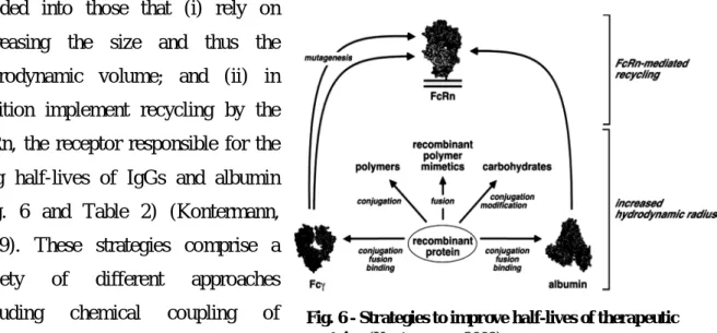
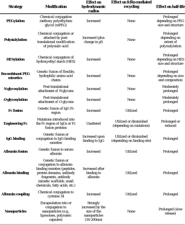
III - Half-life extension strategies 13
III.1 - Strategies to increase the hydrodynamic radius 14
III.1.1 - PEGylation 15
III.1.2 - N- and O-glycosylation 17
III.1.3 - Polysialylation, HESylation and other strategies 18 III.2 - Strategies implementing FcRn-mediated recycling 19 III.2.1 - Half-life extension by binding to albumin 21
III.2.1.1 - Human serum albumin 21
III.2.1.2 - Fusion to albumin 23
III.2.1.3 - Fusion to bacterial albumin-binding domains 24
III.2.1.3.1 - Protein G 25
III.2.1.3.2 - PAB streptococcal cell-surface protein 28 III.2.1.3.3 - MAG and MIG streptococcal cell-surface proteins 30 III.2.1.3.4 - ZAG streptococcal cell-surface protein 31
III.2.1.3.5 - Protein H 32
III.2.1.4 - Fusion to an albumin-binding peptides 34 III.2.1.5 - Fusion to an albumin-binding antibody fragments and other strategies
strategies
35
III.2.2 - Fusion to Ig-binding domains 37
IV - Anti-TNFα VHH as study model 38
IV.1 - Rheumatoid arthritis 39
IV.2 - Ablynx VHH technology for treating rheumatoid arthritis and cancer 42
II - Objectives 45
III - Albumin-binding domain (Zag) from Streptococcus zooepidemicus as a strategy to
improve half-life of therapeutic proteins 47
Brief Introduction 47
Keywords 49
xxii
Abstract 49
Introduction 50
Experimental procedures 51
Materials 51
Cloning of recombinant proteins 52
Expression and purification of proteins 52
ELISA 53
Size Exclusion Chromatography 53
Affinity measurements 53
Neutralization of TNF-dependent cytolytic activity 54
Protein thermal stability and in vitro serum stability 54
Pharmacokinetics 55
Radiolabelling of 99mTc(CO)3-VHH and 99mTc(CO)3-VHH-Zag proteins 55
Partition coefficient 56
Biodistribution studies of 99mTc(CO)3-VHH and 99mTc(CO)3-VHH-Zag 56
Results 56
Expression and purification of VHH and VHH-Zag proteins 56
Binding activity of VHH-Zag against TNFα 57
Binding activity of VHH-Zag to albumins 57
Thermal stability and in vitro serum stability 58
Neutralization of TNFα-dependent cytolytic activity by VHH and VHH-Zag 58 Pharmacokinetic and organ biodistribution of VHH and VHH-Zag 59
Discussion 60 References 62 Abbreviations 65 Acknowledgments 65 Figure legends 65 Tables 67 Figures 70
IV - Biodistribution of a 67Ga-labeled anti-TNF VHH single-domain antibody containing
a bacterial albumin-binding domain (Zag) 81
Brief Introduction 81
Abstract 83
1. Introduction 83
2. Materials and methods 84
2.1. Construction of VHH and VHH-Zag fusion proteins 84
2.2. Expression and purification of VHH and VHH-Zag 84
2.3. Conjugation of p-SCN-Bn-NOTA to VHH and VHH-Zag 84
2.4. Radiolabelling 84
2.5. ELISA assay 85
2.6. DTPA challenge 85
2.7. Biodistribution studies 85
3. Results and discussion 85
xxiii
Acknowledgments 86
References 86
V - Albumin-binding domain (ProtH) from Streptococcus pyogenes, a new approach to
increase half-life of therapeutic proteins 89
Brief Introduction 89 Keywords 91 Capsule 91 Abstract 91 Introduction 92 Experimental procedures 93 Materials 93
Cloning of recombinant proteins 94
Expression and purification of proteins 94
ELISA 95
Size Exclusion Chromatography 95
Protein thermal stability and in vitro serum stability 96
Neutralization of TNF-dependent cytolytic activity 96
Pharmacokinetics 96
Radiolabelling of 99mTc-VHH and 99mTc-VHH-ProtH proteins 97
Partition coefficient 97
Biodistribution studies of 99mTc(CO)3-VHH and 99mTc(CO)3-VHH-ProtH 98
Results 98
Expression and purification of VHH and VHH-ProtH 98
Antigen binding of VHH and VHH-ProtH proteins 99
Binding of VHH-ProtH to serum albumins 99
Thermal stability and in vitro serum stability 100
Neutralization of TNFα-dependent cytolytic activity by VHH and VHH-ProtH 100
Pharmacokinetic properties of VHH-ProtH 100
Biodistribution of 99mTc(CO)3-VHH and 99mTc(CO)3-VHH-ProtH 101
Discussion 102 References 104 Abbreviations 108 Acknowledgments 108 Figure legends 108 Tables 110 Figures 112
VI - Albumin-binding characterization and three-dimensional structure of Zag ABD from
Streptococcus zooepidemicus 121 Brief Introduction 121 Keywords 123 Capsule 123 Abstract 123 Introduction 124
xxiv
Experimental procedures 125
Materials 125
Construction of pT7-Zag 125
Expression and purification of Zag 125
Protein thermal stability 126
ELISA 126
Size Exclusion Chromatography 126
Circular dichroism measurements 127
Expression and purification of 13C 15N-labelled Zag ABD 127
NMR spectroscopy 128
Results 129
Production of Zag 129
Zag ABD thermal stability 129
Binding of Zag ABD to albumin 129
Circular dichroism 130 Zag NMR 130 Discussion 131 References 133 Abbreviations 137 Acknowledgments 138 Figure legends 138 Tables 139 Figures 141
VII - Albumin-binding characterization and in silico structure prediction of protein H
ABD from Streptococcus pyogenes 147
Brief Introduction 147 Keywords 149 Capsule 149 Abstract 149 Introduction 150 Experimental procedures 151 Materials 151
Cloning of ProtH and ProtH domains 151
Expression and purification of ProtH 152
Protein thermal stability 152
ELISA 153
Size Exclusion Chromatography 153
Circular dichroism measurements 153
Structure prediction of ProtH ABD 153
Results 154
Expression and purification of ProtH ABD constructs 154
ProtH ABD thermal stability 154
Binding of ProtH ABD to albumin 155
xxv
ProtH in silico predicted three-dimensional structures 156
Discussion 156 References 158 Abbreviations 161 Acknowledgments 161 Figure legends 162 Tables 163 Figures 164
VIII - Concluding remarks and future perspectives 171
IX - References 181
Figures
I - Introduction 1
Fig. 1 - The molecular mass and half-life of plasma proteins 3 Fig. 2 - Schematic representation of different antibody formats 5 Fig. 3 - Schematic diagram of the VHH domain of a camelid heavy chain antibody 6 Fig. 4 - Representative illustrations of a selection of scaffold proteins applied for generation of novel affinity proteins
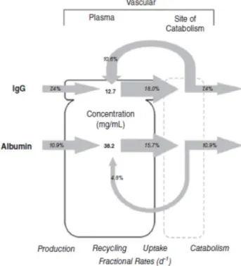
9 Fig. 5 - Effect of FcRn-mediated recycling on IgG and albumin turnover in humans expressed as fractional rates
12 Fig. 6 - Strategies to improve half-lives of therapeutic proteins 13 Fig. 7 - The crystal structure of FcRn and its ligands binding sites 19 Fig. 8 - FcRn mediated, pH-dependent, albumin recycling 20 Fig. 9 - Summary of the ligand binding capacity of HSA as defined by crystallographic studies 22 Fig. 10 - Historical outline of the development of using ABD for half-life extension 25 Fig. 11 - An overview of the streptococcal protein G (SpG) 26 Fig. 12 - Peptide sequence alignment of 16 different GA modules from six proteins and four bacterial species
28 Fig. 13 - Structure of the complex formed by SpG ABD and HSA 29 Fig. 14 - Schematic presentation of protein MAG 31 Fig. 15 - Schematic presentation of protein ZAG 32 Fig. 16 - Schematic presentation of protein H 33 Fig. 17 - Pathophysiological role of cytokines, other mediators and their inhibitors in RA 41 Fig. 18 - Therapeutic effects of Enebrel® 43
III - Albumin-binding domain (Zag) from Streptococcus zooepidemicus as a strategy to improve half-life of therapeutic proteins
47
Fig. 1 - Construction and expression of VHH and VHH-Zag 70 Fig. 2 - Binding of VHH and VHH-Zag to human TNFα 71 Fig. 3 - Binding of VHH-Zag to human, rat and mouse albumin 72 Fig. 4 - Binding affinity measurements 73 Fig. 5 - Formation of VHH-Zag/albumin complexes 74 Fig. 6 - TNFα-neutralization of VHH and VHH-Zag 75 Fig. 7 - In vitro stability in human and mouse serum of VHH and VHH-Zag 76 Fig. 8 - Pharmacokinetic properties 77 Fig. 9 - In vivo stability in mouse serum of VHH and VHH-Zag 78
xxvi
Fig. 10 - Biodistribution of 99mTc(CO)3-VHH and 99mTc(CO)3-VHH-Zag 79
IV - Biodistribution of a 67Ga-labeled anti-TNF VHH single-domain antibody containing a bacterial albumin-binding domain (Zag)
81
Fig. 1 - Binding of the protein conjugates NOTA-VHH and NOTA-VHH-Zag to human, rat and mouse albumin, and human TNFα in ELISA assay
85 Fig. 2 - Binding of 67Ga-NOTA-VHH and 67Ga-NOTA-VHH-Zag proteins to human, rat and mouse albumin, and human TNFα in ELISA
85
V - Albumin-binding domain (ProtH) from Streptococcus pyogenes, a new approach to increase half-life of therapeutic proteins
89
Fig. 1: Construction and production of VHH and VHH-ProtH 112 Fig. 2: Binding of VHH and VHH-ProtH to human TNFα 113 Fig. 3: Binding of VHH-ProtH to human, rat and mouse albumin 114 Fig. 4: Formation of VHH-ProtH/albumin complexes 115 Fig. 5: TNFα-neutralization capacities of VHH and VHH-ProtH 116 Fig. 6: In vitro stability in human and mouse serum of VHH and VHH-ProtH 117 Fig. 7: Pharmacokinetic properties 118 Fig. 8: In vivo stability in mouse serum of VHH and VHH-ProtH 119 Fig.8: Biodistribution of 99mTc(CO)3-VHH and 99mTc(CO)3-VHH-ProtH 120
VI - Albumin-binding characterization and three-dimensional structure of Zag ABD from
Streptococcus zooepidemicus
121
Fig. 1: Construction and expression of Zag ABD 141 Fig. 2: Binding of Zag to human, rat and mouse albumin at pH 6.0 and 7.4 142 Fig. 3: Size exclusion chromatography for Zag/albumin complexes analysis 143 Fig. 4: CD studies of Zag and Zag/HSA binding 144 Fig. 5: 1H-15N HSQC spectrum of Zag ABD 145 Fig. 6: Three-dimensional structure of Zag ABD determined by NMR 146
VII - Albumin-binding characterization and in silico structure prediction of protein H ABD from Streptococcus pyogenes
147
Fig. 1: Construction and production of ProtH ABD and C repeats 164 Fig. 2: Binding of ProtH ABD to human, rat and mouse albumin 165 Fig. 3: Binding of ProtH domains to human serum albumin 166 Fig. 4: Size exclusion chromatography for ProtH/albumin complexes analysis 167 Fig. 5: CD studies of ProtH and ProtH/HSA binding 168 Fig. 6: In silico prediction of ProtH ABD (C1C2C3) structure 169
Tables
I - Introduction 1
Table 1: Non-immunoglobulin scaffolds for generation of new affinity ligands 8 Table 2: Recombinant antibody and antibody/albumin complexes 14
III - Albumin-binding domain (Zag) from Streptococcus zooepidemicus as a strategy to improve half-life of therapeutic proteins
47
Table 1: Binding of VHH-Zag to HSA, RSA and MSA 67 Table 2: Affinities of VHH-Zag for HSA, RSA and MSA by SPR measurements 67 Table 3: Molecular mass and hydrodynamic radius 67 Table 4: Thermal stability of VHH and VHH-Zag 67
xxvii
Table 5: Pharmacokinetic parameters of VHH and VHH-Zag 68 Table 6: Biodistribution of 99mTc VHH 68 Table 7: Biodistribution of 99mTc VHH-Zag 69 Table 8: Organ to blood ratio of 99mTc-VHH and 99mTc-VHH-Zag at 24 h time point 69
IV - Biodistribution of a 67Ga-labeled anti-TNF VHH single-domain antibody containing a bacterial albumin-binding domain (Zag)
81
Table 1: Biodistribution of 67Ga-NOTA-VHH and 67Ga-NOTA-VHH-Zag in CD-1 mice (n=3) at 1h, 4 h and 24 h p.i.
86
V - Albumin-binding domain (ProtH) from Streptococcus pyogenes, a new approach to increase half-life of therapeutic proteins
89
Table 1: Binding of VHH-ProtH to HSA, RSA and MSA 110 Table 2: Molecular mass and hydrodynamic radius 110 Table 3: Thermal stability of VHH and VHH-ProtH 110 Table 4: Pharmacokinetic parameters of VHH and VHH-ProtH 110 Table 5: Organ to blood ratio of 99mTc-VHH and 99mTc VHH-ProtH at 24 h time point 111
VI - Albumin-binding characterization and three-dimensional structure of Zag ABD from
Streptococcus zooepidemicus
121
Table 1: NMR experiments for Zag assignment 139 Table 2: Thermal stability of Zag ABD 139 Table 3: Binding of Zag to HSA, RSA and MSA 139 Table 4: Zag ABD and Zag/albumin complexes molecular mass and hydrodynamic volume 140
VII - Albumin-binding characterization and in silico structure prediction of protein H ABD from Streptococcus pyogenes
147
Table 1: Thermal stability of ProtH and ProtH C repeats 163 Table 2: ProtH ABD/albumin complexes 163 Table 3: Binding of ProtH to HSA, RSA and MSA 163
3
I - Therapeutic Proteins
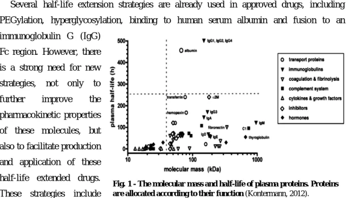
At the end of 2011, approximately 200 biologics were approved for therapeutic applications and more than 600 are in different stages of clinical development as drug molecules (Walsh, 2010; Kontermann, 2011).
Besides monoclonal antibodies (mAbs) and vaccines, which account for more than two-thirds of these products, hormones, growth factors, cytokines, fusion proteins, coagulation factors, enzymes and other proteins are enumerated. With the exception of antibodies and Fc fusion proteins, many of these proteins possess a molecular mass below 50 kDa and for that reason, are rapidly cleared from circulation, presenting short terminal half-lives in the range of minutes to a few hours (Fig. 1). In order to maintain a therapeutically effective concentration over a prolonged period of time, infusions or frequent administrations are performed, or the drug is applied loco-regional or subcutaneously, using a slow adsorption into the blood stream. Half-life extension strategies have therefore become increasingly important to improve the pharmacokinetic (PK) and pharmacodynamic (PD) properties of therapeutic proteins, but also for reasons of compliance (Kontermann, 2012).
Several half-life extension strategies are already used in approved drugs, including PEGylation, hyperglycosylation, binding to human serum albumin and fusion to an immunoglobulin G (IgG)
Fc region. However, there is a strong need for new strategies, not only to further improve the pharmacokinetic properties of these molecules, but also to facilitate production and application of these half-life extended drugs. These strategies include
those that increase the hydrodynamic radius of the drug, thus aiming at reducing the renal clearance and also strategies that implement recycling by neonatal Fc receptor (FcRn), which is responsible for the extraordinary long half-life of IgG molecules and serum albumin. In the past 5 to 10 years, half-life extension strategies experienced a rapid growth in the establishment of novel approaches, including the application of novel hydrophilic polymers,
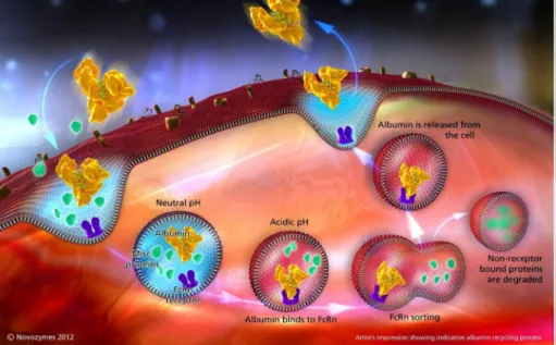
Fig. 1 - The molecular mass and half-life of plasma proteins. Proteins are allocated according to their function (Kontermann, 2012).
4
the generation of recombinant PEG mimic polypeptide chains and the development of several albumin-binding molecules. Furthermore, the half-life of IgG molecules was altered by engineering of the Fc region, which opens new opportunities for the development of next-generation antibody and protein drugs (Kontermann, 2012).
I.1 - Engineered antibody fragments and therapeutic proteins
As one of the most important defences against disease, antibodies are produced by the immune system in response to substances (antigens) that appear to be foreign to the human body. In vertebrates, there are five different classes of antibodies known as IgD, IgA, IgM, IgE and IgG, which differ in their function from the immune system. IgGs are the most abundant immunoglobulins in the blood and the dominant format of therapeutic antibodies (Aires da Silva et al., 2008). These typical antibodies are usually ‘Y’ shaped molecules and are bivalent molecules composed of two identical heavy (H) and two light (L) chains with a molecular mass of approximately 150 kDa (Fig. 2). Monovalent derivates thereof include Fab fragments, which are linked via a flexible region (hinge) to a constant fragment (Fc) region, single-chain Fv (scFv) and single variable domains from human IgGs (domain antibodies, dAbs) or camelid (VHH) and shark (VNARs) single-domain antibodies (sdAbs) (Holliger and Hudson, 2005). These antibody formats are small in size (11-50kDa) and do not possess an Fc region, thus they are not capable of exerting Fc-mediated effector functions. The small size and less complex structure of these molecules compared with IgG facilitate production, e.g. in bacterial expression systems (Kontermann, 2009). In addition, small bivalent derivates (e.g. diabodies) have been generated with the lack of constant domains. Bivalent diabodies are composed of the VL and VH domain connected by a short linker of approximately 5 amino acid residues, leading to the formation of a homodimeric protein. Recombinant antibody fragments also include several bispecific formats, e.g. minibodies, Fab2, as well as multimeric formats (Fig. 2) (Holliger and Hudson, 2005).
5
In comparison with whole antibodies, small antibody fragments such as Fab or scFv exhibit better pharmacokinetics for tissue penetration and also provide full binding specificity because the antigen-binding surface is unaltered. Indeed, the first clinical trials of scFv fragments are likely to be as multivalent reagents, because they exhibit high functional affinity and have been very successful in preclinical studies (Holliger and Hudson, 2005; Hudson and Souriau, 2003). Small size antibody fragments are monovalent and often present fast off-rates and poor retention time on the target (Hudson and Souriau, 2003). Therefore, Fab and scFv fragments have been engineered into multimeric conjugates to increase
functional affinity through the use of either chemical or genetic cross-links. Various methods have been devised to genetically encode multimeric scFvs, of which the most successful design was the simple reduction of scFv linker length to direct the formation of bivalent dimers (diabodies), triabodies or tetrabodies(Holliger and Hudson, 2005). In general, several multivalent antibody conjugates are generated to be monospecific. However, some strategies have also been applied to produce bispecific multimers. These bispecific antibodies have two different binding specificities fused together in a single molecule. Because of dual specificity, they can bind to two adjacent epitopes on a single antigen, thereby improving avidity. Antibody engineering has also been applied to the development of bifunctional antibodies. In contrast to bispecific antibody derivates, bifunctional molecules combine the antigen-binding site of an antibody with a biological function encoded by a linked or fused partner (Hudson and Souriau, 2003). These include radionuclides, cytokines, toxins, enzymes, peptides and proteins. Recently, ibritumomab or Zevalin® (Biogen Idec) (Vose et al., 2007), tositumomab or Bexxar® (GlaxoSmithKline) (Macklis, 2006), both applied to the treatment of
Fig. 2 - Schematic representation of different antibody formats. A variety of antibody fragments are
depicted, including Fab, scFv, single-domain VH, VHH and VNAR and multimeric formats, such as
minibodies, bis-scFv, diabodies, triabodies, tetrabodies and chemically conjugated Fab multimers (sizes given in kDa are approximate) (Holliger and Hudson, 2005).
6
Hodgkin lymphoma, and gemtuzumab or Mylotarg® (Wyeth) (Larson et al., 2002) with therapeutic use against acute myeloid leukemia, are three examples of FDA-approved bifunctional antibodies designed to specifically deliver cytotoxic drugs into cancer cells.
To escape immunosurveillance, many pathogenic viruses have evolved narrow cavities (canyons) in their surface antigens, which bind their target receptors but are poorly accessible to intact antibodies and are thus largely immunosilent. This antibody response is caused by the limited diversity of complementarity-determining region (CDR) loop lengths, which constrains the displayed antigen-binding surfaces to mostly flat or concave topologies (Arndt
et al., 2004). Interestingly, the camelids (camels and llamas) and cartilaginous fish
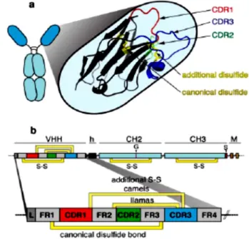
(wobbegong and nurse sharks), have evolved high-affinity single V-domains mounted on an Fc-equivalent constant domain framework as an integral and crucial component of their immune system (Klooster et al., 2007; Stretsov and Nuttal, 2005). In addition to these conventional antibodies, camelids and sharks also produce unusual antibodies composed only of heavy chains. These peculiar heavy
chain antibodies (hcAbs) lack light chains (and, in the case of camelid antibodies, also the CH1-domain). Therefore, the antigen binding site of hcAbs is formed only by a single domain that is linked directly via a hinge region to the Fc-domain. The variable domain is designed VHH for camelid and VNAR for shark single-domain antibodies (Fig. 3). The CDR3 region of these antibodies possesses the extraordinary capacity to form long finger-like extensions that can extend into cavities on antigen, e.g. the active site crevice of enzymes. The CDR3 can form convex extensions that occupy the cleft for the substrate and is often much longer than that of conventional VH domains. The extended CDR3 is usually
stabilized by an additional disulfide bond connecting the CDR3 to the adjacent CDR1 loop Fig. 3 - Schematic diagram of the VHH domain of a camelid heavy chain antibody. a The three CDRs of the
antigen-binding paratope are depicted as colored loops: CDR1 red, CDR2 green, and CDR3 blue. b The canonical disulphide bond connecting framework regions 1 and 3 (FR1 and FR3) in the two β-sheets of the immunoglobulin domain is indicated in yellow. Many camelid antibodies contain an additional disulfide bond (S–S) connecting the CDR3 with the end of the CDR1 (camels) or the beginning of the CDR2 (llamas). h Hinge, M transmembrane domain of membrane isoform, G glycosylation site, S stop codon of secretory isoform (Wesolowski et al., 2009).
7
(common in camel and shark sdAbs) or to the CDR2 loop (common in llama sdAbs). The interface of conventional VH and VL domains is stabilized by hydrophobic interactions, the corresponding residues in llama antibodies are replaced by hydrophilic residues. In shark sdAbs, the CDR2 loop is replaced by two shorter less variable loops (Wesolowski et al., 2009).
In general, camelid VHH and shark VNAR domains are soluble and can be produced as stable in vitro targeting reagents for sensitive diagnostic platforms and nanosensors. Compared to monoclonal antibodies, VHH domains have also demonstrated improved penetration against cryptic (immuno-evasive) target antigens (Holliger and Hudson, 2005). However, for in vivo administration, humanization (or deimmunization) may be crucial to reduce immunogenicity, although llama VHH domains have been claimed to be only minimally immunogenic (Cortez-Retamozo et al., 2004). As it is frequently observed with display technologies, favourable properties (such as good expression, stability and solubility) are co-selected with binding activity of VHH molecules (Holliger and Hudson, 2005).
I.2 - Scaffolds
Antibody size dependence of epitope accessibility can be highly non-linear and some protein surface-exposed structures can be completely obstructed for full size antibodies (Chen
et al., 2010; Chen et al., 2009). Therefore, a large amount of work has been aimed at
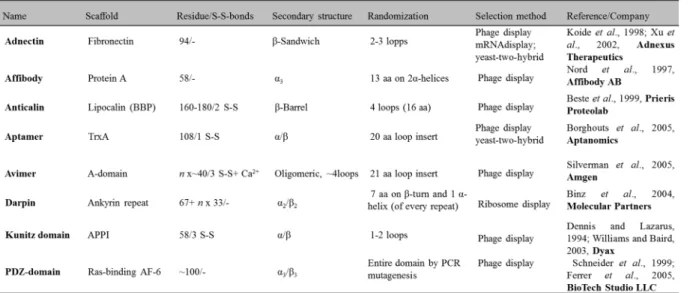
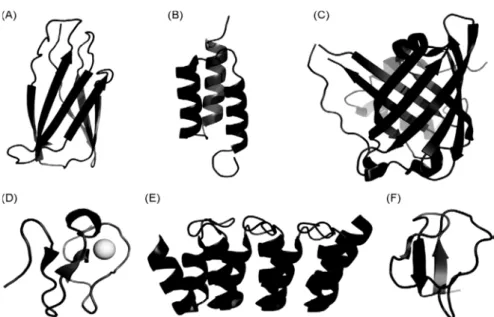
developing novel scaffolds of much smaller size. The term “scaffold” reflects the use of a protein framework that can carry altered amino acids or insertions giving protein variants with entirely novel functions, typically new binding specificities. Such scaffolds are stable, soluble and easy to format, manufacture and express in microbial cell cultures. High stability independent of disulfide bonds is a clear advantage, facilitating high yields in bacterial expression and enabling intracellular applications. There are presently approximately 50 suggested protein scaffolds reported and these have intensely reviewed over the last years (Dimitrov, 2009; Dimitrov and Marks, 2009; Chen et al., 2008; Honegger, 2008; Kolmar and Skerra, 2008; Saerens et al., 2008; Skerra, 2007). Examples of several non-immunoglobulin scaffolds are illustrated in Fig. 4 and Table 1.
8
Table 1: Non-immunoglobulin scaffolds for generation of new affinity ligands (adapted from Grönwall and Ståhl, 2009).
Affibodies are small antibody mimetics (~6 kDa), robust 3-helix proteins, designed based on the Z domain of staphylococcal protein A that were developed for targeted therapy of several diseases, e.g. cancer and inflammatory diseases (Jonsson et al., 2009; Tolmachev et
al., 2007). Affibody® molecules can also be used for protein purification (Nord et al., 2001), enzyme inhibition (“Phusion Hot Start High-Fidelity DNA Polymerase", Finnzymes), research reagents for protein capture and detection (Lundberg et al., 2007; Renberg et al., 2007) and diagnostic imaging (Orlova et al., 2006).
An ABD-fused tumor-targeting construct was derived from a HER2-specific Affibody molecule, which belongs to a class of small protein A-derived antibody mimetics, originally developed for imaging purposes. The original affibody presented a rapid kinetics ideal for imaging, but the uptake in kidneys was very high, which was incompatible with radionuclide therapy that would be a logical extension of the successful tumour targeting obtained. Therefore, Nilsson and co-workers decided to convert the imaging tracer to a candidate for targeted radiotherapy by fusion to ABD (Tolmachev et al., 2007). The albumin association did not compromise the tumour targeting properties. Compared with the non-fused Affibody molecule, kidney uptake was decreased 25-fold and the tumour uptake increased three to five- fold. This combined with retained high specificity for the tumour, allowed for successful radioimmunotherapy using the beta-emitting nuclide 177Lu. Treatment of mice bearing HER2-overexpressing microxenografts completely prevented tumour formation, in contrast to radioactivity coupled to an ABD-fused construct against an irrelevant antigen (Tolmachev et
al., 2007). The HER2-specific Affibody ABY-025 is in clinical development for tumour
9
I.3 - Pharmacokinetics of therapeutic proteins
The basic paradigm of clinical pharmacology is the fact that drug effects, desired as well as undesired, are function of drug concentrations within different organs and tissues in the human body. Thus, drug concentrations are the driving force for the spectrum of drug responses observed in a drug-treated patient (Meibohm, 2012). Pharmacokinetics is the study and characterization of the time course of drug adsorption, distribution, metabolism and excretion (ADME), and provides a quantitative analysis of how living systems handle xenobiotics and biodrugs (Mahmood, 2009). The pharmacokinetic properties, especially their distribution, metabolism and excretion, are influenced by several factors including size, shape, charge, hydrophilicity, interaction with other molecules and cells, and sensitivity to proteolytic degradation (Tang et al., 2004). Similar to conventional small molecule drugs, protein therapeutics is characterized by well-defined properties that form the basis for the design of therapeutic dosing regimens as well as drug delivery strategies. The distribution volume of proteins is determined largely by their molecular weight, physicochemical properties (e.g., charge, lipophilicity), protein binding, and their dependency on active Fig. 4 - Representative illustrations of a selection of scaffold proteins applied for generation of novel affinity proteins. (A) Adnectin, derived from 10th fibronectin type III domain (PDB ID: 1TTG). (B)
Affibody, derived from Z-domain of protein A (PDB ID: 1Q2N). (C) Anticalin, derived from the bilin-binding protein, here a digoxigenin bilin-binding anticalin is illustrated (PDB ID: 1LKE). (D) Avimer, designed from a consensus human A-domain here an A-domain is represented by the LDL receptor ligand-binding module 5 stabilized by a Ca2+ ion (PDB ID: 1AJJ). (E) Darpin, designed from a consensus ankyrin repeat domain, here a HER2-binding darpin with two repeat modules is illustrated (PDB ID: 2JAB). (F) Kunitz domain, derived from the protease inhibitor domain of the amyloid beta protein precursor (PDB ID: 1AJJ). Images were generated using PyMOL software (Grönwall and Ståhl, 2009).
10
transport processes (Meibohm, 2012). Since most therapeutic proteins have high molecular weight and are thus large in size, their apparent volume of distribution is usually small and limited the volume of the plasma or the extracellular space, predominantly because of their limited mobility secondary to impaired passage through biomembranes (Zito, 1997). In contrast, to small molecular drugs, protein transport from vascular space into the interstitial space of tissues is largely mediated by convection rather than diffusion, following the unidirectional fluid flux from the vascular space through paracellular pores into the interstitial tissue space. The subsequent removal from the tissues is accomplished by lymph drainage back into the systemic circulation (Flessner et al., 1997). Another, but much less prominent pathway for the movement of protein molecules from the vascular to the interstitial space is transcellular migration via endocytosis (Reddy et al., 2006).
Engineering antibodies or proteins for therapy must generally exhibit high affinity, discriminating specificity, minimal immunogenicity and low cross-reactivity. Normally, these factors can be addressed by making relatively small changes to the primary protein structure of the molecule. In addition to this, a major consideration in protein engineering is developing one that exhibits optimal pharmacokinetics: appropriate dosing leading to optimal bioavailability, uptake, distribution and clearance in targeted and non-targeted tissues, which will lead to optimal pharmacodynamics (Constantinou et al., 2010; Beckman et al., 2007; Batra et al., 2002). Therapeutic administration requires a balance between prolonged retention at the target site and slow clearance, which can lead to liver accumulation and high radiation exposure of other tissues (Holliger and Hudson, 2005).
Systematic in vivo studies have provided striking confirmation that size is an important parameter in pharmacokinetics and biodistribution of therapeutic proteins. Large IgG molecules specific for tumour surface molecules have been found to penetrate solid tumours only slowly, are non-uniform in their final distribution and have high serum levels and associated toxicities. Conversely, small scFv fragments (30 kDa) are cleared extremely rapidly and have poor tumour retention because of their monovalent binding properties. Some biodistribution studies (Kenanova et al., 2005; Robinson et al., 2005) have independently confirmed that diabodies, because of their small size, are rapidly eliminated through the kidneys, thereby limiting the exposure to the bone marrow, which is most often the dose-limiting organ with intact radiolabeled mAbs.
However, small size antibodies have to be administered by infusion or repeated intravenous bolus injections in order to maintain a therapeutically effective dose over a prolonged period of time, or are restricted to loco-regional treatment. Their rapid elimination
11
is mainly due to renal filtration and degradation, because the kidney generally filters out molecules below 50-60 kDa (Tang et al., 2004). This has led to the development of half-life extension strategies to prolong circulation of these recombinant antibodies in the blood and thus improve administration and pharmacodynamic properties. With an increasing number of small antibody molecules and antibody mimetics developed for clinical applications, strategies to improve pharmacokinetics, especially half-life extension, have become increasingly important, not only with respect to therapeutic efficacy but also regarding the costs of therapy as well as convenience for patients, and compliance (Mahmood and Green, 2005; van de Weert et al., 2005; Tang et al., 2004).
II – Renal clearance and FcRn-mediated recycling
As mentioned in the previous section, due to their small size, some protein therapeutics have to be administered as infusion or repeated intravenous (i.v.) or subcutaneous (s.c.) bolus injections in order to maintain a therapeutically effective dose over a long period of time, or are restricted to loco-regional treatment. A comparison of the half-lives of plasma proteins reveals the threshold for rapid excretion to be in the range of approximately 40-50 kDa, demonstrating that the size of the molecules is one of the determining factors (Fig. 1). The glomerular filtration barrier is formed by the fenestrated endothelium, the glomerular basement membrane (GBM) and the slit diaphragm located between the podocyte foot processes (Tryggvason and Wartiovaara, 2005). The fenestrae between the glomerular endothelial cells presents diameters between 50-100 nm, consequently, allowing free diffusion of molecules. It was suggested that the slit diaphragm represents the ultimate macromolecular barrier, forming an isoporous, zipper-like filter structure with numerous small, 4-5 nm diameter pores and a lower number of 8-10 nm diameter pores (Haraldsoon and Sörensson, 2004; Wartiovaara et al., 2004; Rodewald and Karnovsky, 1974). In addition to size, the charge of a protein contributes to renal filtration. It has been suggested that the proteoglycans of the endothelial cells and the GBM contribute to an anionic barrier, which partially prevents the passage of plasma macromolecules (Tryggvason and Wartiovaara, 2005). Therefore, the size of a therapeutic protein, that is, its hydrodynamic radius, and also its physicochemical properties, that is, charge, represent starting points in order to improve half-life. Interestingly, of all the plasma proteins, two kinds of molecules, serum albumin and IgGs, exhibit an extraordinary long half-life in humans. Thus, human serum albumin (HSA) has a half-life of 19 days and immunoglobulins (IgG1, IgG2 and IgG4) have half-lives in the