Iso latio n and culture o f um bilical
ve in me se nchymal ste m ce lls
1Centro de Terapia Celular (CEPID-FAPESP), Fundação Hemocentro de Ribeirão Preto, Centro Regional de Hemoterapia, Hospital das Clínicas, Faculdade de Medicina de Ribeirão Preto, Universidade de São Paulo, Ribeirão Preto, SP, Brasil
2Divisão de Hematologia e Hemoterapia, Departamento de Clínica Médica, Faculdade de Medicina de Ribeirão Preto, Universidade de São Paulo, Ribeirão Preto, SP, Brasil
D.T. Covas1,2, J.L.C. Siufi1, A.R.L. Silva1 and M.D. O rellana1
Abstract
Bone marrow contains a population of stem cells that can support hematopoiesis and can differentiate into different cell lines including adipocytes, osteocytes, chondrocytes, myocytes, astrocytes, and tenocytes. These cells have been denoted mesenchymal stem cells. In the present study we isolated a cell population derived from the endothelium and subendothelium of the umbilical cord vein which possesses morphological, immunophenotypical and cell differentia-tion characteristics similar to those of mesenchymal stem cells isolated from bone marrow. The cells were isolated from three umbilical cords after treatment of the umbilical vein lumen with collagenase. The cell population isolated consisted of adherent cells with fibroblastoid morphology which, when properly stimulated, gave origin to adipo-cytes and osteoadipo-cytes in culture. Immunophenotypically, this cell popu-lation was found to be positive for the CD29, CD13, CD44, CD49e, CD54, CD90 and HLA-class 1 markers and negative for CD45, CD14, glycophorin A, HLA-DR, CD51/61, CD106, and CD49d. The charac-teristics described are the same as those presented by bone marrow mesenchymal stem cells. Taken together, these findings indicate that the umbilical cord obtained from term deliveries is an important source of mesenchymal stem cells that could be used in cell therapy protocols.
Co rre spo nde nce D.T. Covas
Fundação Hemocentro de Ribeirão Preto
Av. Tenente Catão Roxo, 2501 14051-140 Ribeirão Preto, SP Brasil
E-mail: dimas@ fmrp.usp.br
Research supported by FAPESP, CNPq and FUNDHERP.
Received March 2, 2003 Accepted July 14, 2003
Ke y words
•Mesenchymal stem cells
•Umbilical vein
•Adipocytes
•O steocytes
•Cell differentiation
Bone marrow (BM) contains a special population of stem cells able to support he-matopoiesis and to differentiate into differ-ent cell lines (adipogenic, osteogenic, chon-drogenic, myogenic, and cardiomyogenic lines) (1,2). These cells were first described in 1974 by Friedenstein et al. (3), who char-acterized them as fibroblastic stem cells ca-pable of forming colonies (fibroblast colony-forming units, CFU-F). The CFU-F
they can differentiate into at least seven cell types, i.e., osteocytes, chondrocytes, adipo-cytes, tenoadipo-cytes, myotubules, astroadipo-cytes, and stromal cells able to support hematopoiesis (6).
In addition to being present in BM, MSC have been demonstrated to occur in various organs and in the circulating blood of preterm fetuses, where they circulate together with hematopoietic stem cells (7,8). The presence of MSC in umbilical cord blood of term infants is a controversial topic (9). Wexler et al. (10) concluded that umbilical cord blood and peripheral blood with stem cell mobili-zation do not contain MSC.
The objective of the present investiga-tion was to determine the presence of MSC in the vascular endothelium of the umbilical cord vein of infants born at term. Thus, three umbilical cords were obtained from term deliveries after each mother signed a dona-tion form according to a protocol approved by the Research Ethics Committee of HCRP-USP. The umbilical vein was catheterized and washed twice internally with 1X PBS and its distal end was clamped. The vein was then filled with a 1% collagenase solution (Sigma, St. Louis, MO, USA) in PBS and the proximal end was occluded. After incuba-tion at 37ºC for 20 min, the collagenase solution was drained and the cells of the endothelial and subendothelial layers were collected by washing with PBS. The cell suspension was centrifuged at 400 g and the
cell pellet was resuspended in 199 growth medium (Sigma) supplemented with 20% fetal calf serum (HyClone, Logan, UT, USA), 2 mM L-glutamine (Gibco-BRL, Gaithers-burg, MD, USA), 100 U penicillin/strepto-mycin (Sigma), 1X endothelial cell growth factor (Sigma), and 10 ng/ml vascular endo-thelial growth factor (Sigma). Next, the cells were counted and plated onto 25-cm2 culture
bottles (Cellstar, Greiner, Germany) at the concentration of 105/ml. After 4 days of
culture the medium was changed and non-adherent cells were removed. After 3 weeks
with weekly medium changes, fibroblastoid cells became the predominant cells in cul-ture. At that time the culture medium was replaced with α-MEM (Gibco) supplemented with 20% fetal calf serum (HyClone), 2 mM L-glutamine (Gibco), and 100 U peni-cillin/streptomycin (Sigma). When the cells reached confluence they were trypsinized (5 mg trypsin/ml PBS), washed in PBS, resus-pended in 20 ml medium, and replated onto 75-cm2 bottles (Cellstar) for expansion.
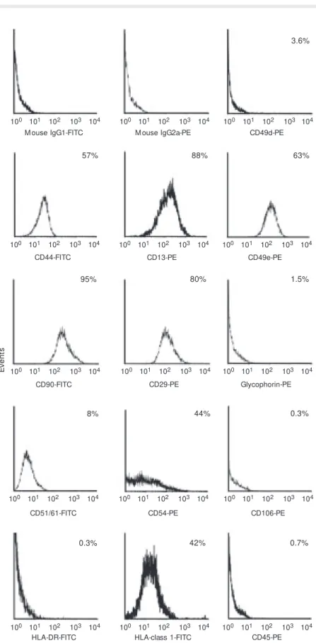
Af-ter expansion, the cells were trypsinized again and analyzed with a flow cytometer (FACsort, BD, San Jose, CA, USA). The following monoclonal antibodies were used: CD13-PE, CD14-CD13-PE, CD29-CD13-PE, CD49d-CD13-PE, CD49e-PE, CD54-CD49e-PE, CD106-CD49e-PE, glycophorin-CD49e-PE, CD44-FITC, CD45-FITC, CD51/61-FITC, CD90-FITC, class 1-FITC, and HLA-DR-FITC (Pharmingen, San Diego, CA, USA).
Assays of adipogenic and osteogenic dif-ferentiation were performed after the third cell passage by plating 104 cells onto
3.6-cm2 plates. The stimulus for adipogenic
car-ried out in parallel to the experiments and stained in the same manner.
After 24 h of culture, two types of adher-ent cells were observed: a more numerous cell population consisting of small flattened cells morphologically similar to the endo-thelial cells (human umbilical vein endothe-lial cells), and a population consisting of a few spindle-shape fibroblastoid cells pre-liminarily identified as MSC. After 1 week of culture, these MSC became the predomi-nant cell type. After the second cell passage the MSC cultures appeared to be homoge-neous and with a high replicative potential. This potential remained unchanged over 20 cell passages when the cells were cultured and maintained at low concentrations. When they reached high confluence, the cells lost their replicative potential and presented mor-phological changes.
In the cytometric analysis, MSC did not present labeling for the hematopoietic line (CD45-, CD14-, glycophorin A-) or for HLA-DR, CD51/61, CD106 (VCAM-1), and CD49d (integrin α4) and were positive for the following adhesion molecules: CD29 (in-tegrin ß1), CD13 (aminopeptidase), CD44 (H-CAM), CD49e (integrin α5), CD54 (ICAM-1), CD90 (Thy 1), and HLA-class 1 (Figure 1).
MSC culture in adipogenic differentia-tion medium led to the appearance, after 7 days, of larger rounded cells presenting nu-merous fat vacuoles in the cytoplasm visual-ized by Sudan III staining. The number of these cells increased continuously up to the 20th day of culture and remained stable for more than two months of culture (Figure 2). The osteogenic stimulus of MSC led to the appearance, after 15 days of culture, of refringent crystals on the cells, better visual-ized by silver nitrate staining. Staining with alkaline phosphatase and silver nitrate per-mitted us to demonstrate the presence of osteocytic differentiation in the induced MSC culture (Figure 2).
In the present study we isolated a cell
E
v
e
n
ts
100 101 102 103 104
M ouse IgG1-FITC
100 101 102 103 104
M ouse IgG2a-PE
100 101 102 103 104
CD49d-PE
100 101 102 103 104
CD44-FITC
100 101 102 103 104
CD13-PE
100 101 102 103 104
CD49e-PE
100 101 102 103 104
CD90-FITC
100 101 102 103 104
CD29-PE
100 101 102 103 104
Glycophorin-PE
100 101 102 103 104
CD51/61-FITC
100 101 102 103 104
CD54-PE
100 101 102 103 104
CD106-PE
100 101 102 103 104
HLA-DR-FITC
100 101 102 103 104
HLA-class 1-FITC
100 101 102 103 104
CD45-PE 3.6%
57% 88% 63%
95% 80% 1.5%
8% 44% 0.3%
0.3% 42% 0.7%
Figure 2. M orphology and dif-ferentiation of umbilical cord vein mesenchymal stem cells (M SC). A,B, Fibroblastoid mor-phological aspect of M SC ob-served by phase microscopy.
Re fe re nce s
1. M inguell JJ, Conget P & Erices A (2000). Biology and clinical utiliza-tion of mesenchymal progenitor cells. Brazilian Journal of M edical and Biological Research, 33: 881-887.
2. Caplan AI & Bruder SP (2001). M esenchymal stem cells: building blocks for molecular medicine in the 21st century. Trends in M olec-ular M edicine, 7: 259-264.
3. Friedenstein AJ, Gorskaja JF & Kulagina NN (1976). Fibroblast pre-cursors in normal and irradiated mouse hematopoietic organs. Ex-perimental Hematology, 4: 267-274.
4. Kuznetsov SA, Friedenstein AJ & Robey PG (1997). Factors required for bone marrow fibroblast colony formation in vitro. British Journal of Haematology, 97: 561-570.
5. Caplan AI (1994). The mesengenic process. Clinics in Plastic Sur-gery, 21: 429-435.
6. M inguell JJ, Erices A & Conget P (2001). M esenchymal stem cells.
Experimental Biology and M edicine, 226: 507-520.
7. Campagnoli C, Roberts IA, Kumar S, Bennett PR, Bellantuono I & Fisk NM (2001). Identification of mesenchymal stem/progenitor cells in human first-trimester fetal blood, liver and bone marrow .
Blood, 98: 2396-2402.
8. Erices A, Conget P & M inguell JJ (2000). M esenchymal progenitor
population derived from the endothelium or subendothelium of the umbilical cord vein with morphological, immunophenotypical and differentiation characteristics similar to those of MSC obtained from BM and origi-nally described by Friedenstein et al. (3). This is the first time that cells with these characteristics isolated from the endotheli-um or subendotheliendotheli-um of the hendotheli-uman endotheli- umbili-cal vein are extensively characterized from an immunophenotypical viewpoint. The im-munophenotypical and morphological pro-file of these cells is the same as that of MSC isolated from BM (11,12). Romanov et al. (13) recently described cells isolated from the endothelium and subendothelium of the umbilical cord morphologically similar to
those isolated here and also showing the ability of adipogenic and osteogenic differ-entiation. Although the biochemical mark-ers used by these investigators were differ-ent from those employed in the presdiffer-ent study, the cell type is probably the same in view of the similarities described, including cell ad-herence and fibroblastoid morphology. Ad-ditional studies are needed for further char-acterization of the pluripotentiality of these cells in view of the pluripotentiality of BM-derived MSC (2). The umbilical cord, in addition to containing hematopoietic stem cells, seems to also be an important source of MSC, a fact indicating the possibility of its use in cell therapy protocols.
cells in human umbilical cord blood. British Journal of Haematology, 109: 235-242.
9. M areschi K, Biasin E, Piacibello W, Aglietta M , M adon E & Faioli F (2001). Isolation of human mesenchymal stem cells: bone marrow versus umbilical cord blood. Haematologica, 86: 1099-1100. 10. Wexler SA, Donaldson C, Denning-Kendall P, Rice C, Bradley B &
How s JM (2003). Adult bone marrow is a rich source of human mesenchymal stem cells but umbilical cord and mobilized adult blood are not. British Journal of Haematology, 121: 368-374. 11. M ajumdar M K, Thiede M A, M osca JD, M oorman M & Gerson SL
(1998). Phenotypic and functional comparison of cultures of mar-row -derived mesenchymal stem cells (M SC) and stromal cells.
Journal of Cellular Physiology, 176: 57-66.
12. Conget PA & M inguell JJ (1999). Phenotypical and functional prop-erties of human bone marrow mesenchymal progenitor cells. Jour-nal of Cellular Physiology, 181: 67-73.