Comparative and Molecular Endocrinology Group, CCMAR, Universidade do Algarve, Faro 8005-139, Portugal
NÁDIA SILVA, VITOR M. BAPTISTA and DEBORAH M. POWER
Expression patterns of MLC isoforms during halibut (Hippoglossus
hippoglossus L.) metamorphosis Abstract
Atlantic halibut is an important commercial fish in the countries of the North Atlantic and is emerging as a promising species for marine cold-water aquaculture. The axial musculature of the developing larvae is the largest and most rapidly growing tissue and during the transition from larval to adult muscle fibre types significant changes in fibre morphology and gene transcription occur. In fact the change in myotome height correlates well with different larval halibut stages. In the present study the spatial and temporal expression of myosin light chain 1 (MLC1), 2 (MLC2) and 3 (MLC3) was studied in metamorphosing halibut by in situ hybridization. As a first step to establishing a role for the thyroid axis in halibut muscle development whole body thyroid hormone (TH) concentrations were also determined. In first feeding larvae MLC1, MLC2A and MLC3 transcripts had a similar distribution and were confined to the muscle fibres of the germinal zones. In pre-metamorphic larvae transcripts were highly expressed throughout the epaxial and hypaxial musculature and expression levels reached a maximum in larvae starting metamorphosis, this change coincided with a significant increase in the concentration of thyroid hormones. By the time larvae reached the metamorphic climax, MLC1, MLC2A and MLC3 expression was still high throughout the musculature but expression was confined to fibres adjacent to the myosepts and to small cells scattered in the musculature, possibly satellite cells. MLC2A was also expressed in the red muscle fibres; no transition between larval and adult MLC isoforms was detected. Key Words: Flatfish, in situ localisation, muscle development, thyroid hormones
Zusammenfassung
Titel der Arbeit: Expressionsmuster von MLC Isoformen während der Metamorphose beim Heilbutt
(Hippoglossus hippoglosus)
Der Atlantikheilbutt ist ein kommerziell wichtiger Fisch für Länder des Nordatlantiks und entwickelt sich zu einer viel versprechenden Spezies für die Aquakultur. Die axiale Muskulatur sich entwickelnder Larven stellt das größte und am stärksten wachsende Gewebe dar. Während der Transformation von den larvalen zu adulten Muskelfasertypen treten signifikante Änderungen der Fasermorphologie und der Genexpression auf. Die Myotomgröße korreliert gut mit verschiedenen Stadien der Heilbuttlarven. In der vorliegenden Untersuchung wurde die lokale und temporale Expression der Gene der leichten Myosinketten 1, 2 und 3 (MLC1, MLC2, MLC3) während der Metamorphose mittels in situ Hybridisierung dargestellt. Als erster Schritt um die Rolle der Schilddrüsenhormon-Achse in der Muskelentwicklung des Heilbutts zu beleuchten, wurden auch die Gesamtkörpergehalte an Schilddrüsenhormonen (TH) bestimmt. Im frühen Larvenstadium (Stadium 5) haben MLC1, MLC2A und MLC3 Transkripte eine ähnliche Verteilung und sind auf Muskelfasern der Keimzonen begrenzt. In prä-metamorphen Larven werden hohe Transkriptionsraten in der epaxialen und hypaxialen Muskulatur gefunden. Die Expression erreicht ein Maximum in den Larven zu Beginn der Metamorphose; gleichzeitig tritt eine signifikante Zunahme der Konzentration der Schilddrüsenhormone auf. Bis Larven den metamorphen Höhepunkt erreichen, ist die Expression von MLC1, MLC2A und MLC3 in der Muskulatur hoch, aber die Expression ist begrenzt auf Fasern nahe den Myosepten und auf kleine Zellen, die in der Muskulatur verstreut sind, vielleicht Satellitenzellen. MLC“A wird auch in den roten Muskelfasern exprimiert; kein Umschalten von larvalen zu adulten MLC Isoformen wird beobachtet.
Schlüsselwörter: Plattfisch, in situ Lokalisation, Muskelentwicklung, Schilddrüsenhormone
Introduction
The importance of the thyroid hormones (THs); thyroxin (T4) and triiodothyronine
(T3), in vertebrate development is well established (POWER et al., 2001). In fish, THs
are involved in the transition of larvae to juveniles, the most dramatic manifestation of which is flatfish metamorphosis. TH treatment stimulates flatfish metamorphosis and the transformation from larvae to juvenile of a variety of teleost species although the mechanism by which it acts still remains to be clearly established (DE JESUS et al.,
1998; SOLBAKKEN et al., 1999). In developing fish larvae the axial musculature is the largest and most rapidly growing tissue and the transition from larval to adult muscle fibre types occurs gradually but appears to accompany metamorphosis raising questions about the role of THs. In fact THs have been associated with the developmental transition of myosin isoforms during flounder metamorphosis (KEIKUZE et al., 1993). The functional unit of myosin in vertebrate adult fast skeletal muscle is composed of two heavy chains and four associated light chains; two regulatory myosin light chains (MLC2) and two alkali light chains (MLC1 and MLC3) (THIÉBAUD et al., 2001). In the present study, the ontogeny and distribution of regulatory and alkali light chain myosin was studied using in situ hybridization and related to whole body TH concentrations and thyroid histology.
Material and methods
The Atlantic halibut larvae, used in this study, were raised at Fiskey, Iceland. Larval rearing was carried out using standard commercial production routines. The larvae were reared at 10-11 ºC, under constant light conditions (24L) and fed live artemia. and were sampled at regular intervals from the age of 260 D° through to the end of metamorphosis 800ºD. The T4 and T3 content of whole larvae were assessed by
radioimmunoassay (RIA) of larval extracts. Frozen larvae were extracted in methanol, reextracted in 50µl methanol, 200µl chloroform and 100µl barbital buffer, centrifuged (3,000 rpm for 30 min at 4oC). Then, the upper phase removed, lyophilized, reconstituted in assay buffer and assayed. Assays for both T3 and T4 were performed
using a double-antibody method under equilibrium conditions. Free hormone was separated from the bound hormone using precipitation with a second antibody. Larvae for histology and in situ hybridization were fixed in paraformaldehyde (PFA, 4%) overnight at 4oC, decalcified in EDTA pH 8 when necessary, embedded in low melting point paraffin and sagittal and transverse section prepared and mounted on APES treated glass slides. In order to study thyroid gland development, sectioned larvae were stained using the Cleveland-Wolfe trichrome method, after dewaxing and rehydrating the sectioned material. Digoxygenin-labelled riboprobes for MLC1, MLC2A and MLC3 were prepared by in vitro transcription of plasmid DNA containing PCR fragments encoding halibut MLC1, MLC2A and MLC3. Riboprobe synthesis was carried out using linearized DNA, 20U of the appropriate RNA Polymerase in transcription buffer (Promega, USA) with 1µl of Digoxygenin-RNA labeling mix (Roche Diagnostics, Germany). Hybridization was carried out overnight at 58ºC using 2 µg µl-1
of riboprobe in hybridization solution (50% formamide, 4xSSC, 0.1% torula RNA, 0.01% Heparin, 1× Denhart’s, 0.1% Tween 20, 0.04% CHAPS). To remove non-specifically bound probe, high stringency washes were carried out. Detection of hybridized probe was done using anti-digoxigenin-AP Fab fragments (1:600, Roche Diagnostics) and colour detection was carried out using as substrate the chromagens NBT and BCIP. Control experiments were performed by treating samples with RNase prior to hybridization and/or by omitting riboprobe from the reaction. Sections were analyzed using a microscope (Olympus BH2) coupled to a digital camera (Olympus DP11).
Results
Histological studies of the thyroid gland indicate that its activity appears to accompany the change in concentration of whole body THs measured by RIA which accompanies halibut development. In first feeding larvae (Fig. 1A) thyroid follicles are visible in the loose connective tissue of the pharynx at the insertion of the gill bars, they are infrequent, small and composed of a flattened unicellular epithelial cell layer surrounding a colloid-filled center. In halibut larvae following the yolk-sack period, vesicles are evident in the periphery of the colloid, and instances are found where the colloid is absent from follicles, suggesting increased thyroid activity. Furthermore, the number and size of thyroid follicles and their apparent activity continually increases until the start of metamorphosis (Fig. 1B) when follicles are numerous of a large diameter and frequently lack colloid, indicative of high thyroid activity. After the climax of metamorphosis thyroid follicles are still abundant but are less active. The variation in the concentration of T3 and T4 measured by RIA of whole halibut larval
extracts accompanies the activity cycle of the thyroid follicles. Prior to metamorphosis T4 (2 pg mg-1 wet weight, n=6) and T3 (0.5pg mg-1 wet weight, n=6) is fairly constant.
Larval T4 (6 pg mg-1 wet tissue) and T3 (2 pg mg-1 wet tissue) content peaks at 610 D°
just around the start of metamorphosis. After this, the T4 and T3 content of larvae
declines.
Fig. 1: Cleveland-Wolfe trichrome staining of sagital sections of Atlantic halibut (Hippoglossus hippoglossus) larvae showing the development of thyroid follicles (arrowheads). (A) first feeding (stage 5); (B) start of metamorphosis (stage 8); colloid, c; epithelial cell layer, e; follicles lacking colloid, ∗. Scale bars indicates 20µm (Färbung nach Cleveland-Wolfe: Entwicklung der Schilddrüsenfollikel in Sagitalschnitten von Heilbuttlarven (Hippoglossus hippoglossus) (Pfeilspitzen). (A) frühes Stadium (Stadium 5); (B) Start der Metamorphose (Stadium 8); Kolloid c; epitheliale Zellschicht, e; Follikel ohne Kolloid, *. Maßstab 20 µm)
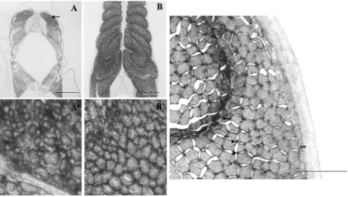
At first feeding (Fig. 2 A) MLC1, MLC2A and MLC3 are expressed and have a similar pattern of distribution, with the hybridization signal being most intense at the periphery, of the dorsal and ventral areas where the germinal zones occur (Fig. 2 A’). The expression levels reached a maximum in white muscle at the beginning of metamorphosis (Fig. 2 B, B’) and all three transcripts are highly expressed throughout the epaxial and hypaxial musculature. By the time larvae reached the metamorphic climax (Fig. 2 C), MLC1, MLC2A and MLC3 expression is still high but expression is confined to fibres adjacent to the myosepts and to small cells scattered in the white
muscle. MLC2A is also expressed in the red muscle fibres. A provisional analysis of MLC expression by RT-PCR revealed that there is a gradual increase in the expression up until stage 9 after which it declines. Moreover, there did not appear to be a transition in MLC isoforms during the larval to juvenile transition.
Fig. 2: Determination of temporal and spatial expression of, MLC2a by means of in situ hybridization with DIG labeled riboprobe in halibut larvae sections. (A) Stage 5, arrow indicates germinal zones; (A’) detail of the germinal zone in (A); (B) stage 8; (B’) detail of (B); (C) stage 10, arrow indicates fibers staining adjacent to the myosept; arrowheads indicate positive staining cells; RM, red muscle fibers. Scale bars indicates 200µm (Darstellung der temporalen und lokalen Expression von MLC2a mittels in situ Hybridisierung mit DIG-markierten Riboproben in Schnitten von Heilbuttlarven. (A) Stadium 5, Pfeile zeigen Keimzonen; (A´) Details der Keimzonen aus (A); (B) Stadium 8; (B´) Detail von (B); (C) Stadium10, Pfeil deutet auf Fasern nahe der Myosepten; Pfeilspitzen zeigen positive gefärbte Zellen; RM, rote Muskelfasern, Maßstab 200µm)
Discussion
Associated with the radical change in the volume and organisation of axial muscle which occurs during fish larval development there is a shift in the distribution of myosin isoforms expressed. The ontogeny of thyroid tissue development and activity coincides with the change in muscle which accompanies metamorphosis. The present observations are consistent with the notion that the change in MLC gene expression may be driven by thyroid hormones. Further work will be required to establish if THs act directly or indirectly on gene encoding muscle proteins to change in musculature during metamorphosis. The functional consequence for growth and survival of the changes in musculature which accompany development and metamorphosis requires much more study. As muscle is of key importance for aquaculture production, it will be of importance to establish a better understanding of how muscle development is regulated by hormones.
Acknowledgements
This work was funded by European Union project CT 96-1442, ARRDE-Q5Rs-2002-01192 and Portuguese Ministry of Science and Technology – Pluriannual funding to CCMAR. We thank Heiddis Smáradóttir, (Fiskey, Iceland) for providing the animal samples.
References BANDMAN, E.; ROSSER, B.W.C.:
Evolutionary significance of myosin heavy chain heterogeneity in birds. Microscopy Research and Technique 50 (2000), 473-491
BASS, J.J.; SHARMA, M.; OLDHAM, J.; KAMBADUR, R.:
Muscle growth and genetic regulation. In: Ruminant Physiology: Digestion, Metabolism, Growth & Reproduction (PB CRONJE, Ed) CABI Publishing (2000), 227-236
DE JESUS, E.G.T; TOLEDO, J.D.; SIMPAS, M.S.:
Thyroid hormones promote early metamorphosis in grouper (Epinephelus coioides) larvae. Gen. Comp. Endocrinol. 112 (1998) 1, 10-16
KEIKUZE, Y., TAKANO-OHMURO H., OBINATA T., INUI Y.:
Effect of Thyroid hormone on developmental transition of myosin light chains during flounder metamorphosis. Gen. Comp. Endocrinol. 93 (1994), 321-326
POWER, D.M.; LLEWELLYN, L.; FAUSTINO, M.; NOWELL, M.A.; BJORNSSON, B.T.; EINARSDOTTIR, I.E.; CANÁRIO, A.V.M.; SWEENEY, G.E.:
Thyroid hormones in growth and development of fish. Comp. Biochem. Physiol. C-Toxicol. Pharmacol.
130 (2001) 4, 447-459
SOLBAKKEN, J.S.; NORBERG, B.; WATANABE, K.; PITTMAN, K.:
Thyroxine as a mediator of metamorphosis of Atlantic halibut, Hippoglossus hippoglossus. Environmental Biology of Fishes 56 (1999) 1-2, 53-65
THIÉBAUD, P.; RESCAN, P.Y.; BARILLOT, W.; RALLIÈRE, C.; THÉZE, N.:
Developmental program expression of myosin alkali light chain and skeletal genes in the rainbow trout
Oncorhynchus mykiss. Biochimica et Biophysica Acta 1519 (2001), 139-142
Corresponding Author
DEBORAH M. POWER
CCMAR, Universidade do Algarve
Campus Gambelas
8005-139 Faro, Portugal E-Mail: [email protected]