Braz. Arch. Biol. Technol. v.59: e16160147, Jan/Dec 2016
1
Vol.59: e16160147, January-December 2016 http://dx.doi.org/10.1590/1678-4324-2016160147
ISSN 1678-4324 Online Edition
BRAZILIAN ARCHIVES OF BIOLOGY AND TECHNOLOGY
A N I N T E R N A T I O N A L J O U R N A L
Histological Observation and Expression Patterns of
antimicrobial peptides during Fungal Infection in Musca
domestica (Diptera: Muscidae) Larvae
Xiu Jiangfan
1,
Wang Tao
1, Wang Yu
1,2, Wu Jianwei
1,3*, Guo Guo
1, Zhang Yingchun
1,
Shang Xiaoli
31 School of Basic Medical Sciences, Guizhou Medical University, Guiyang 550004, China; 2 Guizhou Provincial Center for Disease Control and Prevention, Guiyang 550004, China; 3 School of Biology & Engineering, Guizhou Medical University, Guiyang 550004, China.
ABSTRACT
Housefly, Musca domestica, has a complicated immune system. However, its underlying operating mechanism remains elusive. Candida albicans is a major pathogen affecting humans by causing deep infection fungous disease, but it is non-symbiotic in houseflies. To investigate the C. albicans infection process in housefly, the changes in morphological and histological and expression patterns of antimicrobial peptide were monitored to indicate the
insect’s response to fungal infection. The results showed that scattered brown spots were comprising melanized encapsulation and encapsulated fungal cells were initially observed at the inner side of larvae’s body wall 3 h post-infection (PI). Between 6 and 36 h PI, the whole body of larvae was densely covered with the brown spots, which then gradually disappeared. The majority had disappeared at 48 h PI. Some fungi colonized in the gaps between the body wall and the muscle layer, as well as among muscle fibers of the muscle layer at 12 h PI and hyphal was observed at 18 h PI. These fungi colonized distribution changed from a continuous line to scattered spots at 24 h PI and virtually disappeared at 48 h. The results of quantitative PCR analysis revealed that in coordination with the variation during the infection, the expression levels of four antimicrobial peptides were up-regulated. In conclusion, C. albicans infection in M. domestica larvae involved the following stages: injection, infection, immune response and elimination of the pathogen. The rapid response of antimicrobial peptides, melanized encapsulation and agglutination played a vital role against the pathogenic invasion.
Key words: Musca domestica, larvae, Candida albicans, infection, innate immunity
*Authors for correspondence:[email protected]
INTRODUCTION
Insects lack the adaptive immune responses typical of vertebrates; thus, they are heavily dependent on their innate immune system to guard against pathogen invasion (Hoffmann 2003). Vector insects, with their susceptibility to human pathogens, which can carry a great variety of pathogens and cause many diseases, hence lead to serious damages to human health, and their immune response mechanism is highly homologous to the mammalian innate immune response (Cirimotich et al. 2011; Stokes et al. 2015). In what way the insect innate immune system responds to human pathogens has become a recent focus of research. Many studies have investigated the host–pathogen interaction between insects and pathogen; for example, the interaction between the insects Galleria mellonella, Drosophila melanogaster, silkworm and Tribolium castaneum and the pathogens Candida albicans, Cryptococcus neoformans, Pseudomonas aeruginosa, Escherichia coli, Staphylococcus aureus and Streptococcus pneumoniae (Altincicek et al. 2008; Fuchs et al. 2010; Chamilos et al. 2011; Wang et al. 2013). Recent research indicates that the insect immune system can be activated by the alien pathogens. When the insects are infected by the invading pathogens, the pattern recognition proteins/receptor (PRPs) in insects would firstly recognize and combine with pathogen-associated molecular pattern (PAMPs) in the pathogens, and then initiate the activation and regulation of a series of innate immune response (Ohta et al. 2006). On the one hand, the hemolymph cell served as the major cellular immune response triggering cytophagocytosis and melanization tubercle formation (Lavine and Strand 2002; Lu 2008; Wu et al. 2015). On the other hand, immune effector molecules such as antimicrobial peptides (AMPs) and lysozymes synthesized and secreted by fat body cells (i.e. similar to mammals’ liver), hemocytes and other cells trigger humoral immune responses (Lemaitre and Hoffmann 2007). The innate immune response in insects is divided into humoral immunity and cellular immunity. They function together to kill and eliminate the pathogens via phagocytosis, nodulation, encapsulation, coagulation, and melanization.
Houseflies are present worldwide and considered to be an important medical insect which can carry and transmit over 100 human pathogens and zoonotic
agents, but they rarely to be infected (Scott et al. 2014). It has been demonstrated that housefly larvae were prone to induce the generation of AMPs such as attacin, cecropin, defensin and diptericin by means of heat shock, ultraviolet exposure and needle piercing (Wang et al. 2006; Liang et al. 2006; Dang et al. 2010; Liu et al. 2011). Once the adult housefly is infected with Beauveria bassiana, hemocyte density changes and fungi number in the hemolymph are synchronized (Mishra et al. 2015). The published genome sequences analysis of housefly indicated that compared with Drosophila melanogaster, the genome contains a rich resource of shared and novel protein coding genes, a significantly higher amount of repetitive elements, and substantial increases in copy number and diversity of both the recognition and effector components of the immune system, including a large number of genes encoding PAMPs, immune-related signalling pathways, immune effector molecules and antimicrobial peptides (Scott et al. 2014). Moreover, many specific peptides with strong antibacterial effects are now being discovered in houseflies, including MAF-1, Muscin and MDAP-2 (Fu et al. 2009, 2015; Pei et al. 2014; Yang et al. 2015). These results indicate that houseflies are gifted with a complicated innate immune system that has likely adapted to the complex living environment, particularly to the pathogens these houseflies carry and transmit. However, the mechanisms underlying the operation of this unique innate immune system remains elusive, and the interactions between houseflies and pathogens require further studies.
C. albicans is an opportunistic fungus that can cause deep fungal infections and severely harm human health (Miceli et al. 2011). However, it is an exogenous and non-symbiotc fungus in a housefly (Phoku et al. 2014), furthermore, there was no direct evidence that Candida albicans is a natural pathogen of M. domestica. In our preliminary study, all the larvae which injected with 210 nl PBS buffer were able to complete their life cycle, and in the meanwhile, the larvae injected with an equal volume of the fugal suspension (i.e. approximately 2 × 10⁴ CFU of C. albicans) were controlled with
Process of Fungal infection in M. domestica larvae
Braz. Arch. Biol. Technol. v.59: e16160147, Jan/Dec 2016
3
infection process and broaden the understanding of the insect’s immune response to fungal infections.
MATERIAL AND METHODS
House fly rearing
Houseflies were bred in the Modern Pathogenic Biology Laboratory, Guizhou Medical University (Guiyang, China) (Fu et al. 2009). Their larvae were raised on an artificial diet comprising bran and water and routinely reared at 26–28°C with 70–80% relative humidity and a photoperiod of 12 h: 12 (Light: Dark) h for up to the third-instar larval stage.
C. albicans culturing
The Candida albicans (ATCC10231) used in infection experiments was stored at our laboratory. C. albicans was cultivated in Sabouraud dextrose agar (SDA) and its cell suspension was prepared by inoculating a single colony in Sabouraud dextrose broth (SDB) at 37°C for 12 h with agitation (Kelly and Kavanagh 2011). Fungus entering the logarithmic growth phase was selected for infection experiments. C. albicans cells solution were centrifuged at 4000 g/min and washed and re-suspended in phosphate buffered saline (1 × PBS 0.01M, Solarbio, China) and standardized to 1 × 10⁸
CFU/ml.
Infection experiments
Each experimental group comprised 200 randomly chosen third-instar larvae of appropriate weight (26 ± 0.5mg). The infected group (IC group) larvae were injected with 210 nl suspension, i.e. approximately 2 × 10⁴ CFU of C. albicans at the
10th segment of segmental venter of larvae using the Auto-Nanoliter Injector (Nanoject II Nanoliter Injector, Drummond Scientific Co., Broomall, USA) under a stereomicroscope (SMZ25, Nikon, Japan) (Khalil et al. 2015). After injection, these larvae were raised on sterilized bran and water. At 3, 6, 12, 18, 24, 36 and 48 h post-infection (PI), the larvae were collected. Untreated larvae were also collected (blank control group, C group), and larvae injected with an equal volume of PBS buffer were used as the negative control group (PBS group).
Morphological and histological observations
Larvae were collected at the indicated time points. Morphology was observed using a stereoscopic microscope (SMZ25, Nikon, Japan). Afterembedding in Tissue-Tek optimal cutting temperature compound (OCT, Sakura Finetek, Netherlands), the larvae were transferred to a microtome cryostat (MEV3.01, SLEE, Germany). Histological sections of 10 μm were removed and subjected to periodic acid Schiff (PAS) staining (Okada et al. 2013), observed and photographed using a microscope (Eclipse Ci-S, Nikon, Japan) and NIS-Elements System (NIS-Elements, Nikon Instruments Inc., USA).
qPCR analyses of AMP genes expression
Total RNA was isolated from 6-8 larvae using TRIzol® Reagent (TRIzol® Reagent, Invitrogen,USA) strictly following the manufacturer’s protocol. The concentration and purity level of samples was detected by NanoDrop (ND1000, Thermo Scientific, USA) and agarose (1%) gel electrophoresis. Then, 1 μg of total RNA was reverse-transcribed in 10 μl reaction volumes using PrimeScript RT (PrimeScriptTM RT reagent Kit with gDNA Eraser, Takara Bio, Japan) with random hexamer and oligo dT primers. After synthesis, cDNA was diluted 1: 10 and used in quantitative polymerase chain reaction (qPCR) analysis. Each reaction was performed using the SYBR Premix (SYBR Premix Ex TaqTM II, Takara Bio, Japan), and reactions were performed using the real-time PCR system (ABI PROSM 7300, Applied Biosystems, USA). Reaction mixtures were incubated for 30 s at 95°C, followed by 40 cycles of 5 s at 95°C and 34 s at 60°C. Primers for attacin, cecropin, defensin, diptericin and reference (rps18) genes were selected on the basis of previous studies on housefly immune responses (Wang et al. 2009; Fleming et al. 2014). The primers were designed using Primer 5.0 software (Primer PREMIER Version 5.00, PREMIER Biosoft International 2000). The primers used for qPCR are listed in Table 1. For relative expression analysis, CT values for calibrator conditions (i.e. larvae untreated at each indicated time points, C group) and treatment groups (i.e. PBS and IC groups) were calculated using the 2−ΔΔCT method (Pfaffl et al. 2002). Log2
analysed to determine differences between groups (P < 0.05).
Table 1 Primer sequences for qPCR
Gene Access no. Sense Sequence (5′-3′) Antisense Sequence (5′-3′)
rps18 KC424479.1 GCGTGACGATTTGGAACGCTTGA TTCTTGGATACACCGACAGTGCGA attacin DQ062744.1 CGAATTCATGTTCTCTAAATC CTCTAGATTAAAAATCATGACTT cecropin ES608437.1 GGACAAAGTGAAGCTGGATGGTTG GCTGGGCCACACCAATAGTTTGA defensin ES608345.1 AAATTTCGTCCATGGAGCTGACGC ACCGCTCAACAAATCGCAAGTAGC diptericin ES608652.1 AGTGCAACATTTGTGGTTGCCGAC GCCATAACCTGCTGTGGCATCA
RESULTS
Morphological observations
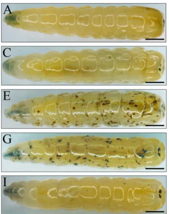
Morphological observations revealed no changes under the epidermis of the larvae in the C and PBS groups (Fig. 1A, Fig. 1B). However, under the body wall of the larvae in the IC group, many scattered brown spots were observed PI. At 3 h PI these spots were scattered and distributed at the inner side of larvae’s body wall (Fig. 1C), and at 6 h PI, the spots had substantially increased in both number and size (Fig. 1D). At 12, 18 and 24 h PI, the entire body was densely covered by brown spots of various sizes (Figs. 1E, 1F and 1G). At 36 h PI, the quantity of these spots had significantly decreased and their colour had faded (Fig. 1H). Majority of the spots had disappeared at 48 h PI (Fig. 1I).
Figure 1 Morphology observation of housefly larvae after Candida albicans injection. A Blank control group:
untreated larvae. B PBS negative control: larvae were injected with 210 nl PBS buffer. C-I Infection group: the larvae were injected with 210 nl suspension of C.
albicans (2 × 10⁴ CFU). C 3 h post infection (PI), D 6 h PI, E 12 h PI, F 18 h PI, G 24 h PI, H 36 h PI and I 48 h PI. The arrow indicated the brown spots under the body wall of the larvae. (A–I) Scale Bar = 1.0 mm.
Histological observations
Process of Fungal infection in M. domestica larvae
Braz. Arch. Biol. Technol. v.59: e16160147, Jan/Dec 2016
5
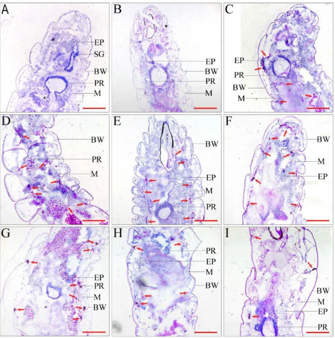
Figure 3 Histopathological identification of C. albicans in housefly larvae (PAS stain, ×200). A Blank control group: untreated larvae. B PBS negative control: larvae were injected with equal volume PBS buffer. C-I Infection group: larvae were injected with C. albicans larvae. C 3 h post-infection (PI), D 6 h PI, E 12 h PI, F 18 h PI, G 24 h PI, H 36 h PI and I 48 h PI. Proventriculus (PR), body wall (BW), epiploon (EP), muscle (M), arrows indicate C. albicans.
Bar = 100 μm
In all infected larvae, many scattered brown spots were observed. These spots were dark in colour because of the accumulation of melanin. Another plane of histological sections revealed that these spots were comprising melanized encapsulation and encapsulated agglutination of fungal cells, spread among the coelom, muscle tissue and the inner side of the body wall (Fig. 4). Between 3 and 12 h PI, we observed a considerable number of C.
Process of Fungal infection in M. domestica larvae
Braz. Arch. Biol. Technol. v.59: e16160147, Jan/Dec 2016
7
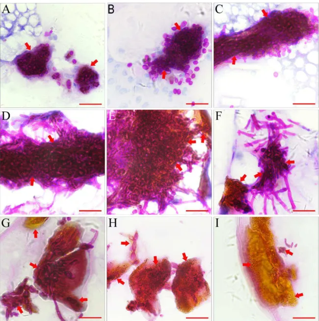
Figure 4 Histopathological identification of melanized encapsulations. A 3 h post-infection (PI), melanized encapsulations encysted yeast from fungal cells. B 6 h PI similar to 3 h PI. C At 12 h PI, melanized encapsulations encysted yeast from fungal cells. D At 18 h PI, melanized encapsulations encysted yeast and hyphae from fungal cells. E 24 h PI similar to 18 h PI. F At 36 h PI, melanized encapsulations encysted hyphae from fungal cells. G At 48 h PI, melanized encapsulations encysted hyphae from fungal cells. H At 48 h PI, melanized encapsulations encysted limited amount of hyphae from fungal cells. I At 48 h PI, empty encapsulations. Arrows indicate melanized encapsulations.
Bar = 50 μm
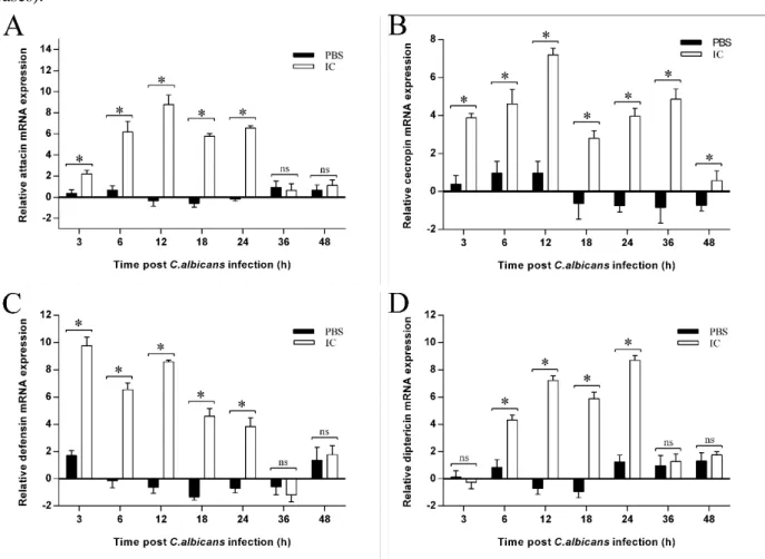
qPCR analyses of AMP genes expression. qPCR was used to assay the temporal patterns of mRNA from attacin, cecropin, defensin and diptericin at respectively challenged 3rd-instar larva at 3, 6, 12, 18, 24, 36 and 48 h after C. albicans infection. Comparing with the PBS group, the results revealed that in coordination with the variation during the infection process, the expression levels of four antimicrobial peptides were up-regulated, but their expression patterns
expression level slowly decreased (P < 0.05, in all cases).
Figure 5 AMP genes expression pattern in housefly larvae with C. albicans infection. In the IC group, the larvae were injected with 210 nl suspension of C. albicans (2 × 10⁴ CFU). In the PBS group, the larvae were injected with equal volume PBS buffer. The C group was untreated. Collecting samples (n = 4 larvae per time point) at 3, 6, 12, 18, 24, 36 and 48 h post-infection. The C group was set as calibrator conditions, compared with the PBS and IC groups. For relative expression analysis, CT values for calibrator conditions (i.e. larvae untreated at each indicated time points, C group) and treatment groups (i.e. PBS and IC groups) were calculated using the 2−ΔΔCT method. Log2-fold change in expression ratios of the target genes attacin, cecropin, defensin and diptericin was compared with the calibrator conditions using the reference gene rps18. A attacin, B cecropin, C defensin, D diptericin. Data was presented as mean ± SE. Significance levels: * P < 0.05; ns, not significant
DISCUSSION
Insects lack the adaptive immune responses typical of vertebrates; thus, they are heavily dependent on their innate immune system to defend against pathogenic invasion. Knowledge on how the insect innate immune response fights infection may aid the understanding of the first step in the human host–pathogen interaction (Wang et al. 2013; Singh et al. 2014; Wu et al. 2014; Khalil et al. 2015). Many models and techniques of insect infection have been successfully established, including injury, rolling and ingestion methods. The rolling method involves the infection of the epithelial cell surface and the ingestion method involves the
Process of Fungal infection in M. domestica larvae
Braz. Arch. Biol. Technol. v.59: e16160147, Jan/Dec 2016
9
infection to achieve controllable infection and minimize mechanical injury by a precise and quantitative inoculation (Khalil et al. 2015). In our preliminary study, the third-instar housefly larvae infection model was built using minimally invasive injection methods. The previous results have indicated that all the larvae of the PBS group were able to complete their life cycle, and in the meanwhile, the larvae of the IC group were controlled with the ratio of approximately 50%. In this study, morphological and histological investigation revealed no significant difference between larvae that were not inoculated and those that were inoculated with PBS (Figs.1 and 2). Although the qPCR results revealed a slight difference in AMP expression between the PBS and C groups, no significant differences were observed. This fluctuation may have been caused by the activation of a weak immune response by the micro wound on the larvae somatic layer caused by injection.
We observed that antibacterial peptides were synthesized in response to microorganism infestation. The housefly genome contains more antibacterial peptide genes than other insect genomes, and such genes are present in the form of a gene family in the genome (Scott et al. 2014; Tang et al. 2014). It was previously reported that in houseflies challenged by microorganisms, antibacterial peptides such as attacins, cecropins, defensins and diptericins were significantly up-regulated. These rapid changes in expression were coordinated in several genes (Wang et al. 2009; Liu et al. 2011; Fleming et al. 2014). In this study, all four antibacterial peptides (attacins, cecropins, defensins and diptericins) were simultaneously up-regulated 6–24 h PI, and their expression parrterns were consistent with those of our previous experiments and some other previouslty published manuscripts (Wang et al. 2009; Fleming et al. 2014). In D. melanogaster AMP expression is regulated by two signal transduction pathways: the Toll and IMD signal pathways. The Toll pathway regulates the response to gram-positive bacterial and fungal infections. The IMD pathway regulates the response to gram-negative bacterial infection (Cho et al. 2012; Kleino and Silverman 2014). Even though the Toll and IMD pathways have separate components and preferential target genes, but some antimicrobial peptide are expressed following the stimulation of one of the two aforementioned pathways (Hultmark 2003; Brennan et al. 2004, Royet et al. 2005, Tanji et al. 2005). We found that
the expression of attacin, cecropin, defensin and diptericin varied 3 to 48 h PI. For instance, cecropin and defensin were up-regulated at 3 h PI, whereas attacin and diptercin were up-regulated later, and in particular, diptercin was not up-regulated until 6 h PI. The rapid response of cecropin and defensin suggested that because houseflies were infected with C. albicans, they may preferentially respond to the Toll pathway. Other studies have shown that the Toll and IMD pathways can be cooperative and may contribute to the activation of each other (Lemaitre and Hoffmann 2007; Tanji et al. 2007, Xiong et al. 2015). Defensin and cecropin are predominantly regulated by the Toll pathway, and attacin and diptericin are predominantly regulated by the IMD pathway during the C. albicans infection. Cecropin is reported to possess fungistasis activity in vitro (Hultmark 2003); therefore, the maintenance of high cecropin expression throughout infection process may have contributed to the control of infection.
under the epidermis of larvae within 3 h of infection with C. albicans. The brown spots were Melanized encapsulations surrounding aggregated C. albicans mycothallus. Throughout infection, similar structures were found, and some empty encapsulations without mycothallus were found 48 h PI. Our results suggested that encapsulation and agglutination may play an important role in the housefly innate immune response against C. albicans.
Housefly larvae, like some other insects, have a mixocoel, and humoral hemocyte concentrated in the last segments and scattered around tissues and organs. In this study, C. albicans mycothallus colonized in the gaps between the body wall and the muscle layer, as well as among muscle fibers of the muscle layer of larvae. We speculated that C. albicans was injected into the hemocoel at the 10th segment of the segmental venter and was quickly disseminated through the blood stream during muscle contractions to the entire body. C. albicans was able to persist between the inner side of the body wall and muscle tissue layers because of the low concentration of hemocytes in these spaces. In these environments, C. albicans was able to develop from the yeast to hyphal form (Huang 2012; Mayer et al. 2013). Our data are consistent with previous research reporting the formation of pseudomycelium by Candida tropicalis mycothallus when hemocyte numbers were reduced in G. mellonella (Mesa-Arango et al. 2013). Some reports suggested that insect blood cells were constantly regenerated and kept dynamically changing. In Locusta migratoria, the hemocytes and hematopoietic tissue mutually assist each other to clear invading pathogens from the circulation (Duressa et al. 2015). In housefly, hemocyte number varied during fungal infection (Mishra et al. 2015).
According to these results, we infer that because a great deal of fungal spores was injected into the coelom of housefly larvae, a fraction was transported to the gaps of tissues. Because there are fewer hemocytes in these locations than in the haemocoel, fungus which stayed in these locations was not eliminated immediately by the immune system. And then began to colonize at these locations. With the regeneration hemocytes and other immune molecule such as AMPs were transported to these gaps during blood circulation, these rest fungus were gradually cleared. This may explain why the mycothallus distribution changed
from a continuous line to scattered spots and then disappeared.
The immune response should be carefully viewed as a double edged sword. Although it protected the larvae from pathogen invasion, excessive immune response could also cause self injury. Similar to the vertebrate immune system, the insect immune system is also regulated by complicated control mechanisms (Cerenius et al. 2008). A great number of immune regulators are involved in these processes, including serine proteinase inhibitors (Serpin). In Tenebrio molitor (Jiang et al. 2009) and D. melanogaster (Ligoxygakis et al. 2002; Fullaondo et al. 2011), serpins can regulate the immune response through the Toll signal pathway and prophenoloxidase-activating system. Li discovered at least 11 serpins in housefly, including a gene homologous to serpin27A of D. melanogaster (Li et al. 2015). In this study, at 48 h PI, the scattered brown spots gradually disappeared and empty Melanized encapsulations were observed and mycothallus colonization and AMP expression were reduced, indicating that mechanisms negatively regulating the immune response had begun to play their roles following the elimination of C. albicans. At 3 and 48 h PI, the expression level of 4 AMPs experienced up-regulated and then gradual recovery suggested that the balance, which provides protection for developing insects, permitting qualitatively normal inflammatory responses and protection against infection, occurred in Musca domestica as it did in vertebrates. However, the mechanisms underlying melanized encapsulation, coagulation and AMP expression during C. albicans infection remain to be determined. Thus, further study into fungal infection of the housefly is warranted.
CONCLUSIONS
In conclusion, we studied the morphology and histology of C. albicans infected M. Domestica larvae between 3 and 48 h PI. The infection involved a series of stages, including injection, infection, immune response and elimination of the fungal pathogen, and the housefly immune response was observed to involve the up-regulation of AMP, melanized encapsulation and agglutination.
ACKNOWLEDGMENTS
Process of Fungal infection in M. domestica larvae
Braz. Arch. Biol. Technol. v.59: e16160147, Jan/Dec 2016
11
critical review of this manuscript. All experiments in this study were performed in the Modern Pathogenic Biology Laboratory and the Center for basic medical science of Guizhou Medical University. This study was funded by National Natural Science Foundation of China (81360254, 81560337), National Science and technology support program Foundation of China (2011BAC06B12) and Science and Technology Foundation of Guizhou Province (LH [2014]7076, gzwjkj2014-2-100).
REFERENCES
Altincicek B, Knorr E, Vilcinskas A. Beetle immunity: identification of immune-inducible genes from the model insect Tribolium castaneum. Dev Comp
Immunol. 2008; 32:585-595.
Brennan CA, Anderson KV. Drosophila: the genetics of innate immune recognition and response. Annu Rev
Immunol. 2004; 22:457-483.
Cao XH, Zhou MH, Wang CL, Hou LH, Li YY, Chen LY. Musca domestica pupae lectin improves the immunomodulatory activity of macrophages by activating nuclear factor-kappa B. J Med Food. 2012; 15:145-151.
Cerenius L, Lee BL, Soderhall K. The proPO-system: pros and cons for its role in invertebrate immunity.
Trends Immunol. 2008; 29:263-271.
Chamilos G, Samonis G, Kontoyiannis, DP. Drosophila
melanogaster as a model host for the study of
microbial pathogenicity and the discovery of novel antimicrobial compounds. Curr Pharm Design. 2011; 17:1246-1253
Cho IH, Jeon JW, Paek SH, Kim DH, Shin HS, Ha UH, et al. Toll-like receptor-based immuno-analysis of pathogenic microorganisms. Anal Chem. 2012; 84:9713-9720.
Cirimotich CM, Ramirez JL, Dimopoulos G. Native microbiota shape insect vector competence for human pathogens. Cell Host Microbe. 2011; 10:307-310.
Dang XL, Wang YS, Huang YD, Yu XQ, Zhang WQ. Purification and characterization of an antimicrobial peptide, insect defensin, from immunized house fly (Diptera: Muscidae). J Med Entomol. 2010; 47:1141-1145.
Duressa TF, Vanlaer R, Huybrechts R. Locust cellular defense against infections: sites of pathogen clearance and hemocyte proliferation. Dev Comp
Immunol. 2015; 48:244-253.
Dudzic JP, Kondo S, Ueda R, Bergman CM, Lemaitre B. Drosophila innate immunity: regional and functional specialization of prophenoloxidases. BMC Biol. 2015; 13:81.
Fu P, Wu JW, Guo G. Purification and molecular identification of an antifungal peptide from the
hemolymph of Musca domestica (housefly). Cell Mol
Immunol. 2009; 6:245-251. doi:
10.1038/cmi.2009.33
Fu P, Wu JW, Gao S, Guo G, Zhang Y, Liu JThe recombinant expression and activity detection of MAF-1 fusion protein. Sci Rep-Uk. 2015; 5:14716. Fleming A, Kumar HV, Joyner C, Reynolds A, Nayduch
D. Temporospatial fate of bacteria and immune effector expression in house flies fed
GFP-Escherichia coli O157: H7. Med Vet Entomol. 2014;
28:364-371.
Fuchs BB, Eby J, Nobile CJ, El Khoury JB, Mitchell AP, Mylonakis E. Role of filamentation in Galleria
mellonella killing by Candida albicans. Microbes Infect. 2010; 12:488-496.
Fullaondo A, Garcia-Sanchez S, Sanz-Parra A, Recio E, Lee SY, Gubb D. Spn1 regulates the GNBP3-dependent Toll signaling pathway in Drosophila
melanogaster. Mol Cell Biol. 2011; 31:2960-2972.
Harding CR, Schroeder GN, Reynolds S, Kosta A, Collins JW, Mousnier A, Frankel G. Legionella
pneumophila pathogenesis in the Galleria mellonella
infection model. Infect Immun. 2012; 80:2780-2790. Hoffmann JA. The immune response of Drosophila.
Nature. 2003; 426:33-38.
Huang GH. Regulation of phenotypic transitions in the fungal pathogen Candida albicans. Virulence. 2012; 3:251-261.
Hultmark D. Drosophila immunity: paths and patterns.
Curr Opin Immunol.2003; 15:12-19.
Jiang R, Kim EH, Gong JH, Kwon HM, Kim CH, Ryu KH, et al. Three pairs of protease-serpin complexes cooperatively regulate the insect innate immune responses. J Biol Chem. 2009; 284:35652-35658. Kelly J, Kavanagh K. Caspofungin primes the immune
response of the larvae of Galleria mellonella and induces a non-specific antimicrobial response. J Med
Microbiol. 2011; 60: 189-196.
Khalil S, Jacobson E, Chambers MC, Lazzaro BP. Systemic bacterial infection and immune defense phenotypes in Drosophila melanogaster. Jove-J Vis
Exp. 2015; 99:e52613.
Kleino A, Silverman N. The Drosophila IMD pathway in the kactivation of the humoral immune response. Dev
Comp Immunol. 2014; 42:25-35.
Lavine MD, Strand MR. Insect hemocytes and their role in immunity. Insect Biochem Molec. 2002; 32:1295-1309.
Lemaitre B, Hoffmann J. The host defense of Drosophila
melanogaster. Annu Rev Immunol. 2007;
25:697-743.
Li DX, Liang YL, Wang XW, Wang L, Qi M, Yu Y, Luan YY. Transcriptomic analysis of Musca
domestica to reveal key genes of the Prophenoloxidase-activating system. G3-Genes Genom Genet. 2015; 5:1827-1841.
from the housefly (Musca domestica), and its expression in Escherichia coli. Dev Comp Immunol. 2006; 30:249-257.
Ligoxygakis P, Pelte N, Ji CY, Leclerc V, Duvic B, Belvin M, et al. A serpin mutant links Toll activation to melanization in the host defence of Drosophila. Embo J. 2002; 21:6330-6337.
Liu FS, Sun LL, Tang T, Wang LN. Cloning, sequence analysis and induced expression of attacin-2 gene in housefly (Musca domestica). Acta Entomologica
Sinica. 2011; 54:27-33. Chinses.
Lu HS. Principles of insect immunology. Shanghai Scientific and Technical Publishers, Shanghai, China; 2008. Chinses.
Matsumoto Y, Miyazaki S, Fukunaga DH, Shimizu K, Kawamoto S, Sekimizu K. Quantitative evaluation of cryptococcal pathogenesis and antifungal drugs using a silkworm infection model with Cryptococcus neoformans. J Appl Microbiol. 2012; 112:138-146. Mayer FL, Wilson D, Hube B. Candida albicans
pathogenicity mechanisms. Virulence. 2013; 4:119-128.
Mesa-Arango AC, Forastiero A, Bernal-Martinez L, Cuenca-Estrella M, Mellado E, Zaragoza O. The non-mammalian host Galleria mellonella can be used to study the virulence of the fungal pathogen Candida
tropicalis and the efficacy of antifungal drugs during
infection by this pathogenic yeast. Med Mycol. 2013; 51:461-472.
Miceli MH, Diaz JA, Lee SA. Emerging opportunistic
yeast infections. Lancet Infect Dis. 2011;
11:142-151.
Mishra S, Kumar P, Malik A. The effect of Beauveria
bassiana infection on cell mediated and humoral
immune response in house fly, Musca domestica L. Environ Sci Pollut R. 2015; 22:15171-15178. Nayduch D, Cho H, Joyner C. Staphylococcus aureus in
the house fly: temporospatial fate of bacteria and expression of the antimicrobial peptide defensin. J
Med Entomol. 2013; 50:171-178.
Okada M, Hisajima T, Ishibashi H, Miyasaka T, Abe S, Satoh T. Pathological analysis of the Candida
albicans-infected tongue tissues of a murine oral
candidiasis model in the early infection stage.[J].
Arch Oral Biol. 2013; 58:444-450.
Onfelt Tingvall T, Roos E, Engstrom Y. The IMD gene is required for local cecropin expression in
Drosophila barrier epithelia. Embo Rep. 2001;
2:239-243.
Pfaffl MW, Horgan GW, Dempfle L. Relative expression software tool (REST) for group-wise comparison and statistical analysis of relative expression results in real-time PCR. Nucleic Acids
Res. 2002; 30:e36.
Pei ZH, Sun XN, Tang Y, Wang K, Gao YH, Ma HX. Cloning, expression, and purification of a new antimicrobial peptide gene from Musca domestica larva. Gene. 2014; 549:41-45.
Phoku JZ, Barnard TG, Potgieter N, Dutton MF. Fungi in housefly (Musca domestica L.) as a disease risk indicator-A case study in South Africa. Acta Trop. 2014; 140:158-165.
Royet J, Reichhart JM, Hoffmann. Sensing and signaling during infection in Drosophila. Curr Opin Immunol. 2005; 17:11-17.
Scott JG, Warren WC, Beukeboom LW, Bopp D, Clark AG, Giers SD, et al. Genome of the house fly, Musca
domestica L., a global vector of diseases with
adaptations to a septic environment. Genome Biol. 2014; 15:466.
Singh S, Reese JM, Casanova-Torres AM, Goodrich-Blair H, Forst S. Microbial population dynamics in the hemolymph of Manduca sexta infected with
Xenorhabdus nematophila and the
entomopathogenic nematode Steinernema carpocapsae. Appl Environ Microb. 2014;
80:4277-4285.
Stokes BA, Yadav S, Shokal U, Smith LC, Eleftherianos I. Bacterial and fungal pattern recognition receptors in homologous innate signaling pathways of insects and mammals. Frint Microbiol. 2015; 6:19.
Tang T, Li X, Yang X, Yu X, Wang JH, Liu F, Huang DW. Transcriptional response of Musca domestica larvae to bacterial infection. Plos One. 2014; 9:e104867.
Tang HP. Regulation and function of the melanization reaction in Drosophila. Fly. 2009; 80:105-111. Tanji T, Hu XD, Weber ANR, Ip YT. Toll and IMD
pathways synergistically activate an innate immune response in Drosophila melanogaster. Mol Cell Biol. 2007; 27:4578-4588.
Tanji T, Ip YT. Regulators of the Toll and Imd pathways in the Drosophila innate immune response. Trends
Immunol. 2005; 26:193-198.
Tokura A, Fu GS, Sakamoto M, Endo H, Tanaka S, Kikuta S, et al. Factors functioning in nodule melanization of insects and their mechanisms of accumulation in nodules. J Insect physiol. 2014; 60:40-49.
Wang JL, Zhang Q, Tang L, Chen L, Liu XS, Wang YF. Involvement of a pattern recognition receptor C-type lectin 7 in enhancing cellular encapsulation and melanization and melanization due to its carboxyl-terminal CRD domain in the cotton bollwork,
Helicoverpa armigera. Dev Comp Immunol. 2014;
44:21-29.
Wang JX, Zhao XF, Liang YL, Li L, Zhang W, Ren Q, et al. Molecular characterization and expression of the antimicrobial peptide defensin from the housefly (Musca domestica). Cell Mol Life Sci. 2006; 63:3072-3082.
Wang Y, Jin XB, Zhu JY, Zeng AH, Chu FJ, Yang XR, et al. Expression pattern of antibacterial genes in the
Musca domestica. Sci China Ser C. 2009;
Process of Fungal infection in M. domestica larvae
Braz. Arch. Biol. Technol. v.59: e16160147, Jan/Dec 2016
13
Wang Y, Li DD, Jiang YY, Mylonakis E. Utility of Insects for Studying Human Pathogens and Evaluating New Antimicrobial Agents. In: Vilcinskas A, editor. Yellow biotechnology I: insect biotechnologie in drug discovery and preclinical research. Springer-Verlag Berlin; 2013. pp 1-25. Wu GQ, Li M, Liu Y, Ding Y, Yi YH. The specificity of
immune priming in silkworm, Bombyx mori, is mediated by the phagocytic ability of granular cells.
J Insect Physiol. 2015; 81:60-68.
Wu MH, Sugimura Y, Iwata KV, Takaya N, Takamatsu D, Kobayashi M, et al. Inhibitory effect of gut bacteria from the Japanese honey bee, Apis cerana
japonica, against Melissococcus plutonius, the causal
agent of European foulbrood disease. J Insect Sci. 2014; 14:129.
Xiong GH, Xing LS, Lin Z, Saha TT, W CS, Jiang HB et al. High throughput profiling of the cotton
bollworm Helicoverpa armigera
immunotranscriptome during the fungal and bacterial infections. BMC Genomics. 2015; 16:321.
Yang X, Tang T, W YL, Liu X, Cao XR, N ZH, et al. Gene cloning, expression profiling and anti-microbial assay of Muscin, a new antianti-microbial peptide in the housefly (Musca domestica). Acta
Entomologica Sinica. 2015; 58:617-624. Chinses.
Ohta M, Watanabe A, Mikami T, Nakajima Y, Kitaini M, Tabunoki H, et al. Mechanism by which Bombyx
mori hemocytes recognize microorganisms: direct
and indirect recognition systems for PAMPs. Dev Comp Imm