The Influence of Tubule Density and Area
of Solid Dentin on Bond Strength of Two
Adhesive Systems to Dentin
Marcelo GianniniVRicardo M. Carvalhob/Luis R. M. MartinsV Carlos T. S. Dias^/David H. Pashley^
Purpose: To determine the correiation between the tubule density (TD) and the area occupied by soiid dentin (ASD) with the bond strength of one conrentionai and one self-etching adhesive system to dentin. Materials and Methods: The crown of extracted human third moiars was transversally sectioned with a di-amond saw to expose either superficial, middle, or deep dentin. The three groups cf dentin surfaces were randomly divided and bonded with either Clearfil Liner Bond 2V (LB) or Prime & Bond 2,1 (PB) adhesive systems according to manufacturer's directions. Resin composite buildup crowns (10,0 mm high) were in-crementally constructed on the bonded surfaces and the teeth stored in water at 37''C. After 24 h of stor-age, the teeth were vertically, serially sectioned in both x and y directions to obtain several bonded sticks of approximately 0.7 mm^ cross-sectional area. Each stick was tested in tension in a EMIC DL-500 tester at 0.5 mm/min until faiiure. After testing, the dentin side of the fractured specimen was gently abraded with a lOOQ-grit SiC paper, etched with 37% phosphoric acid for 15 s and allowed to air dry. SEM micro-graphs at lOOOX and 4000X magnification were taken to permit calculation of the TD (number of tubules/ mm^) and ASD {% of total area) at the site of fracture. Correlation between TD and ASD with the bond strength data was performed by iinear regression. Aii statistical anaiysis was done with a - 0.05. Results; Overail bond strength (MPa) for LB was 26.0 ± 10,2, and 42.6 ± 15.2 for PB. There was a signifi-cant direct relationship between bond strength and ASD for both materiais (r^ = 0,20, p < 0.05 and r^ = 0.66, p < 0,01, respectively for LB and PB). PB bond strength dropped significantly as the TD increased (r^ = 0,63, p < 0.05), while LB was not sensitive to TD {fi '^ 0.05, p > 0.05). Mean bond strength of PB was significantly higher than LB for both superficial and middle dentin (p < 0.05), while there was no signifi-cant difference for deep dentin (p > 0,05),
Conclusion: Regional variations in TD and ASD may modify bond strength of both conventional and self-etching adhesive systems. Bonding sites with larger ASD seem to yieid higher bond strengths regardiess of the type of adhesive system used.
J Adhesive Dent 2001:3:315-324. Submitted tor publication: 16.05.01: accepted for pubiication: 14.09.01.
Assistant Professor. Department of Restorative Dentistry, Piraci-caba Sctiooi of Dentistry. University of Campinas, PiraciPiraci-caba. SP, Brazil.
Assistant Professor, Department of Operative Dentistry, Bauru School of Dentistry, University of Sao Pauio, Bauru. SP. Brazil. Adjunct Professor, Department of Restorative Dentistry, Piracicaba School of Dentistry, University of Campinas. Piracicaba. SP, Brazii. Assistant Professor, Department ot Statisticai Sciences. School of Agricuiture Luiz de Queiroz. University of Sao Paulo, Piracicaba. SP, Brazii.
Regent's Professor. Department of Oral Bioiogy and Maxiliofacial Pathology. Schooi of Dentistry. Medical College ci Georgia, Au-gusta. GA. USA.
Feprintrequests: Dr. (Marcelo Giannini. Depto. de Odontologia Restauradora, FOP UNICAMP, Av. Limeira 901, Piracicaba, SP. 13414-018, Brazii. Tei: *55-19-430-5340, Fax: 55 19 430-5218. e-mait: giannini'ëitop.unicamp.br
H
igh-quality hybrid layers require optimai infiltra-tion of adhesive monomers into the demineral-ized dentin surface. It has been demonstrated that higher bond strength to dentin is achieved by a combination of micromechanical retention provided by resin tag formation into the dentinal tubules,Giannini étal
brid layer formation into the intertubular dentin, and surface adhesion,^ Due to the wide variation in the morphoiogicai characteristics of dentin as a function of depth, Pashley et al^^ proposed a math-ematical model to predict bond strength values to dentin according to regionai variances in the sub-strate. That model predicted that in superficial dentin, the iarger surface area occupied by inter-tubular dentin favors the contribution of hybrid la-yer formation to the total bond strength. Conversely, deep dentin contains a much larger surface area occupied by dentinal tubules, therefore favoring the contribution of resin tags to the total adhesion. That model predicted that deep dentin wouid pro-vide higher bond strengths than superficial dentin. In that study, however, the authors assumed that each bonding mechanism would be ideally achieved, such as fuliy infiltrated hybrid layers and resin tags that were properiy hybridized with the laterai walls of the dentinal tubules, disregarding other factors that could interfere with the ideal bonding such as the high water content of deep dentin.
Dentin is a dynamic substrate,i'' and its morpho-iogicai and functional characteristics are determi-nants of the quality of resin-dentin bonds achieved with adhesive agents.^° Sclerotic, caries-affected and deep dentin have been considered unfavorable substrates for bonding.s.22,23 Lower bond strengths have been reported for deep dentin using earlier, (ess hydrophilic bonding agents.^ Increased wet-ness in deep dentin has been held responsible for diluting the resin monomers, thereby compromising adhesion.16 More recent work continues to demon-strate lower bond strength in deep dentin using more hydrophilic adhesive agents.^^'^^ Suzuki and Finger^^ demonstrated that the lower bond strengths usualiy observed in deep dentin were more related to the amount of soiid dentin at the site of bonding than to the intrinsic wetness of dentin.
Among several factors that may interfere with the quality of bonding, the type of adhesive systems used is of great importance. Systems that employ a separate acid etching step are apparently more sensitive to the dentin characteristic depth than are self-etching systems.is
Most of the studies that investigated the effects of dentin depth on bond strength have simpiy iden-tified dentin surfaces as having originated from superficial, middle, or deep dentin. Due to the ana-tomy of the pulp, it is likely that dentin previously classified as middle or superficiai dentin may, in
fact, be deep dentin or vice-versa. When using con-ventional shear or tensile testing, the problem may be even worse, because the large bonding area used with these tests may include dentin regions that are representative of different depths in one singie specimen. The microtensile technique mini-mizes this problem by using much smaller bonding areas with less variance of the substrate within each specimen. Additionally, smaller bonding areas facilitate a more profound analysis of the dentin surface at the site of bonding.i^ The small surface area allows SEM observation of the bonded site tc more fully characterize the dentin substrate.
The purpose of this study was to test the bond strength of two adhesive systems to different dentin depths and to correlate the bond strength values with the tubuie density and the area occu-pied by solid dentin at the site of bonding. The null hypothesis tested here was that bond strength is not influenced by the characteristics of the sub-strate regardless of the type of adhesive system used.
MATERIALS AND METHODS
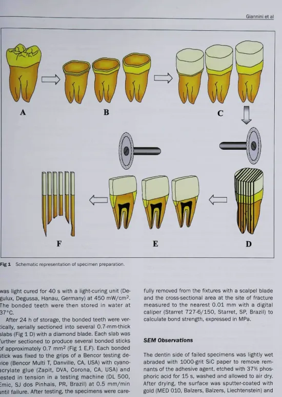
Nineteen extracted, caries-free human third moiars that were stored for no longer than 3 months were used in this study. The crowns of the teeth were transversally sectioned with a diamond blade (PC 10, Imptech-Equilan, Diadema, SP, Brazil) under water irrigation just beneath the deepest occlusal fissure (n = 6), in the middle of the crown (n = 7), or next to the cemento-enamel junction (n = 6) to ex-pose areas of superficial, middle, or deep dentin, respectively (Fig 1 A, B). The exposed dentin sur-faces were wet-polished with 600-grit SiC paper to create a standard smear layer before being bonded with the adhesive systems.
Three superficial, 4 middle, and 3 deep dentin surfaces were bonded with Prime & Bond 2.1 adhe-sive system according to manufacturer's instruc-tions, following etching with 36% phosphoric acid for 15 s and rinsing. Clearfil Liner Bond 2V adhe-sive system was applied to 3 superficial, 3 middle, and 3 deep dentin surfaces also according to man-ufacturer's instructions. The composition, applica-tion steps and manufacturers of the materiais used are described in Table 1. After bonding, the entire dentin surfaces received several layers of Z-100 resin composite (Table 1) to buiid up a crown ap-proximately 10.0 mm in height (Fig 1 C). Each layer
Giannini et al
Fig 1 Schematic representation of specimen preparation.
was light cured for 40 s with a light-curing unit (De-gulux, Degussa, Hanau, Germany) at 450 mW/cm^. The bonded teeth were then stored in water at 37°C.
After 24 h of storage, the bonded teeth were ver-tically, serially sectioned into several 0.7-mm-thick slabs (Rg 1 D) with a diamond blade. Each slab was further sectioned to produce several bonded sticks of approximately 0.7 mm2 {Fig 1 E,F). Each bonded stick was fixed to the grips of a Bencor testing de-vice (Bencor Multi T, Danville, CA, USA) with cyano-acrylate glue (Zapit, DVA, Corona, CA, USA) and tested in tension in a testing machine (DL 500, Emic, SJ dos Pinhais, PR, Brazil) at 0.5 mm/min until failure. After testing, the specimens were
care-fully removed from the fixtures with a scalpel blade and the cross-sectional area at the site of fracture measured to the nearest 0.01 mm with a digital caliper (Starret 727-6/150. Starret, SP, Brazil) to calculate bond strength, expressed in MPa.
SEM Observations
The dentin side of failed specimens was lightly wet abraded with 1000-grit SiC paper to remove rem-nants of the adhesive agent, etched with 37% phos-phoric acid for 15 s, washed and allowed to air dry. After drying, the surface was sputter-coated with gold (MED 010, Balzers. Balzers. Liechtenstein] and
Gianninietal
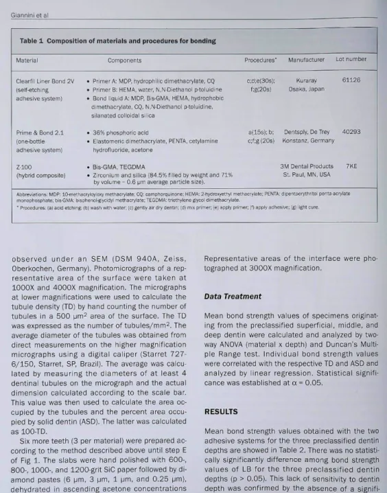
Table 1 Composition of materials and procedures for bonding
Material
Clearfil Liner Bond 2V (self-etchihg adhesive system]
Primes Bond 2,1 (one-öottle adhesive system)
Z-100 (hybrid composite)
ABbreviations: MDP: lCHneth morophüsphate; bis-GMA: bi " Procédures' (a) add etching
Components
• Primer A: MDP, hydrophiiic dimethacryiate, CQ • Primer B: iHEMA, water, N,N-DJethahoi p-toiuidine • Bond iiquid A: MDP, Bis-GMA, HEMA, hydrophobic
dimethacryiate, CQ, N.N-Diethanol p-toluidine. silanated coiloidsl siiica
• 36%phosohorioacid
• Elastomeric dimethacryiate, PENTA, cetylamine hydrofluoride, acetone
. Bis-GMA, TEGDMA
• Zirconium and Siiioa (84,5% filled by weight and 71% by volume - 0,6 gm average particie size).
aoiylnyloiy metliBcrylate; CQ: camphorquinone: HEMA; 2-hydroxyBthyl m phenolfilycidyl methacrylate; TEGOMA: triettiylene glycol dimeüiacrylstf (b| wash with water: |c) gently ail dry dertin: (d) mix pnmer: le) apply pr
Procedures' c;d;e(30s]: f:g(20s) a(15s): b; c:f;g (20s) äthaciylate: PENTA
Manufacturer Lot number
Kuraray 61126 Osaka, Japan
Dentsply, DeTrey 40293 Konstani, Germany
3M Dental Products 7KE St, Paul, MN, USA
dipentaerythritol perita-acrylate
mer; (f) apply auhesive; (g) light cure.
Observed under an SEM (DSiVl 940A, Zeiss, Oberkochen, Germany). Photomicrographs of a rep-resentative area of the surface were taken at lOOOX and 4000X magnification. The micrographs at lower magnifications were used to calculate the tubuie density (TD) by hand counting the number of tubuies in a 500 |jm^ area of the surface. The TD was expressed as the number of tubuies/mm^. The average diameter of the tubules was obtained from direct measurements on the higher magnification micrographs using a digital caiiper {Starret 727-6/150, Starret, SP, Brazil), The average was oaicu-lated by measuring the diameters of at least 4 dentinai tubules on the micrograph and the actual dimension calculated according to the scale bar. This vaiue was then used to caicuiate the area oc-cupied by the tubuies and the percent area occu-pied by solid dentin (ASD), The latter was calculated as 100-TD,
Six more teeth (3 per materia!) were prepared ac-cording to the method described above until step E of Eig 1. The siabs were iiand poiished with 600-, 800-, 1000-, and 1200-grit SiC paper followed by di-amond pastes (6 urn, 3 \¡<r\, 1 |jm, and 0.25 (jm), dehydrated in ascending acetone concentrations (30%, 50%, 70%, 90% and 100%), critical-point dried (CPD 030, Balzers, Balzers, Liechtenstein), sputter-coated with gold and examined under SEM.
Representative areas of the interface were pho-tographed at 3000X magnification.
Data Treatment
Mean bond strength vaiues of specimens originat-ing from the preclassified superficiai, middle, and deep dentin were calculated and analyzed by twc-way ANOVA (material x depth) and Dunoan's Multi-ple Range test. Individual bond strength values were correlated with the respective TD and ASD and analyzed by iinear regression, Statisticai signifi-cance was established at a ^ 0.05,
RESULTS
Mean bond strength values obtained with the two adhesive systems for the three preclassified dentin depths are showed in Table 2, There was no statisti-oally significantly difference among bond strength values of LB for the three preclassified dentin depths (p > 0,05). This iack of sensitivity to dantin depth was confirmed by the absence of a signifi-cant relationship between bond strength and TD (R2 = 0.05, p > 0,05, Fig 2) However, when individ-ual bond strength values were correlated with their
Table 2 Average microtensile bond strength (MPa) of Clearfil Liner Bond 2V and Prime & Bond 2.1 to superfi-cial, middle, and deep dentin
Adhesive System Superficiai Middie Deep
Ciearfil Liner Bond 2V 29.9 ± 15.1 (n-S S Prime & Bond 2.1 61.7 ± 12.4 ¡n-S
24.3 ±9.5 (n-9P S 41.1 ±5.9 ln-121
23.9 ±10.6 NS 25.6 ±7.4
Differences Beween malerials are indicaled by S = significant (p < 0.05¡ oc NS = nonsignificanl (D > O.OS). Sa enees Between äentin defjths (p i 0.05).
case letters indicate no significant differ.
Fig 2 Clearfil Liner Bond 2V. Regression analysis of bond strength vs tubule density.
50 1
45
4 0
3 5
-
302 5
2 0
-
15-
10-
5-
0-= 0.0508
0 10000 20000 30000 40000 50000
T D = number of tubule /
respective ASD, linear regression showed a weak, but significant relationship. There was a tendency for LB bond strength to increase as the area of solid dentin increased (R^ = 0.2, p < 0.05, Fig 3).
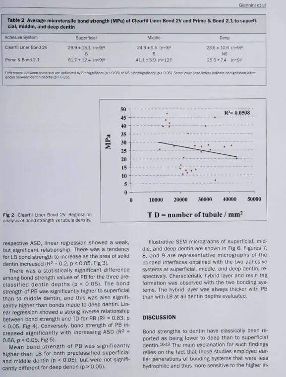
There was a statisticaily significant difference among bond strength vaiues of PB for the three pre-classified dentin depths (p < 0.05). The bond strength of PB was significantly higher to superficial than to middie dentin, and this was also signifi-cantly higher than bonds made to deep dentin. Lin-ear regression showed a strong inverse relationship between bond strength and TD for PB (R^ - 0.63, p < 0.05, Fig 4). Conversely, bond strength of PB in-creased significantly with increasing ASD {R^ = 0.66, p< 0.05, Fig 5].
Mean bond strength of PB was significantly higher than LB for both preclassified superficial and middle dentin (p < 0.05), but were not signifi-cantly different for deep dentin (p > 0.05).
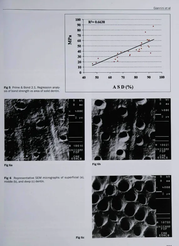
Illustrative SEM micrographs of superficiai, mid-dle, and deep dentin are shown in Fig 6. Figures 7, 8. and 9 are representative micrographs of the bonded interfaces obtained with the two adhesive systems at superficial, middle, and deep dentin, re-spectively. Characteristic hybrid layer and resin tag formation was observed with the two bonding sys-tems. The hybrid layer was always thicker with PB than with LB at aii dentin depths evaluated.
DISCUSSION
Bond strengths to dentin have classically been re-ported as being iower to deep than to superficial dentin.is-15 The main explanation for such findings relies on the fact that those studies empioyed ear-lier generations of bonding systems that were less hydrophilic and tbus more sensitive to the higher
Giannini et al
50- 45-40 35 30- 25-
20- 15-
10-5 O
40
í= 0.2066
50 60 70 90 100
ASD (%)
analysis of bond strength vs area of solidFlg 3 Clearfil Liner Bond 2V. Regression dentin.at 100
90 80 70 60 50-1 40 30 20 10 0
R ^ 0.6309
0 10000 20000 30000 40000 50000 60000
T D = number of tubule /
Fig 4 Prime & Bond 2 . 1 . Regression analy-sis of bond strength vs tubule density.trinsic v^etness of deep dentin. Hovi^ever, more re-oent studies aiso reported iov^ier bond strength to deep dentin using current, more hydrophilic adhe-sive systems.^5'^^ In one of these studies.^^ the au-thors evaluated the bond strength of one acetone-based and one self-etching system to different re-gions of dentin {ie, periphery, center, or pulp horn) either with or without simulated pulpal pressure. The acetone-based system was very sensitive to both dentin depth and pulpal pressure, while the
self-etching system bonded homogeneously in any situation. The authors explained their findings by the fact that enhanced permeability - resulting from the separate etching step with the acetone-based system - increased the surface wetness and may have compromised the bonding with that sys-tem because of the overwet phenomenon,20 The same phenomenon did not occur with the self-etch-ing system because its mild etchself-etch-ing action permits smear plugs to remain within the dentinal tubules,
Giannini étal
Fig 5 Prime & Bond 2.1. Regression analy-sis of bond strength vs area of solid dentin.
100 1
R ^ 0.6638
80 90 100
A S D
(%)
Fig 6a Fig 6b
Fig 6 Representative SEM micrographs of superfioial (a), middle (b), and deep (c) dentin.
Fig 6c
Giannini et al
Fig 7 Representative bonûed interfaces of Ciearfii Liner Bond 2V (a¡ and Prime & Bond 2.1 (b] at superficial dentin (CR- compos-ite resin, BA- bonding agent, HL- hybrid layer, D- dentin).
Fig S Representative bonded interfaces of Ciearfii Liner Bond 2V (al and Pnme & Bond 2,1 (b) at middie dentin (CR- composite resin, BA- bonding agent, HL- hybrid layer, D- dentin).
Fig 9 Representative bonded interfaces of Ciearfii Liner Bond 2V (a) and Prime & Bond 2,1 (b) at deep dentin (CR- composite resin, BA- bonding agent, HL- hybrid iayer, D- dentin).
Giannini étal
thus reducing the permeability and surface wet-ness. The other study,^^ however, did not use simu-lated pulpal pressure and also demonstrated that bond strengths decreased with dentin depth for all the adhesive systems used in their study, in our study, simuiated puipai pressure was not used and the surface wetness was exciusively determined by the operator according to manufacturer's instruc-tions,
Suzuki and Eingeri'' pointed out that bond strengths to dentin are more related to the availabii-ity of soiid dentin at the site of bonding than to other factors, such as surface wetness. This appar-ently was the case when we analyzed our data from the PB adhesive system. The bond strength of PB decreased significantly as the TD increased and the ASD availabie for bonding decreased as well (Figs 4 and 5). The higher bond strengths to more superfi-cial dentin can be explained by the fact that more intertubular dentin is availabie for hybrid layer for-mation, this being the main bonding mechanism re-sponsible for increased bond strength to dentin,^ Theoretically, bond strengths shouid be higher to deep dentin whenever resin tags can be firmly bound (hybridized) to the lateral walis of the de-mineralized dentinal tubules.^i However, we cannot rule out the fact that, even without simulated pulpal pressure, deep dentin is more porous and retains more water within ¡ts eniarged tubule openings, which may preclude adequate lateral bonding of the resin tags. Moreover, the wetter and more porous deep dentin is more likely to result in the overwet phenomenon,^f which may entrap air within the blisters or within the dentinal tubuies, also possibly compromising the poiymerization of the resin bonding agent.15 |n that respect, whiie our study did not confirm the theoreticai possibility of achieving higher bond strengths in deep dentin, others have shown that deep dentin produced bond strengths that were either not different than super-ficial dentinas or even higher^ for some adhesive systems. It seems reasonable to admit that bond strengths to deep dentin can be higher than to su-perficial dentin. However, ideai bonding to deep dentin is largely dependent on the adequate bond-ing of resin tags within the wetter and iarger denti-nal tubules. This makes bonding to deep dentin more technique-sensitive and iargely dependent on the ability of the operator to properly control the surface moisture and application technique for each adhesive system.
Our anticipated nuli hypothesis was only partially
confirmed. It is evident from our findings that the seif-etching system (LB] was less sensitive to dentin depth and TD than was PB (Table 2, Fig 2). The in-sensitivity of self-etching systems to surface vari-abies such as dentin depth, intrinsic wetness, and presence, absence, or thickness of smear layer has been previously reported,^'^^-^^ Apparently, be-cause seif-etching systems bond to the most super-ficial iayer of dentin and do not completely remcve smear piugs, the intrinsic wetness of dentin is less iikelyto interfere with bonding because the perme-abiiity of the tubules is reduced. Since the bonding mechanism of these systems relies on resin infiltra-tion into the solid dentin underneath the smear layer, ¡t is expected that bond strengths should be higher when more intertubuiar dentin is available. Indeed, we found a weak, but significant direct rela-tionship between bond strength of LB and the ASD ¡Eig 3). For seif-etching adhesive systems, resultant bond strengths are more iargely dependent on hy-brid layer formation than on resin tag retention, if we apply the modeling approach proposed by Pash-iey et al,11 the contribution of hybrid layer to the total adhesion increases from the pulp to the per-iphery.
The stronger relationship between bond strength and both TD and ASD observed for PB can be ex-plained by the wider range of values of both para-meters. The bond strength of PB ranged from as low as 16,37 MPa [for a TD of 58,105 tubuies/ mm^) to a maximum of 86,98 MPa (for a TD of 10,472 tubules/mm^). The percentage of ASD ranged from approximateiy 45% for the deepest dentin up to approximately 94% for the most super-ficiai. These same values for LB had a much smailer range. Bond strength vaiues ranged from 6,64 MPa (for a TD of 34,290 tubules/mm^) to 46.96 MPa (for a TD of 14,864 tubules/mm^j. The ASD varied from approximately 55% (one singie specimen. Fig 6a) up to approximately 93% for deep and superficiai dentin, respectively. The value range of both bond strengths and TD for LB were smaller than for PB; this may have reduced the power of regression anaiysis to identify a stronger interaction between bond strength and TD.
Our overall range of number of tubules per mm^ is within the range of values usually reported in the iiterature,2'^'5i3 For both PB and LB, most of the specimens were located within the range of 20,000 to 40,000 tubules per mm^. These are more repre-sentative of middle than of very superficial or very deep dentin. Our flat dentin surfaces exposed for
Giannini étal
bonding were obtained by transversally sectioning the crowns at three different distances from the ce-mento-enamel junction towards tbe cusps. The sec-tions were preclassified as being deep, middle, and superficial dentin, respectively. Although our at-tempt to expose dentin at different depths was suc-cessfui, the irregular anatomy of both dentai pulp and peripheral ename! does not permit exposure of large areas of very deep or very superficial dentin. Therefore, care must be taken when interpreting data of bond strength of resins to different dentin depths, particularly when iarge bonding areas are used such as in tbe conventionai shear or tensile tests. The microtensiie technique offers the possi-bility of employing a much smaiier bonding area, thus reducing the variabiitty of the substrate on the site of bonding. This aiso aiiows for a more realistic SEM analysis of the susbtrate to which the bond was made.
CONCLUSIONS
The results of this work demonstrated that the bond strength of adhesive systems to dentin was dependent on the microstructure of the substrate at the site of bonding. This was more evident with the acetone-based system than with the seif-etch-ing system.
ACKNOWLEDGMENTS
Tîie adhesive systems used in (his study were generously supplied by Kuraray Company. Osaka. Japan, and Dentsply Ind. e Com. Ltda., Petrópolis, RJ, Brazil. The authors are indebted (o Dr. E.W. Kitajima (NAP..MEPA/ESALQ-USP) for lechnlcal electron mi-croscopy support. This study was supported, in part, by grants DE 06427 from (he NIDCR. USA. and 300481/95-0 from CNPq. Braal.
REFERENCES
1. Burrow MF. Tskakura H, Nakajima M, Inai M, Tagami J, Takatsu T The influence of age and depth of dentin on bond-ing. Dent Mater 1994:10:241.246.
2. Carrigan PJ, Morse DR, Fürst ML, Sinai iH. A scanning eiec-tron miorosoopic evaiuation of hurnan dentinai tubules ac-cording to age and location. J Endod 1984:10:359-363. 3. Fernandes CAO . Estudo comparativo da resistencia adesiva
à dentina superficial e profunda testada simultáneamente, empregando-se deis sistemas adesivos. Thesis, Bauru School ot Dentistry, USP, Bauru, SP, Brazil, 2000.
4. Fosse G, Saele PK, Eide R. Numerical density and distribu-tional pattern of dentin tubuies. Acta Odont Scand 1992: 50:201-210.
5. Garberoglio R. Bránnstróm M. Scanning eiectron microscopic investigation of human dentinal tubules. Archs Oral Biol 1976;21:355-362.
6. Gwinnett AJ. Quantitative contribution of resin infiltration/hy-bridizstion to dentin bonding. Am J Dent 1993:6:7-9. 7. McCabe JF, Rusby S. Dentine bonding agents - characteristic
bond strength as a function of dentine depth. J Dent 1992; 20:225-230.
8. Makajima M, Sano H, Burrow MF, Tagami J, Yoshiyama M, Ebisu S, Ciuochi B, Russeli CM, Pashley DH. Tensiie bond strength and SEM evaiuation of caries-effected dentin using adhesives. J Dent Res 1995:74:1679-1688.
9. Nery S, McCabe JF, Wassell RW. A comparative study of three dental adhesives. J Dent 1995:23:55-61.
10. Pashley DH, Carvaiho RM. Dentine permeabiiity and dentine adhesion. J Dent 1997:25:335-372.
11. Pashiey DH, Ciucchi B. Sano H, Carvaiho RM, Russen CM. Bond strength versus dentine structure: a modeiling ap-proach. Archs Orai Biol 1995:40:1109-1118.
12. Pashiey DH, Ciucchi B, Sano H, Yoshiyama M, Can/aihc RM. Adhesion testing of dentin bonding agents. A review. Dent Mater 1995:11:117-125.
13. Pashley DH. Clinical correlations of dentin structure and func-tion. J Prosth Dent 1991;66:777-7S1.
14. Pashley DH. Oentin: a dynamic substrate - a review. Scanning Microsc 1989,3:161-74.
15. Pereira PNR, Okuda M. Sano H, ïoshikawa T Burrow MF, Tagami J. Effect of intrinsic wetness and regional differehce cn dentin bond strength. Dent Mater 1999:15:46-53. 16. Prati C, Pashiey DH. Dentin wetness, permeability and
thick-ness and bond strength of adhesive systems. Am J Dent 1992:5:33-38.
17. Suzuki T. Finger WJ. Dentin adhesives: site of dentin vs. Bonding of composite resins. Dent Mater 1988:4:379-383. 18. Tagami J, Tao L, Pashley DH. Correlation among dentin depth,
permeability, and bond strength of adhesive resins. Dent Mater 1990:6:45-50.
19. Tao L. Pashiey DH. Shear bond strengths to dentin: effects of surface treatments, depth and position. Dent Mater 1988: 4:373-378.
20. Tay FR, Gwinnett AJ, Wei SHY. The overwet phenomenon: a soannmg eiectron microscopic study of surfaoe moisture in tlie acid-conditioned, resin-dentin interface. Am J Dent 1996:9:109-114.
21. Tay FR. Sano H, Can^^alho RM, Pashiey EL, Pashley DH. An ul-trastructurai study of the infiuence of aoidity of self-etching primers and smear iayer thickness on bonding to intact dentin. J Adhesive Dent 2000:2:83-98.
22. Yosliiyama M, Carvalho RM, Sano H, Horner J, Brewer PD, Pashiey DH. interfacial morphology and strength cf bonds made to superficiai versus deep dentin. Am J Dent 1995: 8:297-302.
23. Voshiyama M. Sano H, Ebisu S. Tagami J. Ciucchi B, Carvalho RM, Johnson MH, Pashley DH. Regional strengths of bonding agents to cervical scierotio root dentin. J Dent Res 1996: 75:1404-1413.
![Fig 7 Representative bonûed interfaces of Ciearfii Liner Bond 2V (a¡ and Prime & Bond 2.1 (b] at superficial dentin (CR- compos- compos-ite resin, BA- bonding agent, HL- hybrid layer, D- dentin).](https://thumb-eu.123doks.com/thumbv2/123dok_br/16218079.712423/8.601.20.587.8.248/representative-bonûed-interfaces-ciearfii-liner-prime-superficial-bonding.webp)
