Ana Carolina Maito Villela-Rosa(a) Mariane Gonçalves(a)
Iara Augusta Orsi(a) Paola Kirsten Miani(a)
(a)Department of Dental Materials and Prosthodontics, Ribeirão Preto School of Dentistry, Universidade de São Paulo - USP, Ribeirão Preto, SP, Brazil.
Corresponding author: Mariane Gonçalves
Department of Dental Materials and Prosthodontics, Ribeirão Preto School of Dentistry, University of São Paulo Av. do Café, s/n - Monte Alegre Ribeirão Preto - SP - Brazil CEP: 14040-904
E-mail: [email protected]
Received for publication on Oct 14, 2010 Accepted for publication on Dec 17, 2010
Shear bond strength of self-etch and
total-etch bonding systems at different
dentin depths
Abstract: The purpose of this study was to evaluate the dentin shear bond strength of four adhesive systems (Adper Single Bond 2, Adper Prompt L-Pop, Magic Bond DE and Self Etch Bond) in regards to buccal and lingual surfaces and dentin depth. Forty extracted third molars had roots removed and crowns bisected in the mesiodistal direction. The buc-cal and lingual surfaces were ixed in a PVC/acrylic resin ring and were divided into buccal and lingual groups assigned to each selected adhe-sive. The same specimens prepared for the evaluation of supericial den-tin shear resistance were used to evaluate the different depths of denden-tin. The specimens were identiied and abraded at depths of 0.5, 1.0, 1.5 and 2.0 mm. Each depth was evaluated by ISO TR 11405 using an EMIC-2000 machine regulated at 0.5 mm/min with a 200 Kgf load cell. We performed statistical analyses on the results (ANOVA, Tukey and Scheffé tests). Data revealed statistical differences (p < 0.01) in the adhesive and depth variation as well as adhesive/depth interactions. The Adper Single Bond 2 demonstrated the highest mean values of shear bond strength. The Prompt L-Pop product, a self-etching adhesive, revealed higher mean values compared with Magic Bond DE and Self Etch Bond adhesives, a total and self-etching adhesive respectively. It may be concluded that the shear bond strength of dentin is dependent on material (adhesive system), substrate depth and adhesive/depth interaction.
Descriptors: Dentin; Adhesives; Dental Etching.
Introduction
The mechanism for adhesive bonding to dental substrates, especially to dentin, has been extensively studied in the last few decades. The es-tablishment of effective interlocking occurs when the adhesive penetrates into the intratubular and intertubular dentin so that resin penetration into the conditioned dentin results in the formation of intratubular resin tags and a “hybrid layer.”1 Micromechanical attachment is the most
im-portant mechanism of resin adhesion to dentin. However, there are sev-eral factors that can affect hybrid layer and resin tag formation, such as acid-etching, the moisture conditions of the substrate, and adhesive and dentin depth.2
During dentin acid-etching, the mineral content of the dentin surface is removed, and the collagen ibrils remain supported by water.3 The
and provides stability to the collagen, thus enabling greater iniltration of adhesive monomers.4
Howev-er, some studies have found that the resin monomer iniltration may be incomplete even when the “wet” bonding technique is used,5 suggesting that another
factor affect hybridization, the composition of adhe-sive systems.
After decades of evolution, adhesives may in-clude different formulations and, consequently, their bond values may vary in relation to dental substrate.6 Currently there is a tendency to simplify
bonding procedures by mixing the prime and bond functions. In addition, acid monomers are currently used in primers, which introduced the self-etching adhesive concept.7
Due to different factors that affect the bond strength of composite resin and dentin, it is impor-tant to evaluate the inluence of the adhesive system, the type of acid-etching and the dentin depth as they relate to the shear bond strength of the dentin-com-posite resin interface at the same dentin locations.
Materials and Methods
This study was approved by the Ethics Research Committee of Ribeirão Preto School of Dentistry (University of São Paulo, Brazil). Forty
noncari-ous extracted human third molars had their roots removed 2 mm beyond the enamel/cementum junc-tion and their crowns bisected longitudinally in a mesiodistal direction with a nº 3072 diamond bur in a water-cooled high speed turbine to create two similar halves (buccal and lingual). The half surfac-es were set in 12 mm high PVC cylinders (Tigre S/A, Joinvile, Brazil) as holders (1.5 cm diameter, 1.0 cm high) using cold-cured acrylic resin (Clássico, São Paulo, Brazil), and a polytetraluoroethylene (Tef-lon) mold developed for this experiment was used to place their central region in the third part of the ring. The telon mold produced a step-off of 0.5 mm between the dental surface and the acrylic resin, which was needed to access the most supericial de-sired dentin for the experiment.
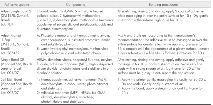
The two groups (40 buccal and 40 lingual) were then randomly reorganized into four groups (n = 10) corresponding to the four adhesive systems tested with a microhybrid resin composite. The systems were the following: Adper Single Bond 2 (ASB2) / Z-250, Adper Prompt L-pop (APLP) / Z-250, Magic Bond DE (MBDE) / Fill Magic and Self Etch Bond (SEB) / Fill Magic. The adhesives, the composition and the bonding procedures are shown in Table 1, according to manufacturer’s provided information.
Table 1 - Dentin adhesive systems tested.
Adhesive systems Components Bonding procedures
Adper Single Bond 2 (3M ESPE, Sumaré, Brazil)
Lot: 7LH
Ethanol, water, Bis-GMA, 5 nm silane treated colloidal silica, 2-hydroxyethyl methacrylate, glycerol 1, 3 dimethacrylate, methacrylate functional copolymer of polyacrylic and polyitaconic acids and diuretane dimethacrylate
After etching, rinsing and drying, apply 2 coats of adhesive while massaging in over the entire surface for 15 s. Dry gently to evaporate the solvent. Light-cure for 10 s
Adper Prompt L-Pop
(3M ESPE, Sumaré, Brazil)
Lot: 287452
A: Phosphate mono and di-hema, dimethacrylate, camphorquinone, substituted aromatical amine and substituted phenol
B: water, hydroxyethyl methacrylate, methacrylate polycarbonic acid and substituted phenol
Mix A and B blisters, according to the manufacturer’s recommendation; the adhesive must be massaged in over the entire surface for greater effect while applying pressure for 15 s; reapply until the appearance of a glossy surface; remove excess solvent until a fine film is formed. Light-cure for 10 s
Magic Bond DE (Vigodent S/A, Rio de Janeiro, Brazil) Lot: 001/07
HEMA, dimethacrylates, neopentyl fluoride, acrylate fluoride, adhesive monomer (MEP), highly dispersed silicon dioxide, photoinitiators and stabilizers in an alcoholic solution
After etching, rinsing and drying, apply adhesive and gently massage in for 10 s; apply a stream of air. Avoid very fine coats with a strong stream of air. Light-cure for 20 s. The surface must be glossy; if not, repeat the application
Self Etch Bond (Vigodent S/A, Rio de Janeiro, Brazil) Lot: 002/07
1: Hema, copolymer, adhesive monomer (MEP), dimethacrylate, alcohol, water, photoinitiators and stabilizers
2: Adhesive monomer (MEP), HEMA, bis-GMA, alcohol, dimethacrylates, microfiller, photoinitiators and stabilizers
1. Apply the primer gently, massaging the cavity for 20-30 s. Do not wash. Gently apply a stream of air
To obtain the required dentin circular shape area with 3 mm diameter, the surfaces were wet abraded in a horizontal polisher (model PFL, Fortel Ind. e Com. Ltda., São Paulo, Brazil) with 150, 320, 400 and 600 grit abrasive papers (Norton, Ind. Com. Ltda., São Paulo, Brazil), to expose and standard-ize the required bond area, the so-called “supericial dentin.”
For all groups, the wet bonding technique was applied and the excess moisture was gently removed using an absorbent paper, gently applied to the den-tin surface.8
The adhesive was applied and then light-cured, according to manufacturer’s recommendations using a photocuring unit (Ultraled XP; Dabi Atlante S/A, Ribeirão Preto, Brazil), with a light output not less than 500 mW/cm². The specimens were individually ixed in a metallic clamping device (developed at the Houston Biomaterials Research Center and manu-factured at the Precision Workshop at Ribeirão Pre-to School of Dentistry of University of São Paulo, Brazil) thereby keeping the dentin surface parallel to a lat base. A split-bisected polytetraluoroethylene jig was positioned on the tooth/resin block surface, thus providing an inverted conical centralized cavity (3-mm diameter at base and 5-mm high) with the smaller diameter corresponding to the demarcated 3-mm-diameter-bonding site. The composite resins were inserted into the matrix in three steps, each polymerized for 20 s and applied in a conical shape 4-mm high. As the matrix cavity illed, the specimen was removed from the clamping device.
After 24 hours of storage in distilled water at 37°C, the specimens (combined PVC ring/dentin surface/composite resin cylinder) were tested for
shear bond strength using an Emic universal testing machine (MEM-2000 Model, São José dos Pinhais, Brazil) at a crosshead speed of 0.5 mm/min and a 200 Kgf load cell. After testing, fractured specimens were observed with an optical microscope (Nikon, Model 86786, Tokyo, Japan) at a magniication of 20x to assess failure modes, which were classiied as adhesive, cohesive or mixed. Adhesive failure oc-curred at the specimen/adhesive interface, cohesive failure occurred in the material or the substrate with no damage to the interface and mixed failure simul-taneously involved the interface and the material or substrate. After the analysis, the 10 identiied speci-mens were again stored in distilled water and refrig-erated.
To evaluate the inluence of dentin depth on shear bond strength, the samples were identiied and abraded 0.5 mm four times until reaching a depth of 2 mm, measured by digital calipers. After each 0.5 mm abrasion the same dentin surface prepara-tion protocol described above was performed, and the specimens were stored again in distilled water and were refrigerated. A schematic illustration of specimen preparation and details about the experi-ment are presented in Figure 1.
The adhesive shear bond strength values were recorded in kgf/cm² and were converted into MPa. Means and standard deviations were calculated, and data were analyzed by ANOVA. Multiple com-parisons were done using the Tukey test.
Results
Table 2 reveals the analysis of variance that showed a statistically signiicant difference (p < 0.01) between the adhesive systems and dentin
Source of variation SS df MM F p value
Depth 913.8391 4 228.4598 8.16 0.0035%*
Adhesive 10697.5645 3 3565.8547 127.36 0.00001%*
Adhesive x depth 768.2980 12 64.0248 2.29 1.0027%*
Residual I 5039.5483 180 27.9975
Residual II 4513.4688 180 25.0748
Total variation 22519.6250 399
*Significant level at 1%.
depths, as well as an adhesive x dentin depth inter-action. The means of the variation factors are given in Tables 3 and 4. Figure 2 shows the interaction be-tween the adhesive and dentin depth factors.
Discussion
The results from this study reveal that dentin shear bond strength depends on adhesive and dentin depth. Consequently, shear bond strength also
de-Figure 1 - Flowchart of the study shear bond strength test. A: Tooth sectioning; B: Tooth section embedding; C: Abrading and polishing of dentin; D: Measuring of dentin depth; E: Specimens at different depths; F: Acid etching; G: Washing; H: Drying with absorbent paper; I: Applying adhesive systems; J: Metallic deposits to build up the specimens; K: Light-curing; L: Finished specimen; M: shear bond strength test.
A B C
D
F G H I
J K L M
E
Superficial dentin 0.5 mm dentin 1.0 mm dentin
pends on the adhesive and dentin depth interaction. It is important to consider the composition and the substrate treatment by adhesive factor and, therefore, self or total etching. Different studies re-port that the chemical composition of adhesive sys-tems determines clinical success.9 Polyacrylic acid in
ASB2 and APLP adhesives promotes chelation with calcium and the formation of hydrogen bridges with dentin components; it may be the signiicant factor resulting in higher shear bond strength values.
Another component that may be responsible for the high bond strength values is the 5 nm sili-ca nanoiller incorporated at 10% weight in ASB2 adhesive. These particles may have a role in the formation of a resin ilm that stabilizes the hybrid layer.10 The intermediate layer of the adhesive iller
promotes an elastic zone that improves the capacity to accommodate contractile forces during composite resin polimerization.11 In addition, the nanoillers in
ASB212 are smaller, which improves its wettability
and penetration compared with bigger illers such as the silic dioxide in the MBDE and the microillers in SEB.
Regarding adhesive and substrate treatment, there are potentially similar bond strength results between self and total etching adhesives.13 On the
other hand, one study14 showed that conventional
adhesive demonstrates better performance than the self-etching adhesive when applied according to the original prescription and with previous acid etching.
The results of this study showed that SEB (Vigo-dent), a two-step self-etching adhesive, had the low-est shear bond strength values, while APLP (3M), a one-step self-etching adhesive, demonstrated a better performance than the total etching adhesive, MBDE.
In this case, in addition to adhesive composi-tion, which has already been discussed, the eficacy of self-etching adhesives may be an important fac-tor responsible for the higher mean bond strength values of APLP compared to SEB. The PLP is at pH 1.0 and is thus considered, in terms of etching aggressiveness, a strong self-etching adhesive.15 SEB
is at pH 5, according to the manufacturer informa-tion, and is thus considered to be mild self-etching. These results suggest that low bond strength val-ues for SEB may be related to the weak etching of the dentin substrate. The incomplete iniltration of some self-etching adhesives may occur because of the etching potential reduction of acid monomers in the hybrid layer base or because of noncured hy-drophilic components due to bonding area deterio-ration when these self-etching adhesives are used.16
However, one study demonstrated that a smear lay-er removal step aftlay-er etching and before adhesive
ap-Table 3 - Means for adhesive variation.
Dental adhesive Mean MPa (SD) Critical value
Adper Single Bond 2 18.7612 (± 7.24)a
1.94189 Magic Bond DE 10.1532 (± 4.72)b
Adper Prompt L-Pop 12.9430 (± 6.10)c
Self Etch Bond 4.40270 (± 2.67)d
Different letters indicate statistical difference.
Table 4 - Means for depth variation.
Dentin depth (mm) Mean MPa (SD) Critical value
Superficial 13.67225 (± 7.4)a
2.30717 0.5 12.39263 (± 7.92)a
1.0 11.94600 (± 7.82)a
1.5 10.47975 (± 6.98)b
2.0 9.33450 (± 6.41)b
The same letters indicate statistical similarity.
Shear
B
o
nd
Stre
ngth
(MP
a)
Dentin Depth (mm) Surface 0.5
0 5 10 20
15 25
1.0 1.5 2.0
Adper Prompt L-Pop Adper Single Bond
Self Etch Bond Magic Bond DE
plication could produce a more durable and realistic bond to dentin.17 On the other hand, some studies18
have also shown that APLP produces an etching ef-fect that approached that of acid etching with phos-phoric acid at 35%, suggesting its eficacy and adhe-sive potential.
The preservation of hydroxyapatite in the hybrid layer may serve as a receptor for additional chemical bonding because the calcium has chemical bonding potential with the monomers usually present in self-etching adhesives, in addition to protecting collagen against hydrolysis.19
Regarding dentin depth, the bond strength val-ues decreased as depth increased. The statistical analysis revealed signiicant differences between two groups of dentin depth, one including the su-pericial, 0.5 mm and 1.0 mm depths, and the other including the 1.5 mm and 2.0 mm depths.
Similar studies have been performed describ-ing the inverse relationship between dentin poros-ity (which increases with depth) and shear bond strength;20 these studies showmorphologic
structur-al variation of dentin that affects dentin bonding.21-23
As depth increases, there is an increase in dentin tubules and minor hybridization with a greater num-ber of tags or larger tags. Because the tags can con-tribute approximately one-third of the shear bond strength,24 minor adhesive resistance is expected at
deep dentin because it is an area that has less solid dentin and thus lower bonding values regardless of the type of adhesive used.22
In general, bond strength decreases as depth in-creases; however, these values demonstrated several points of variation. This may be explained by the morphology and structural features in the same
den-tal areas of the dentin substrate.20 These substrate
features include moisture variation and tubule ori-entation beyond the degree of tubular obstruction due to calciication by the stimuli that the tooth has experienced, and these differences can explain why the same adhesive system may demonstrate different bond strength values when applied to different re-gions of dentin substrate.25 Thus, it is important to
diagnose each tooth individually to choose the ap-propriate treatment.
The adhesive and dentin depth interaction re-vealed statistical differences, consistent with a study26 that concluded that bond strength is affected
by the adhesive system, the substrate or both. Some studies27 have observed that bond strength is
affect-ed by dentin depth depending exclusively on adhe-sive composition.
The dental surface variation did not inluence shear bond strength. A comparison of the results obtained in this study with those of other studies may be dificult because a large amount of studies are conducted to evaluate only one dental surface, usually the buccal, and do not evaluate the dental surface as a variation factor.28
Conclusion
Based on these indings, it may be concluded that bond strength is affected by adhesive and dentin depth. The dental adhesive systems had a signii-cant inluence on shear bond strength. The ASB2 demonstrated the highest mean values and the SEB had the lowest for all dentin depths evaluated. The dentin depth adversely affected the bonding mecha-nism. The dental surface did not affect shear bond strength at the dentin-resin interface.
References
1. Nakabayashi N, Kojima K, Mashuara E. The promotion of adhesion by the infiltration of monomers into tooth substrates. J Biomed Mater Res. 1982 May;16(3):265-73.
2. Tay FR, Gwinnett AJ, Pang KM, Wei SH. Variability in microleakeage observed in a total-ecth wet bonding tech-nique under different handling conditions. J Dent Res. 1995 May;74(5):1168-78.
3. Jacobsen T, Soderholm KJ, Garcea I, Mondragon E. Calcium leaching from dentin and shear bond strength after etching
with phosphoric acid of different concentrations. Eur J Oral Sci. 2000 Jun;108(3):247-54.
4. Moll K, Haller B. Effect of intrinsic and extrinsic moisture on bond strength to dentin. J Oral Rehabil. 2000 Feb;27(2):150-65.
6. Garone-Netto N, Eduardo CP, Youssef MN, Vieira GF, Carvalho RCR, Russo EA, et al. Adesividade em dentística. In: Busato ALS. Dentística. Filosofia, conceitos e prática clínica. São Paulo: Artes Médicas; 2005. p. 125-46.
7. Watanabe I, Nakabayashi N. Bonding durability of photo-cured Phenyl-P in TEGDMA to smear layer-retained bovine dentin. Quintessence Int. 1993 May;24(5):335-42.
8. Gwinnett AJ. Moist versus dry dentin: its effect on shear bond strength. Am J Dent. 1992 Jun;5(3):127-9.
9. Salz U, Bock T. Adhesion performance of new hydrolytically stable one-component self-etching enamel/dentin adhesives. J Adhes Dent. 2010 Feb;12(1):7-10.
10. Cardoso PE, Carrilho MRO, Francci CE, Perdigao J. Micro-tensile bond strengths of one bottle dentin adhesives. Am J Dent. 2001 Feb;14(1):22-4.
11. Miyazaki M, Ando S, Hinoura K, Onose H, Moore BK. In-fluence of filler addition to bonding agents on shear bond strength to bovine dentin. Dent Mater. 1995 Jul;11(4):234-8. 12. Erickson RL. Surface interactions of dentin adhesive materials.
Oper Dent. 1992;Suppl 5:81-94.
13. Paradella TC, Fava M. Bond strength of adhesive systems to human tooth enamel. Braz. Oral Res. 2007 Jan-Mar;21(1):4-9. 14. Carvalho APMC, Turbino ML. Can previous acid etching
increase the bond strength of a self-etching primer adhesive to enamel? Braz. Oral Res. 2009 Apr-Jun;23(2):169-74. 15. Tay FR, Pashey DH. Aggressiveness of contemporary
self-etching systems. I: Deph of penetration beyond dentin smear layers. Dent Mater. 2001 Jul;17(4):296-308.
16. Carvalho RM, Chersoni S, Frankenberg R, Pashey DH, Prati C, Tay FR. A challenge to the convencional widsdom that simultaneous etching and resin infiltration always occurs in self-etch adhesives. Biomaterials. 2005 Mar;26(9):1035-42. 17. Van Meerbeek B, Peumans M, Verschueren M, Gladys S,
Braem M, Lambrechts P, et al. Clinical status of ten dentin adhesive systems. J Dent Res. 1994 Nov;73(11):1690-702. 18. Van Meerbeek B, De Munck J, Yoshida Y, Inoue S, Vargas
M, Vijay P, et al. Buonocore memorial lecture. Adhesion to
enamel and dentin: current status and future challenges. Oper Dent. 2003 May-Jun;28(3):215-35.
19. Yoshida Y, Nagakane K, Fukuda R, Nakayama Y, Okazaki M, Shintani H, et al. Comparative study on adhesive performance of functional monomers. J Dent Res. 2004 Jun;83(6):454-8. 20. Toba S, Veerapravati W, Shimada Y, Nikaido T, Tagami J.
Micro-shear bond strength of adhesive resins to coronal dentin versus the floor of the pulp chamber. Am J Dent. 2003 Sep;16 Spec Nº:51A-6A.
21. Akagawa H, Nikaido T, Takada T, Burrow MF, Tagami J. Shear bond strengths to coronal and pulp chamber floor den-tin. Am J Dent. 2002 Dec;15(6):383-8.
22. Giannini M, Carvalho RM, Martins LR, Dias CT, Pashley DH. The influence of tubule density and area of solid dentin on bond strength of two adhesive systems to dentin. J Adhes Dent. 2001 Winter;3(4):315-24.
23. Sattabanasuk V, Shimada Y, Tagami J. The bond of resin to different dentin surface characteristics. Oper Dent. 2004 May-Jun;29(3):333-41.
24. Gwinnett AJ. Quantitative contribution of resin infiltration/ hybridization to dentin bonding. Am J Dent. 1993 Feb;6(1):7-9.
25. Marshall Jr GW, Marshall SJ, Kinney JH, Balooch M. The dentin substrate: structure and properties related to bonding. J Dent. 1997 Nov;25(6):441-58.
26. Toledano M, Osorio R, Ceballos L, Fuentes MV, Fernandes CA, Tay FR, et al. Microtensile bond strength of several ad-hesive systems to different dentin depths. Am J Dent. 2003 Oct;16(5):292-8.
27. Lopes GC, Perdigao J, Lopes MF, Vieira LC, Baratieri LN, Monteiro Jr S. Dentin bond strengths of simplified adhe-sive: effect of dentin depth. Compend Contin Educ Dent. 2006 Jun;27(6):340-5.