UNIVERSIDADE FEDERAL DO CEARÁ
FACULDADE DE FARMÁCIA, ODONTOLOGIA E ENFERMAGEM PROGRAMA DE PÓS-GRADUAÇÃO EM ODONTOLOGIA
DOUTORADO EM ODONTOLOGIA
CAMILA DE ATAIDE E FERRAZ
ESTUDO DA VIABILIDADE DE MEIOS ALTERNATIVOS DE PRESERVAÇÃO E PROTEÇÃO DA INTERFACE ADESIVO / DENTINA
CAMILA DE ATAIDE E FERRAZ
ESTUDO DA VIABILIDADE DE MEIOS ALTERNATIVOS DE PRESERVAÇÃO E PROTEÇÃO DA INTERFACE ADESIVO / DENTINA
Tese apresentada ao Programa de Pós-Graduação em Odontologia da Universidade Federal do Ceará, como requisito parcial à obtenção do título de Doutor em Odontologia. Área de concentração: Clínica Odontológica. Orientador: Prof. Dr. Sérgio Lima Santiago. Coorientador: Profa. Dra. Monica Yamauti.
CAMILA DE ATAIDE E FERRAZ
ESTUDO DA VIABILIDADE DE MEIOS ALTERNATIVOS DE PRESERVAÇÃO E PROTEÇÃO DA INTERFACE ADESIVO / DENTINA
Tese apresentada ao Programa de Pós-Graduação em Odontologia da Universidade Federal do Ceará, como requisito parcial à obtenção do título de doutor em Odontologia. Área de concentração: Clínica Odontológica.
Aprovada em: ___/___/______.
BANCA EXAMINADORA
________________________________________ Prof. Dr. Sérgio Lima Santiago (Orientador)
Universidade Federal do Ceará (UFC) _______________________________________
Prof. Dr. Carlos Augusto de Oliveira Fernandes Universidade Federal do Ceará (UFC) _________________________________________
Prof. Dr. Alejandro Pedro Ayala Universidade Federal do Ceará (UFC) _________________________________________
Profª. Drª. Larissa Marinho Azevedo de Lavôr Universidade Christus (UNICHRISTUS) _________________________________________
A Deus, por sempre estar comigo.
Ao Tiago, meu grande amor e companheiro de todas as horas, pelo apoio e por estar sempre ao meu lado, mesmo quando estivemos há um oceano de distância. Sem você não teria tido forças para trilhar todo esse caminho.
Aos meus pais, pelo apoio e amor incondicional.
A meu avô Edgar, por ter iniciado, com bravura, o ciclo que finalizo hoje.
AGRADECIMENTOS
A Deus, pelo dom da vida, por me guiar pelos caminhos certos e por permitir a realização de mais esta conquista.
À Universidade Federal do Ceará, representada por seu Magnífico Reitor Prof. Dr. Henry de Holanda Campos.
Ao Programa de Pós-graduação em Odontologia da Universidade Federal do Ceará, representado por seu coordenador Prof. Dr. Vicente de Paulo Aragão Sabóia.
À CAPES, pela concessão da bolsa de doutorado no programa de Pós-graduação e do programa de Doutorado-Sanduiche dos quais fiz parte.
Ao corpo docente do Programa de Pós-Graduação em Odontologia da Universidade Federal do Ceará, por todo o aprendizado adquirido nesta etapa.
À Faculdade de Farmácia, Odontologia e Enfermagem, em nome de sua diretora, Profa. Dra. Lidiany Karla Azevedo Rodrigues.
Ao Prof. Dr. Sérgio Lima Santiago, por aceitar a minha orientação, por todas as contribuições para a realização do presente trabalho e pela atenção com que sempre me tratou.
À Profa. Drª Monica Yamauti, pela orientação e, principalmente, por todo o incentivo e amizade. Obrigada por ter me dado a oportunidade de sentir que o Mundo não é tão grande assim e que somos todos iguais. Obrigada por me dar a oportunidade de descobrir o quanto amo e admiro meu País, apesar de todas as dificuldades pelas quais passamos. O amor e a alegria que nós brasileiros sentimos e demonstramos pela vida e pelos outros é único. Realmente acredito que “O universo conspira a favor do Bem”.
Ao Prof. Dr. Carlos Augusto de Oliveira Fernandes, meu querido professor e amigo. Obrigada por todos os momentos compartilhados.
carinho com que sempre me tratou e pelas valiosas contribuições que fez ao meu trabalho.
Aos professores da Faculdade de Ciências da Saúde da Universidade Rey Juan Carlos - URJC, Madri-Espanha, em especial às professoras Laura CeballosGarcía e Victória Fuentes Fuentes por terem me recebido de braços abertos.
Aos professores da Faculdade de Química da Universidade Rey Juan Carlos - URJC, Madri-Espanha, em especial aos professores Rafael, Raul, Victoria, Maria, Antônio e Pepe, pelo respeito com que me trataram e pelo espírito de equipe que me permitiram vivenciar.
Aos professores participantes da banca examinadora, pelo tempo dispendido e pelas valiosas colaborações com o meu trabalho. Cada um de vocês teve um papel especial na minha formação. Admiro muito cada um de vocês. E, para mim, é uma honra tê-los como meus avaliadores nesse momento.
À Força Aérea Brasileira - FAB, representada pelo comandante Francisco Casarino Ten Cel Aviador, Cmt da BAFZ, por me mostrar novos horizontes.
A minha família, em especial ao meu avô Edgar e avó Socorro, por sempre torcerem pelo meu sucesso e felicidade. À Livramento por toda a dedicação e amor que tem por mim. Ao tio Felipe e à tia Rogéria, por serem meus segundos pais. Obrigada por todas as orações e por todo o carinho. À Luciana e Duga, por serem, para mim, os irmãos que não tive. Obrigada por todo o apoio e incentivo. À Edla, pela amizade e por ter me ajudado sempre que precisei. Vocês moram no meu coração.
À amiga-irmã Nathalie Cerqueira Ciarlini por toda a amizade de uma vida. Obrigada por sempre estar presente e torcendo por mim.
À amiga Jacqueline de Santiago Nojosa, pela amizade e companhia nas muitas horas de laboratório e congressos.
Às amigas Jessica e Maria Cura Peña pelo acolhimento e por todos os momentos vividos na Espanha.
Aos colegas de Pós-Graduação da UFC, UFMG e URJC. Obrigada pela convivência.
Aos colegas da turma de doutorado pelos bons momentos vividos durante o curso.
A todos os funcionários da Faculdade de Farmácia, Odontologia e Enfermagem da UFC, pela atenção com que sempre me trataram.
Aos meus pacientes, por compreenderem minha ausência durante meu afastamento para a realização do Doutorado-Sanduiche, por me apoiarem e torcerem pelo meu sucesso.
Aos irmãos de armas e funcionários civis da FAB, pela presença, apoio e incentivo. Com vocês aprendi que juntos somos mais fortes e resistimos a qualquer dificuldade.
“Mesmo quando tudo parece desabar, cabe a mim decidir entre rir ou chorar, ir ou ficar, desistir ou lutar; porque descobri, no caminho incerto da vida, que o mais importante é o decidir.”
RESUMO
Após microtração, os valores de resistência união foram estatisticamente menores para o grupo SB em relação aos demais grupos em ambos os tempos (p<0.05). (Capítulo 3) Foi possível sintetizar n-HA pelos três métodos testados. As partículas dos dois primeiros métodos apresentaram DRX semelhantes aos da n-HA comercial. Após MET e EDS, observou-se que as partículas sintetizadas apresentaram arranjo morfológico semelhante a prismas, diferindo das comerciais, que são esferas regulares de tamanhos diferentes. Portanto, conclui-se que o uso de β-TCP e DA como alternativas de proteção e preservação da interface adesivo/dentina é promissor. Além disso, partículas de n-HA sintetizada pelos três métodos utilizados podem vir a ser partículas bioativas que proporcionem estabilidade e maior longevidade às restaurações adesivas.
Palavras-chave: Análise Espectral Raman. Clorexidina. Nanopartículas. Propriedades Físicas.
ABSTRACT
after 1 year was higher than those obtained by the control group (p <0.05) (Chapter 1). It was possible to incorporate β-TCP and DA to the adhesive system (degree of conversion, p> 0.05). SEM results and micro-Raman mapping and analysis demonstrated differences in β -TCP and DA distribution at the adhesive interface when incorporated into adhesive systems. For the SB group, the resin tags and the hybrid layer was homogeneously formed. In the SB-DA group, the chlorhexidine particles remained concentrated at the top of the hybrid layer and diffused into the dentinal tubules with the adhesive. At the adhesive interfaces of the SB-βTCP group, bioactive particles did not penetrate the hybrid layer and all of them agglomerated in the adhesive layer. After microtensile test, adhesive strength values were statistically lower for the SB group than for the other SB-DA and SB-βTCP groups (p <0.05) (Chapter 2). It was possible to sinthesize n-HA particles by the three synthetic methods tested. The particles synthesized by the first two methods presented similar X-ray diffraction test peaks pattern to those of the control (commercial n-HA). Concerning the spatial morphological arrangement, the synthesized particles presented a shape similar to prisms, different from the control, which presented a spatial arrangement in the shape of regular spheres and of different sizes (Chapter 3). Therefore, it is concluded that the alternative means of preservation and protection of the adhesive /dentin interface proposed in this work did not impair the physical and mechanical properties of the adhesive systems studied. The incorporation of bioactive particles of β-TCP into adhesive system may be able to preserve bonding stability overtime.
Key-words: Chlorhexidine. Nanoparticles. Physical Properties. Raman Spectrum Analysis.
SUMÁRIO
1 INTRODUÇÃO GERAL... 14
2 PROPOSIÇÃO ... 19
2.1 Objetivo Geral ... 20
2.2 Objetivos Específicos ... 20
3 CAPÍTULOS ... 21
3.1 CAPÍTULO 1: The use of β tricalcium phosphate nanoparticles in dentin adhesive restoration: characterization and an in vitro treatment evaluation 23 3.2 CAPÍTULO 2: The use of micro-Raman spectroscopy to investigate the adhesive interface 40 3.3 CAPÍTULO 3: Synthesis and characterization of nano-hydroxiapatite for use in restorative Dentistry 56
4 CONCLUSÃO GERAL... 71
REFERÊNCIAS ... 74
ANEXO A – APRECIAÇÃO DO COMITÊ DE ÉTICA EM PESQUISA ... 80
ANEXO B – MANUAL DE NORMALIZAÇÃO PARA DEFESA DE DISSERTAÇÃO DE MESTRADO E TESE DE DOUTORADO NO FORMATO ALTERNATIVO DO PROGRAMA DE PÓS-GRADUAÇÃO EM ODONTOLOGIA UNIVERSIDADE FEDERAL DO CEARÁ... 82
ANEXO C – NORMAS DO PERIÓDICO COLLOIDS AND SURFACES. B, BIOINTERFACES... 90
ANEXO D – NORMAS DO PERIÓDICO JOURNAL OF BIOMEDICAL MATERIALS RESEARCH PART B: APPLIED BIOMATERIALS... 109
14
15
1 INTRODUÇÃO
A diminuição da viabilidade das restaurações adesivas devido a falhas na interface adesivo/dentina é considerada o principal motivo de diminuição da efetividade e durabilidade dessas restaurações, sendo a interface adesiva a região mais frágil e passível de falhas (SPENCER et al., 2010). A durabilidade reduzida, o aumento da frequência de substituições de restaurações e a necessidade de restaurações cada vez mais complexas para reabilitação da saúde e função dos elementos dentários proporcionam elevados custos biológicos e econômicos aos pacientes e, nos casos dos serviços públicos, à sociedade como um todo.
Clinicamente, essas falhas que ocorrem na interface adesiva refletem a ocorrência de fraturas, de restaurações ou de dentes, e em cáries secundárias (DEMARCO et al., 2012). Variáveis como erros no plano de tratamento, pouca habilidade e treinamento do operador, características inerentes ao próprio material restaurador ou ao paciente, bem como limitações socioeconômicas, também podem afetar negativamente a longevidade das restaurações (DEMARCO et al., 2012). Assim, na presença de fendas, o surgimento de cáries secundárias pode comprometer a integridade das restaurações adesivas (SHINOHARA et al., 2006). Apesar de já ter passado por toda uma evolução, a Odontologia estética restauradora ainda encontra limitações no que se refere a um bom selamento marginal entre estrutura dentária e material restaurador (VAN MEERBEEK et al., 2010; VAN MEERBEEK et al., 2003; HASHIMOTO et al., 2002) e por esse motivo a busca por um material que promova selamento ideal vem sendo tema de diversas pesquisas (VAN MEERBEEK et al., 2010; DE MUNCK et al., 2005; PEUMANS et al., 2005).
Estudos revelam a persistência de remanescentes bacterianos nas paredes de preparos cavitários, mesmo após a remoção de tecido cariado (UDAY MOHAN et al., 2016; PRABHAKAR et al., 2015). Isso pode interferir na qualidade de união entre materiais adesivos e a estrutura dentária e, além disso, pode aumentar a infiltração marginal (BENGTSON et al., 2008; SOARES et al., 2008).
16
remineralização ou a deposição mineral em tecidos desmineralizados anteriormente expostos (ABUNA et al., 2016; NEDELJKOVIC et al., 2015; NIU et al., 2014; OSORIO et al., 2014; TAVASSOLI-HOJJATI et al., 2014; TOLEDANO et al., 2014; SAURO et al., 2013; PROFETA et al., 2013; PROFETA et al., 2012; COMBES, REY. 2010; KARLINSEY et al., 2010; TAY, PASHLEY, 2009; TAY, PASHLEY, 2008; TAY, PASHLEY, 2002). Assim, é de relevante importância, e ainda um grande objetivo a ser alcançado, o desenvolvimento de produtos que sejam incorporados aos materiais restauradores, ou que sejam aplicados sobre a superfície a ser restaurada para desinfecção ou pré-tratamento dentinário, que tenham função antibacteriana e remineralizadora.
Agentes antibacterianos podem ser benéficos como agentes desinfectantes, aplicados na superfície dentinária, previamente aos procedimentos restauradores adesivos (ABU NAWAREG et al., 2016; HASS et al., 2016), ou como agente antimicrobiano incorporados aos sistemas adesivos (TEKÇE et al., 2016; HASSAN et al., 2014; MANSO et al., 2014; TAVASSOLI-HOJJATI et al., 2014; BRESCH et al., 2010; CARRILHO et al., 2007). Em ambas as situações, o uso de tais agentes tem como objetivo eliminar ou diminuir o depósito de bactérias aderidas às paredes dentinárias (CAMILOTTI et al., 2013; BENGTSON et al., 2008; SAY et al., 2004).
A clorexidina a 2% tem sido utilizada de forma efetiva como agente desinfectante, reduzindo a microflora bacteriana residual presente nos tecidos dentais, inclusive com ação sobre Streptococcus mutans (MEHDAWI et al., 2009). Alguns autores também têm relatado sua ação adicional em reduzir a degradação das interfaces adesivo-resina por proporcionar a inativação de enzimas metaloproteinases da matriz extracelular (MMPs) e catepcinas ativadas após a aplicação do condicionamento ácido à dentina, como primeiro passo da aplicação de sistemas adesivos de condicionamento total (TEKÇE et al., 2016; TJADERHANE, 2015; HASSAN et al., 2014; BRESCH et al., 2010; HIRAISHI et al., 2008; CARRILHO et al., 2007; PASHLEY et al., 2004).
A discrepância entre a profundidade de dentina condicionada (com fibrilas colágenas expostas) e a profundidade de penetração do sistema adesivo na camada híbrida (PASHLEY et al., 2011; SPENCER, SWAFFORD, 1999) também é outro fator que pode contribuir para a degradação da interface adesiva (SPENCER et al., 2014; PASHLEY et al., 2011; SPENCER et al., 2010; LEUNG et al., 2005).
17
de materiais à base de cálcio-fosfato têm sido adicionados a compósitos e sistemas adesivos experimentais com o objetivo de permitir a liberação desses íons em situações em que houver diminuição do pH (ABUNA et al., 2016; NEDELJKOVIC et al., 2015; NIU et al., 2014; OSORIO et al., 2014; TAVASSOLI-HOJJATI et al., 2014; TOLEDANO et al., 2014; SAURO et al., 2013; PROFETA et al., 2013; PROFETA et al., 2012; COMBES, REY. 2010; KARLINSEY et al., 2010; TAY, PASHLEY, 2009; TAY, PASHLEY, 2008; TAY, PASHLEY, 2002). Na maioria dos estudos experimentais, a remineralização da dentina tem sido em ambiente líquido, o que impede ou dificulta sua aplicação clínica. Novos estudos têm tido importância por proporcionar a remineralização de tecido dentinário em delineamentos mais aplicáveis à prática clínica (ABUNA et al., 2016; NEDELJKOVIC et al., 2015; ZHONG et al., 2015).
O beta fosfato tricálcico (β-TCP) é um cálcio ortofosfato sintético biocompatível e bioativo, β -Ca3(PO4)2, (TAVASSOLI-HOJJATI et al., 2014; BOHNER et al., 2013; DOROZHKIN, 2012; GHOSH et al., 2008; ZHANG et al., 2007), precursor da hidroxiapatita (TAVASSOLI-HOJJATI et al., 2014) que tem sido utilizado como agente de remineralização dentária e, consequentemente, com o objetivo de aumentar a longevidade das restaurações adesivas, já que é capaz de aumentar a concentração de íons cálcio e fosfato (TAVASSOLI-HOJJATI et al., 2014; SAURO et al., 2013; PROFETA et al., 20012; KARLINSEY et al., 2010; COMBES AND REY. 2010; MEHDAWI et al., 2009) na camada híbrida (BESINIS et al., 2016; SAURO et al., 2013). Porém, poucos estudos têm incorporado o β-TCP aos sistemas adesivos.
18
material inerte quando interage com fluidos corpóreos e com compostos à base de cálcio e fosfato, como a n-HA (MARTIN et al., 2004).
Por fim, para se ter certeza de como se pode melhorar a longevidade das restaurações, é necessário conhecer o comportamento dos sistemas adesivos e dos materiais restauradores no interior da interface dente/restauração (KIM et al., 2016; SABER-SAMANDARI et al., 2013; SOARES et al., 2013; ROLLAND et al., 2010). Para esta análise, a espectroscopia micro-Raman pode ser utilizada como método não destrutivo da amostra, permitindo a obtenção de informações detalhadas acerca da composição estrutural e química da superfície de interesse da amostra que se quer avaliar, como por exemplo, a secção transversal da camada híbrida de restaurações adesivas (KIM et al., 2016; SABER-SAMANDARI et al., 2013; SOARES et al., 2013; ROLLAND et al., 2010; WANG et al., 2007; CUSCÓ et al., 1998).
19
20
2 PROPOSIÇÃO
2.1 Objetivo Geral
Avaliar a viabilidade de uso de meios alternativos de preservação e proteção da interface adesivo / dentina e sua influência sobre a resistência de união dessas restaurações, de forma imediata e ao longo do tempo. Além disso, propor a síntese e a aplicação de partículas bioativas à base de cálcio e fosfato que possam ser utilizadas em Odontologia de forma a promover benefícios à longevidade das restaurações, com um menor custo aos pacientes.
2.2 Objetivos Específicos
Nota: Os objetivos específicos ficarão divididos em três tópicos distintos, cada um correspondendo ao objetivo geral de cada um dos Capítulos que compõem esta tese.
Capítulo 1: Sintetizar e caracterizar beta fosfato tricálcico (β-TCP) e avaliar a interface adesiva de sistemas convencionais de dois passos carregados com β-TCP;
Capítulo 2: Caracterizar química e estruturalmente a interface de união de um sistema
adesivo convencional de dois passos carregado com β-TCP e clorexidina, bem como avaliar a resistência de união desses materiais à dentina após 24 h e 1 ano;
Capítulo 3: Sintetizar e caracterizar nano-hidroxiapatita (n-HA) originadas por três vias
21
22
3 CAPÍTULOS
Esta tese está baseada no Artigo 46 do Regimento Interno do Programa de Pós-Graduação em Odontologia da Universidade Federal do Ceará, que regulamenta o formato alternativo para dissertações de Mestrado e teses de Doutorado e permite a inserção de artigos científicos de autoria ou coautoria do candidato, publicados ou ainda não submetidos para publicação em periódicos científicos, escritos no idioma exigido pelo veículo de divulgação. Por se tratar de pesquisas envolvendo seres humanos, ou parte deles, o projeto de pesquisa foi submetido à apreciação do Comitê de Ética em Pesquisa, tendo sido aprovado (Anexo A). Assim, esta tese é composta de três capítulos contendo três artigos científicos que serão submetidos para publicação nos periódicos Colloids and Surfaces. B, biointerfaces; Journal
of Biomedical Materials Research Part B: Applied Biomaterials; e Materials Research,
respectivamente, conforme descrito abaixo:
Capítulo 1:
“Evaluation of the longevity of adhesive systems loaded with beta-tricalcium phosphate (β-TCP)”
Authors: Camila Ferraz; Jacqueline de Santiago Nojosa; Erissandra Lourenço; Silvio
Albuquerque; Ricardo Emilio Ferreira Quevedo Nogueira; Sergio Lima Santiago; Alejandro Pedro Ayala; Monica Yamauti
Capítulo 2:
“Dental adhesive loaded with functional particles: bond strength and micro-Raman imaging of adhesive-dentin interfaces”
Authors: Camila Ferraz; Maria Cura; Laura Ceballos; Sergio Lima Santiago; Alejandro Pedro
Ayala; Monica Yamauti
Capítulo 3:
“Synthesis and characterization of nano-hydroxyapatite for use in restorative Dentistry”
Authors: Camila Ferraz; Laura Ceballos; Antônio Martín; Victoria Morales; Rafael A García;
23
CAPÍTULO 1
Journal: Colloids and Surfaces. B, biointerfaces
Title: Synthesis and characterization of beta-tricalcium phosphate (β-TCP) and evaluation of the adhesive interface of systems loaded with β-TCP
Authors: Camila Ferraz1; Jacqueline de Santiago Nojosa1; Erissandra Lourenço2; Silvio Albuquerque2; Ricardo Emilio Ferreira Quevedo Nogueira2; Sergio Lima Santiago1; Alejandro Pedro Ayala3; Monica Yamauti4*
Affiliations:
1
Departament of Operative Dentistry, Universidade Federal do Ceará, Fortaleza, CE, Brazil 2
Departament of Metallurgical Engineering and Materials, Universidade Federal do Ceará, Fortaleza, CE, Brazil
3
Departament of Physical, Universidade Federal do Ceará, Fortaleza, CE, Brazil 4
Department of Restorative Dentistry, Universidade Federal de Minas Gerais, Belo Horizonte, MG, Brazil
*Corresponding author: Monica Yamauti Assistant Professor
Graduate School of Dentistry, Department of Restorative Dentistry, Universidade Federal de Minas Gerais, Belo Horizonte, MG, Brazil
Avenida Presidente Antônio Carlos, nº 6627, Pampulha, Belo Horizonte – MG,-Brazil, 31270-901
24
ABSTRACT
The objective of the study was to synthesize and characterize beta-tricalcium phosphate (β-TCP) bioactive nanoparticles (β-TCP1, 40 nm and β-TCP2, 23nm) to be incorporated in a commercial dental adhesive, and to evaluate the bonding interface with dentin overtime. Fifteen extracted human third molars have their dentin surfaces exposed and standardized smear layer. They were randomly divided into three groups, according kind of adhesive applied (n=5): SB-CT, control - Adper Single Bond 2; SB-β1, β-TCP1-loaded adhesive system (SB 30% wt of β-TCP1), and SB-β2, β-TCP2-loaded adhesive system (SB 30% wt of β-TCP2). All dentin surfaces were applied the adhesive, restored and stored for 24 h and 1 year at 37°C. Stick specimens with 1.0 mm² of cross-sectional area were evaluated by microtensile bond strength test. Representative specimens of each group for each storage time were prepared for SEM analysis. Micro-Raman spectroscopy analysis of adhesive interfaces were performed. According data of this study, after 24 h of storage, SB-β2 presented higher bond strength values than SB-CT. Within 1 year, SB-β1 and SB-β2 bond strength values were statistically similar and higher than SB-CT. There was a significant decrease on bond strength mean values of SB-CT overtime, but data from SB-β1 and SB-β2 were stable after 1 year. The 24 h SEM SB-CT images indicated that the fractured surfaces presented most dentin protected with resin material. The portions on fractured surface of SB-β1 and SB-β2 presented dentin exposed. However, it could be observed an increase of fractured exposed dentin at different levels, including sound dentin after 1 year. Raman spectra and mapping images shown that for SB-CT group, the resin tags and hybrid layer were homogeneously formed. SB-βTCP clusters could not penetrate into demineralized dentin, and they were confined in the adhesive layer. β-TCP bioactive nanoparticles could be successfully synthesized and incorporated into a commercial dental adhesive. Bonding interfaces of loaded adhesives with dentin showed higher bonding strength than control group after 24h and they remained stable overtime after one year. β-TCP clusters could not penetrate into demineralized dentin, but this fact did not decrease the adhesive strength of this group in 24h or after 1 year.
Keywords: Nanoparticles, remineralization agent, dentin adhesives, dentin bonding, hybrid
25
INTRODUCTION
Adhesive/dentin interface is considered the most fragile link in the composite restoration [1]. The lack of durable and effective dentin adhesives due to failed adhesive interface is a great concern for resin composite restoration durability [1]. Reduced longevity, increased frequency of replacement and the need for a more complex restoration and eventually total tooth loss, means increased biologic and economic costs to patients and to society [1,2].
The main causes of resin restoration failures identified are fracture (restoration or tooth) and secondary caries. Early failures were more closely related to fractures, while studies with long periods of observation showed a trend to find more caries-related failures [3]. Several variables, such as clinical treatment plan, operator ability, patient characteristics, socioeconomic limitations, materials and adhesives features, can affect the resin composite restorations longevity [3].
Shrinkage stress of resin composite polymerization and/or hydrolytic degradation of resin-dentin bond could jeopardize interface integrity [4,5]. Consequently, interfacial gaps could appear. Oral fluids, bacterial enzymes, and bacteria could penetrate into those gaps, leading to recurrent decay, hypersensitivity, pulpal inflammation, and restoration failure [3,5-9]. Moreover, polymers´ hydrolytic degradation, nanoleakage, degradation of the collagen fibrils and intrinsic collagenolytic activity of mineralized dentin contribute to the whole failure of the adhesive interface [4].
Among other factors, nanoleakage occurs due to the discrepancy between the depths of dentin demineralization and adhesive penetration resulting in exposed collagen fibrils at the bottom of the hybrid layer (HL) [10,11]. HL can also undergo degradation [1,7,10,12]. Remineralization of the exposed collagen in the incompletely infiltrated areas in the HL can effectively improve adhesive interface durability [13].
26
Hence, the objective of the study was to synthesize and characterize β-TCP bioactive nanoparticles to be incorporated in a commercial dental adhesive, and to evaluate the bonding interface with dentin overtime.
MATERIAL AND METHODS
Synthesis and characterization of beta-tricalcium phosphate particles
Beta-tricalcium phosphate (β-TCP) particles were synthesized by the aqueous precipitation method from 0.3 M H3PO4, 0.5 M Ca(OH)2, and 0.1 M CH3CH(0H)COOH. The pH value of the solution was adjusted to pH 8 by ammonium hydroxide, NH4OH addition. The precipitated powder was dried at 80°C for 24h and then it was sintered at 1200°C for one hour to obtain β-TCP 1 (40 nm/47.80 m2/g) and 2 (23 nm/83 m2/g) particles [33].
β-TCP particles were characterized by X-ray diffraction (X-ray, Rigaku, DMAXB), infrared spectroscopy (Nicolet 800 spectrometer, associated with a cell MTech PAS), Brunauer-Emmett-Teller (BET) analysis (MicroMetrics ASAP 2000, Londonderry, NH), scanning electron microscopy (SEM, INSPECT S50, FEI, Brno, Czech Republic). To SEM, the sample was secured with double sided carbon tape on a support and coated with a thin carbon layer held Bal-Tec vacuum evaporator. The microscopy observed the morphology of particles. The BET analysis measured different sizes of synthesized β-TCP particles at nanoscale range through nitrogen physical adsorption on solid particles surface.
Preparation of Dental Adhesives Solutions
An etch-and-rise adhesive system, Adper Single Bond 2 TM (SB) (3M ESPE, St. Paul, MN, USA) was used. Adhesive solutions 30% wt β-TCP of two different size particles (β -TCP 1 and 2) were prepared. To get complete dispersion of β-TCP particles, the adhesive mixture was shaken during 1 min in a tube agitator (Vortex Phoenix, Ref. 12446, Phoenix Ind. E Com. De Equips Científicos Ltda, Araraquara, São Paulo, Brazil), in darkness. Employed chemicals and adhesive descriptions are provided in Table 1.
Degree of conversion (DC) of dental adhesives
27
the range of 4000 to 600 cm-1 (8cm-1 resolution), 64 scans and in the transmittance mode. The degree of conversion (DC) was calculated using standard methods that evaluated changes in the ratios of aliphatic (1638 cm-1)to aromatic C=C absorption peaks (1608 cm-1). The uncured and cured states were obtained from the infrared spectra, according to the following equation: DC=100-[(R cured)/(R uncured) x 100], where R=ratio of peak height at 1638 cm-1 and 1608 cm-1.
Tooth Specimen Preparation
Fifteen extracted, caries-free, human third molars were obtained with informed consent, under a protocol approved by the Human Research Ethics Committee of the Institution. Teeth were properly cleaned and were stored in 0.1% (w/v) thymol solution at 4°C. For each tooth, the coronal portion was removed to expose a flat dentin surface using a low-speed diamond saw (IsoMetTM Low Speed Saw, Buehler, Lake Bluff, IL, USA) under water-cooling. Dentin surfaces were exposed and standardized smear layer was created using SiC paper (600-grit), under running water, for 60 s. All of the teeth were randomly divided into three groups (n=5):
- Control group (SB-CT): Application of pure adhesive system Adper Single Bond 2 TM (3M ESPE, St Paul, MN, USA);
- Experimental group 1 (SB-β1): Application of β-TCP1-loaded adhesive system (Adper Single Bond 2 ™ 30% wt of β-TCP1);
- Experimental group 2 (SB-β2): Application of β-TCP2-loaded adhesive system (Adper Single Bond 2 ™ 30% wt of β-TCP2).
All exposed dentin surfaces were prepared for the adhesive procedure according to manufacturer’s instructions (Table 1). The adhesive system was light activated for 10 s using a blue LED light source unit (1200 mW/cm2) (EliparTM FreeLight 2, 3M ESPE, Neuss, Germany). Teeth were incrementally restored with Filtek Z250 XT™ (3M ESPE, St Paul, MN, USA), and each increment was light activated for 40 s. After restorative procedure, all teeth were stored in distilled water for 24 h at 37°C.
Microtensile bond strength evaluation
28
(Instron 3345, Instron Inc., Canton, MA, EUA) with cyanoacrylate resin (Super Bonder Gel, Loctite, São Paulo, Brazil) and were subjected to a tensile load at 0.5 mm/min until failure. Fracture modes of each specimen were determined using a stereomicroscope (Leica Microsystems, Wetzlar, Deutschland, Germany), at 60X magnification. Fracture modes were classified as cohesive in dentin (CD) when they were located exclusively within dentin, cohesive in resin (CR) when they were located exclusively within resin, adhesive (A) when failure occurred at the dentin/adhesive interface, or mixed (M) when modes of failure occurred simultaneously. Each type of failure mode was expressed as a percentage of the total number of specimens in that group. Specimens were stored in distilled water for 1 year at 37°C and subjected to a microtensile bond strength evaluation too.
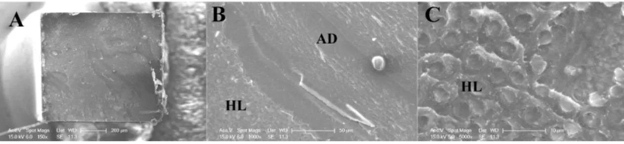
Scaning electronic microscopy - SEM
Three representative specimens of each group for each storage time (24 h and 1 year) were prepared for SEM analysis (INSPECT S50, FEI, Brno, Czech Republic) to study microscopic fracture patterns and the morphology of the debonded interface. Specimens were fixed in a solution of 2.5% glutaraldehyde in 0.1 mol/l sodium cacodylate buffer for 24 hours, rinsed three times in 0.1 mol/l sodium cacodylate buffer. They were then rinsed in distilled water and dehydrated in an ascending ethanol series 30%, 50%, 70%, 80% (for 20 min each), 95% (two times for 15 min) and 100% (two times for 30 min). Afterwards, the specimens were dried with filter paper and then placed for 10 minutes in hexamethyldisilazane (HMDS). Subsequently HMDS was removed and the specimens were placed in contact with filter paper at room temperature for 24 h. They were then positioned on metal stubs with carbon tape, sputter-coated with gold (Q 150T ES, Quorum Tchnologies Ltd, Ashford, Kent, England) and analyzed in a SEM (INSPECT S50, FEI, Brno, Czech Republic).
Micro-Raman spectroscopy analysis of adhesive interfaces
29
(NIH, Bethesda, USA). Spectra were obtained at room temperature and during the time between scanning procedures, the samples were stored in physiological solution with a relative humidity of 100%.
Statistical analysis
Normal distribution of data and homogeneity of variances were evaluated using Kolmogorov-Smirnov and Levene tests. The effects of adhesive level, storage time and their interaction on the mean bond strength were verified by Two-Way ANOVA and Scheffe post hoc test (α=0.05). Premature failure were excluded from the statistical analysis. And the degree of conversion (DC) of dental adhesives were verified by One-Way ANOVA.
RESULTS
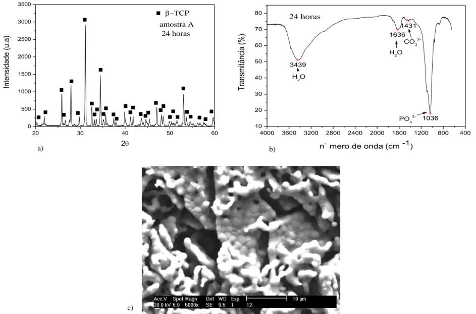
Characterization of β-TCP particles
Crystal phases of β-TCP were observed in X-ray diffractogram and FTIR (Figures 1a and 1b) of calcium phosphate bioceramic powder sintered at temperatures of 1200 ° C for 1 hour. Two different sizes of β-TCP particles were synthesized at nanoscale range: β-TCP-1 and β-TCP-2 with 40 nm and 23 nm of ratio diameter, respectively, according BET data analyses (Table 2). Morphological analysis of β-TCP nanoparticles revealed intertwined fine equiaxial spherical nanoparticles (Figure 1c)
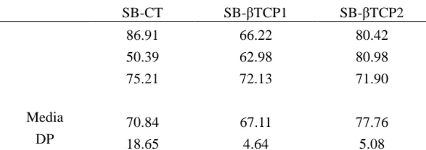
Degree of Conversion (DC) of dental adhesives
One-Way ANOVA did not detect statistically significant difference between DC of all adhesive systems (p=0.64, Table 3). Data from degree of conversion are presented in Table 3.
Microtensile Bond strength, failure mode and SEM images of fractured surfaces
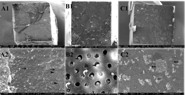
30
failures (Figure 2). The fractured surfaces presented most dentin protected with resin material; in SB-CT, adhesive failure at the top of HL was observed (Figures 2A1-2). The portions on fractured surface of SB-β1 (Figures 2B1-2) and SB-β2 (Figures 2C1-2) where dentin was exposed, it was suggested that particles of β-TCP could be detected (arrows Figures B2 and C2). After 1 year, failure was mostly mixed, however it could be observed an increase of fractured exposed dentin at different levels, including sound dentin (Figures 3A1-2). Wide area of SB-β1 fractured surface was covered with resin material, indicating protection of the adhesive interface (Figures 3B1-2). There was a suggestion of β-TCP particles on small portions of exposed at SB-β2 fractured surface (Figure 3C1 and arrows Figure 3C2).
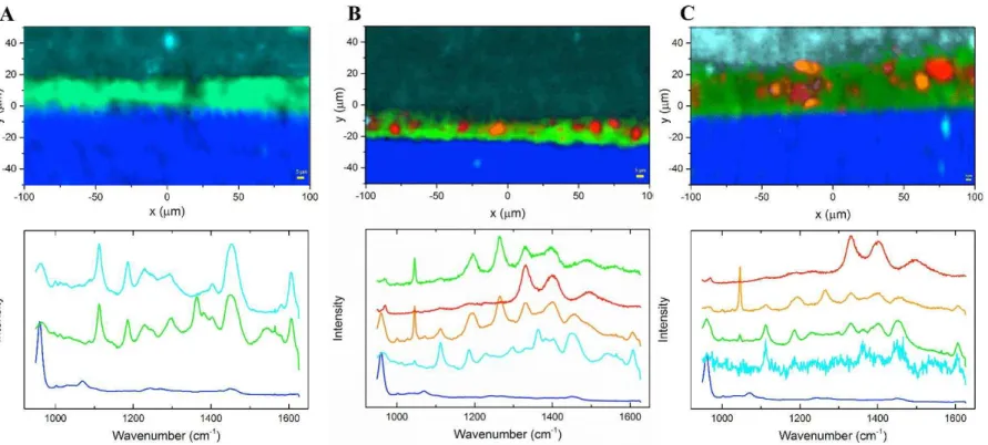
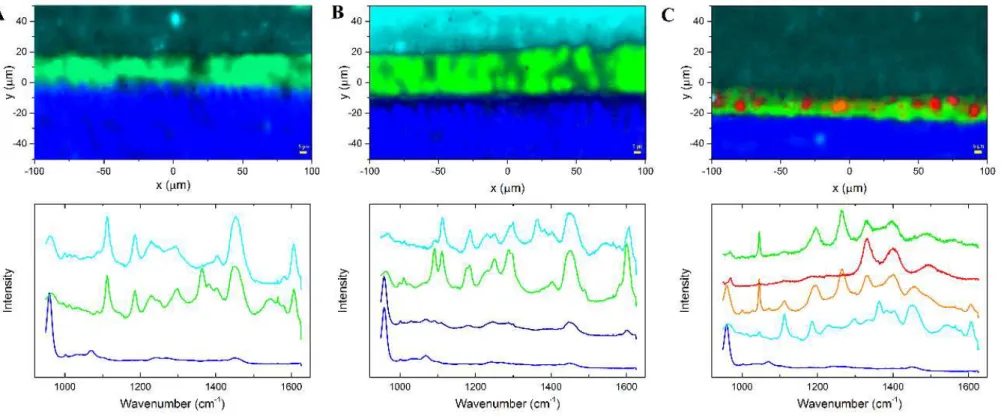
Micro-Raman spectroscopy analysis of adhesive interfaces
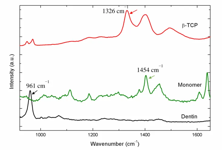
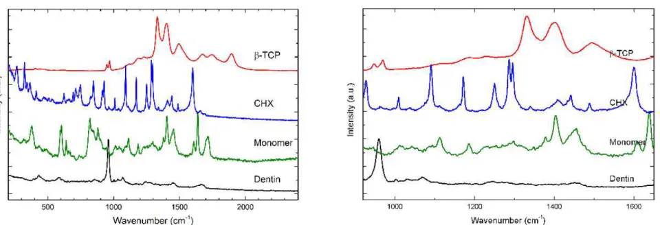
Representative Raman spectra and mapping images were processed at the cross-section interface for all groups. Adhesive polymers infiltrated properly into dentin, however β-TCP nanoparticles agglomerated, forming clusters (Figure 4). For SB-CT group, the resin tags and hybrid layer were homogeneously formed (Figure 4). SB-βTCP clusters could not penetrate into demineralized dentin, and they were confined in the adhesive layer (Figure 4B and 4C). Some small clusters could be observed at the top of the hybrid layer (Figure 4 B and 4C). Representative Raman spectra of pure materials are presents in Figure 5. All spectra were recorded in the region of 700 and 1.900 cm-1. The principal Raman active peaks associates with each pure material were to mineral composed dentin, 961 cm−1 was assigned to dentin mineral phosphate; 1070 cm−1 was assigned to the mineral carbonate. To the dentin collagen matrix features, 1667cm−1 and 1245cm−1 associated with collagen, amide I and amida III, respectivety); to the composed resin BisGMA was assigned the 1609 cm−1raman peck; to the adhesive monomer were assigned 1113, 1454, 1609,1720 cm-1 spectral; and to β -TCP, at 1326, 1395 cm−1.
DISCUSSION
31
Tay and Pashley (2009) [24] affirm that polymerized adhesives containing hydrophilic resin derivatives are also permeable to calcium and hydroxyl ions, which are necessary to remineralize dentin. Mechanical properties of demineralized dentin and/or remineralized dentin can also indirectly reflect the degree of demineralization/remineralization [34], but in this study this test were not performed. It verified that remineralization or large crystal formation within hybrid layers can occur with use of bioactive products like β -tricalcium-phosphate or other calcium/-tricalcium-phosphate materials [35]. Calcium -tricalcium-phosphate is a major component of biological hard tissues, and variants of it, such as octacalcium phosphate (OCP), tri-calcium phosphate (TCP), hydroxyapatite (HAP), dicalcium phosphate dehydrate (DCPD), and tetra-calcium phosphate (Te-CP), are commonly used as bone substitute materials. HAP has been enthusiastically adopted for its superior bone conductivity and rapid osteoinductivity, but its mechanical strength is sufficient to support implant, for example, so it is predominantly used as a coating material for titanium surfaces. The β-TCP current commercial preparations are too expensive, which has limited its clinical application [36].
The β-TCP nanoparticles synthesis was successful in the present study, and it was confirmed with characterization tests. It is possible to produce synthetic β-TCP for use in Dentistry using alternative techniques with similar results with commercial ones [36]. So, according some authors, when nanofillers are added to dental adhesives sometimes they form clusters and agglomerated clusters of fillers. The size values of clusters formed are always above the dimensions of the spaces existing between the demineralized collagen fibers [37]. Filler aggregates may avoid the penetration of the nanoparticles into the collagen network as well as to improve the mechanical properties of the hybrid layer [38,39]. In the bonding process, the demineralized dentin serves as a screener and prevents from resin penetration in underneath dentin by collecting fillers on surface. Cluster formation should be prevented as they could act as flaws, which may induce cracks and cause decreases in bond strength [40]. In this study, it was tested different particles and however, the two β-TCP had been formed clusters, they do not affected the initial bond strength, remaining stable in the long time.
In the present study, it has been very positive results about microtensile and stability when it was incorporated bioactive materials into commercial adhesive system. Moreover, in the majority of experimental studies, dentin remineralization required a liquid environment, which prevent direct clinical application. Further research will play an important role in facilitating dentin remineralization to clinical application [13].
32
β-TCP bioactive nanoparticles could be successful synthesized and could be incorporated in a commercial dental adhesive. Bonding interfaces of loaded adhesives with dentin were higher bond strength values than control group after 24h and they remained stable overtime after one year. Thus, the results presented in this article highlight the need for investigations on the long-lasting performance of bioactive nanoparticles incorporated in commercial dental adhesives applied on dentin trying to innovate with a more accurate clinical method to increase the longevity of adhesive restorations.
REFERENCES
1. P. Spencer, Q. Ye, J. Park, E.M. Topp, A. Misra, O. Marangos, Y. Wang, B.S. Bohaty, V. Singh, F. Sene, J. Eslick, K. Camarda, J. L. Katz, Adhesive/dentin interface: the weak link in the composite restoration, Ann. Biomed. Eng. 38 (2010) 1989-2003. 2. H.Tobi, C. M. Kreulen, H. Vondeling, W. E. van Amerongen, Cost-effectiveness of
composite resins and amalgam in the replacement of amalgam class II restorations. Commun. Dent. Oral Epidemiol. 27 (1999) 137-143.
3. F.F. Demarco, M.B Corrêa, M.S. Cenci, R.R Moraes, N.J. Opdam, Longevity of posterior composite restorations: Not only a matter of materials, Dent. Mater. 28 (2012) 87-101.
4. A. Frassetto, L. Breschi, G. Turco, G. Marchesi, R. di Lenarda, F.R. Tay, D.H. Pashley, M. Cadenaro, Mechanisms of degradation of the hybrid layer in adhesive dentistry and therapeutic agents to improve bond durability-A literature review, Dent. Mater. 32 (2016) e41-e53.
5. I. Nedeljkovic, W. Teughelsb, J.De. Muncka, B. Van Meerbeek, K.L. Van Landuyta, Is secondary caries with composites a material-based problem?, Dent. Mater. 31 (2015) e247-277.
6. A. Jokstad, Secondary caries and microleakage, Dent. Mater. 32 (2016) 11-25.
7. P. Spencer,.Q.Ye, A. Misra, S.E.P. Goncalves, J.S. Laurence, J. S, Proteins, pathogens, and failure at the composite-tooth interface. J. Dent. Res. 93 (2014) 1243-1249. 8. S. Kermanshahi, J.P. Santerre, D.G. Cvitkovitch, Y. Finer, Biodegradation of
resin-dentin interfaces increases bacterial microleakage. J. Dent. Res. 89 (2010) 996-1001. 9. P.E. Murray, L.J. Windsor, T.W. Smyth, A.A. Hafez, C.F. Cox, Analysis of pulpal
reactions to restorative procedures, materials, pulp capping, and future therapies, Crit. Rev. Oral. Biol. Med. 3 (2002) 509-520.
10.D.H. Pashley, F.R. Tay, L. Breschi, L. Tjäderhane, R.M. Carvalho, M. Carrilho, A. Tezvergil-Mutluay, State of the art etch-and-rinse adhesives, Dent. Mater. 27 (2011) 1-16.
11.P. Spencer, J.R. Swafford, Unprotected protein at the dentin-adhesive interface, Quint. Int. 30 (1999) 501-507.
12.D. Leung, D.A. Spratt, J. Pratten, K. Gulabivala, N.J. Mordan, A.M. Young, Chlorhexidine-releasing methacrylate dental composite materials, Biomaterials. 26 (2005) 7145-7153.
13.B. Zhong, C. Peng, G. Wang, L. Tian, Q. Cai, F. Cui, Contemporary research findings on dentine remineralization, J. Tissue. Eng. Regen. Med. 9 (2015) 1004-1016.
33
self-etch adhesives doped with calcium-phosphate fillers and biomimetic analogs of phosphoproteins, J. Dent. 52 (2016) 79-86.
15.L. Niu, W. Zhang, D.H. Pashley, L. Breschi, J. Mao, J. Chen, F.R. Tay, Biomimetic remineralization of dentin, Dent. Mater. 30 (2014) 77-96.
16.R. Osorio, M. Yamauti, S. Sauro, T.F. Watson, M. Toledano, Zinc incorporation improves biological activity of beta-tricalcium silicate resin-based cement, J. Endod. 40 (2014) 1840-1845.
17.S. Tavassoli-Hojjati, M. Atai, R. Haghgoo, S. Rahimian-Imam, S. Kameli, F. Ahmaian-Babaki, F. Hamzeh, M. Ahmadyar, Comparison of various concentrations of tricalcium phosphate nanoparticles on mechanical properties and remineralization of fissure sealants, J. Dent. (Tehran). 11 (2014) 379-388.
18.M. Toledano, E. Osorio, F.S. Aguilera, S. Sauro, I. Cabello, R. Osorio, In vitro mechanical stimulation promoted remineralization at the resin/dentin interface, J. Mech. Behav. Biomed. Mater. 30 (2014) 61-74.
19.S. Sauro, R. Osorio, E. Osorio, T.F. Watson, M. Toledano, Novel light-curable materials containing experimental bioactive microfillers remineralise mineral-depleted bonded-dentine interfaces, J. Biom. Sci. 8 (2013) 940-956.
20.A.C. Profeta, F. Mannoccib, R. Foxtonb, T.F. Watson, V.P. Feitosa, B. De Carlo, R. Mongiorgi, G. Valdré, S. Sauro, Experimental etch-and-rinse adhesives doped with bioactive calcium silicate-based micro-fillers to generate therapeutic resin–dentin interfaces, Dent. Mater. 29 (2013) 729-741.
21.A.C. Profeta, F. Mannocci, R.M. Foxton, I. Thompson, T.F. Watson, S. Sauro, Bioactive effects of a calcium/sodium phosphosilicate on the resin–dentine interface: a microtensile bond strength, scanning electron microscopy, and confocal microscopy study, Eur. J. Oral. Sci. 120 (2012) 353-362.
22.C. Combes, C. Rey. Amorphous calcium phosphates: Synthesis, properties and uses in biomaterials, Acta. Biomater. 6 (2010) 3362-3378.
23.R.L. .Karlinsey, A.C. Mackey,. E.R., Walker, K.E. Frederick, Surfactant-modified β -TCP: structure, properties, and in vitro remineralization of subsurface enamel lesions. J Mater Sci: Mater Med. 21 (2010) 2009-2020.
24.F.R. Tay, D.H. Pashley, Biomimetic remineralization of resin-bonded acid-etched dentin, J. Dent. Res. 88 (2009) 719-724.
25.F.R. Tay, D.H. Pashley, Guided tissue remineralisation of partially demineralised human dentine, Biomarerials. 29 (2008) 1127-1137.
26.F.R. Tay, D.H. Pashley, Dental adhesives of the future, J. Adhes. Dent. 4 (2002) 91-103.
27.M. Bohner, S. Tadier, N. van Garderen, A. de Gasparo, N. Döbelin, G. Baroud, Synthesis of spherical calcium phosphate particles for dental and orthopedic applications, Biomatter. 3(2013) e25103-e25115.
28.S.V. Dorozhkin, Biphasic, triphasic and multiphasic calcium orthophosphates, Acta. Biomater. 8 (2012) 963-977.
29.S.K. Ghosh, S.K. Nandi, B. Kundu, S. Datta, D.K De, S.K. Roy, D. Basu. In vivo response of porous hydroxyapatite and beta-tricalcium phosphate prepared by aqueous solution combustion method and comparison with bioglass scaffolds. J. Biomed. Mater. Res. B. Appl. Biomater. 86 (2008) 217-27.
30.F. Zhang, J. Chang, J. Lu, K. Lin, C. Ning, Bioinspired structure of bioceramics for bone regeneration in load-bearing sites, Acta. Biomater. 3 (2007) 896-904.
34
32.A. Besinis, R. van Noort, N. Martin, The use of acetone to enhance the infiltration of HA nanoparticles into a demineralized dentin collagen matrix, Dent. Mater. 32 (2016) 385-393.
33.J.S.V. Albuquerque, Produção de biocimentos de apatitas nanoméricas aplicados como sistema de liberação controlada de fármacos, Fortaleza, 2012.
34.S. Mai, Y.K. Kim, J. Kim, C.K.Y. Yiu, J. Ling, D.H. Pashley, F.R. Tay, In vitro remineralization of severely compromised bonded dentin. J Dent Res. 89 (2010) 405-410.
35.G. Paolinelis, A. Banerjee, T.F. Watson. An in vitro investigation of the effect and retention of bioactive glass air-abrasive on sound and carious dentine. J Dent. 36 (2008) 214-218.
36.C. Han-Cheol, M. Hori, T. Yoshida, N. Yamada, Y. Komada, Y. Tamaki, T. Miyazaki, Tri-calcium phosphate (ß-TCP) can be artificially synthesized by recycling dihydrate gypsum hardened, Dent. Mater. J. 33 (2014) 845-851.
37.E. Osorio, M. Toledano, M. Yamauti, R. Osorio, Differential nanofiller cluster formations in dental adhesive systems. Microsc Res Tech. 75 (2012) 749-757.
38.J.S Kim, B.H. Cho, I.B. Lee, C.M. Um, B.S. Lim, M.H. Oh, C.G. Chang, H.H. Son. Effect of the hydrophilic nanofiller loading on the mechanical properties and the microtensile bond strength of an ethanol-based one-bottle dentin adhesive. J Biomed Mater Res Part B Appl Biomater 72 (2005) 284-291.
39.K.L. Van Landuyt, J. Snauwaert, J. De Munck, M. Peumans, Y. Yoshida, A. Poitevin, E. Coutinho, K. Suzuki, P. Lambrechts, B. Van Meerbeek. Systematic review of the chemicals composition of contemporarydental adhesives. Biomaterials. 28 (2007) 3757-3785.
35
TABLES
Table 1 - Materials employed in this study and respective manufactures, batch numbers, basic formulation and mode of application.
Material manufacturer batch number
Basic formulation Mode of application
AdperTM Single Bond 2 3M ESPE, St. Paul, MN, USA. N364098BR
Bis-GMA, HEMA,
dimethaclylates, ethanol, water, photoinitiator system, a
methaclylate functional copolymer of polyacrylic, polyitaconic acids
Etch dentin for 15 s and rinse for 30 s. Blot excess water using a cotton pellet or mini-sponge, without air drying, leaving a shiny surface. Apply 2-3 consecutive coats of adhesive for 20 s with gentle agitation and gently air thin for 20 s to evaporate solvent. Light-cure for 10 s.
Filtek Z250 XTTM 3M ESPE, St Paul, MN, USA. 1229100321
Bis-GMA, UDMA, TEGDMA, silanized ceramics and silica
Apply the Filtek Z250 restorative in increments of less than 2.5 mm. Photopolymerize each increment for 20 s.
Attaque gel, Biodinâmica, Ibiporã, PR, Brazil.
2016 01 009 13
Ortho-phosphoric acid 37%; methylparaben; blue dye
(CI52015); thickener and deionized water, pH 1.0.
Apply Attaque gel in the areas to be treated. Time of acting is 15 s for dentin. Rinse off Attaque gel completely for at least 30 s. Dry blowing air (oil free).
Abreviations: Bis-GMA: bis-phenol A diglycidylmethacrylate; HEMA: 2-hydroxyethyl methacrylate; UDMA: diurethane dimethacrylate; TEGDMA: triethyleneglycol dimethacrylate
Table 2 - BET data analyses. Sample Surface
area (m2/g)
Medium particle diameter (nm)
β-TCP1 47.80 40
β-TCP2 83.00 23
Table 3 - Degree of Conversion of dental adhesives
SB-CT SB-βTCP1 SB-βTCP2
86.91 66.22 80.42 50.39 62.98 80.98
75.21 72.13 71.90
Media 70.84 67.11 77.76
DP 18.65 4.64 5.08
Abreviations:SB-CT, Adper Single Bond 2, control; SB-β1,β-TCP1 (40 nm) -loaded adhesive system (SB 30% wt of β-TCP1), and SB-β2,
β-TCP2 (23 nm)-loaded adhesive system (SB 30% wt of β-TCP2).
Table 4: Bond strength results (MPa±SD) from all groups measured after different storage periods
SB-CT SB-B1 SB-B2
24 hours 29.81±8.14 Aa 35.10±11.86 Ab 39.59±11.21 Ab
1 year 25.29±11.24 Ba 34.90±12.89 Ab 39.75±9.79 Ab
36
20 30 40 50 60
0 500 1000 1500 2000 2500 3000 3500 CP In te n si d a d e (u .a ) 2 amostra A 24 horas
4000 3600 3200 2800 2400 2000 1600 1200 800 400 10 20 30 40 50 60 70 80 3439 1636 1431 1036 T ra ns m itâ nc ia (% )
nْ mero de onda (cm -1)
PO4 3-H
2O
H2O CO3 2-24 horas
a) b)
c)
37
Figure 2 - The 24 h SEM images of all three groups showed representative mixed failures; 2A1-2) SB-CT, adhesive failure at the top of HL was observed; 2B1-2) The portions on fractured surface of SB-β1; and 2B1-2) SB-β2 where dentin was exposed, it was suggested that particles of β-TCP could be detected (arrows Figures B2 and C2).
Figure 3 - After 1 year, failure was mostly mixed, however it could be observed an increase of fractured exposed dentin at different levels, including sound dentin (Figures 3A1-2). Wide area of SB-β1 fractured surface was covered with resin material, indicating protection of the adhesive interface (Figures 3B1-2). There was a suggestion of β-TCP particles on small portions of exposed at SB-β2 fractured surface
38
Figure 4 - Representative Raman spectra and mapping images were processed at the cross-section interface for all groups; 4A) SB-CT control group. For SB-CT group, the resin tags and hybrid layer were homogeneously formed; 4B) SB-β1 group. SB-βTCP clusters could not penetrate into demineralized dentin, and they were confined in the adhesive layer. In all groups, adhesive polymers infiltrated properly into dentin,
however β-TCP nanoparticles agglomerated, forming clusters (red); 4C) SB-β2 group. Some small clusters could be observed at the top of the
39
Figure 5 - Representative Raman spectra of pure materials are shown. All spectra were recorded in the region of 700 and 1.900 cm-1. The principal Raman active peaks associates with each pure material were to mineral composed dentin, 961 cm−1 was assigned to dentin mineral phosphate; to the composed resin BisGMA was assigned the 1609 cm−1raman peak; to the adhesive monomer were assigned 1113, 1454, 1609,1720 cm-1 spectral; and to β-TCP, at 1326, 1395 cm−1.
961 cm
−140
CAPÍTULO 2
Journal: Journal of Biomedical Materials Research Part B: Applied Biomaterials
Title: Dental adhesive loaded with functional particles: bond strength, SEM and
micro-Raman imaging of adhesive-dentin interfaces
Authors: Camila Ferraz1; Maria Cura2; Laura Ceballos2; Sergio Lima Santiago1; Alejandro Pedro Ayala3; Monica Yamauti4
Affiliations:
1
Departament of Operative Dentistry, Universidade Federal do Ceará, Fortaleza, CE, Brazil
2
Departament of Stomatology, Health Sciences Faculty, Universidad Rey Juan Carlos University, Madrid, Spain
3
Departament of Physical, Universidade Federal do Ceará, Fortaleza, CE, Brazil 4
Department Restorative Dentistry, Universidade Federal de Minas Gerais, Belo Horizonte, MG, Brazil
*Corresponding author: Monica Yamauti Assistant Professor
Graduate School of Dentistry, Department of Restorative Dentistry, Universidade Federal de Minas Gerais, Belo Horizonte, MG, Brazil
Avenida Presidente Antônio Carlos, nº 6627, Pampulha, Belo Horizonte - MG, Brazil, 31270-901
41
ABSTRACT
The aim of this study was to reveal structural and chemical profile of the adhesive interface using total-etch adhesive solutions loaded with functional (diacetate chlorhexidine – DA) and bioactive (tricalcium phosphate - β-TCP) particles, and to evaluate the bond strength of adhesive to sound dentin overtime (one year). Physical properties including microtensile bond strength, SEM, and micro-Raman spectral analysis were measured. Fifteen extracted, caries-free, human third molars had their dentin surfaces exposed. They were randomly divided into three groups (n=5). In SB-CT was applied pure adhesive system Adper Single Bond 2 (SB); SB-DA was applied DA-loaded SB, and SB-βTCP was applied loaded SB. All exposed dentin surfaces were prepared for the adhesive procedure, restored and stored in distilled water for 24 h at 37°C. Stick specimens with cross-section dentin of 1.0 mm² were evaluated for microtensile bond strength after 24 h and 1 year. Three representative specimens of each group were prepared for SEM and Raman spectroscopy analysis. The micro-Raman and SEM results indicated that adhesive system compounds diffuse differently into hybrid layer. For SB, the resin tags and hybrid layer were homogeneously formed. In the SB-DA group, chlorhexidine particles were concentrated on top of hybrid layer and spread on the adhesive. At SB-βTCP interface, particles could not penetrate into hybrid layer, and they were all accumulated in the adhesive layer. As bond strength after One-Way ANOVA and Scheffe post hoc test for 24 h and one year results, there were statistically significant difference for mean bond strength (p<0.05). The bond strength of control group (SB) was significantly lower than those of DA (p<0.05) and of SB-βTCP (p<0.05), and there were no differences between bond strength of SB-DA and SB-βTCP groups (p=0.580) for all times. Thus, the use of adhesives loaded with functional and bioactive particles is a promising material to obtain stability and longevity of adhesive of restorations interfaces.
Key Words: chlorhexidine, dental adhesive, dentin bonding, interface, micro-Raman
42
INTRODUCTION
Longevity of adhesive restorations relies, among other factors, on quality of hybrid layer and on collagen matrix integrity1-4 at the adhesive interface. To increase restorations longevity, it is essential to understand the behavior of dentin, adhesives and restorative materials at the interface.5-8 Functional and bioactive agents act protecting exposed collagen fibers against bacterial contamination residues. They also remineralizing caries affected or acid-demineralized dentin, and also inhibiting endogenous matrix metalloproteinases (MMPs), cysteines and cathepsins activation. 1,9-17
The incorporation of bioactive particles, such as antimicrobials and potentially remineralizing agents, to adhesives has been a common attempt to obtain better quality and stability of restorations interfaces.18-20
Chlorhexidine (CHX) and beta-tricalcium phosphate (β-TCP) are, respectively, antibacterial/functional and remineralizing agents. They can be incorporated in adhesives, fissure sealants and glass ionomers cements 1,11,15,16,21-22,24 or applied as treatment before adhesive application. 23,25 CHX could also present an important role in preservation of resin-dentin bonds by inhibiting the collagenolytic activity of host-derived enzymes.1,14,16,21,24,26
The β-TCP is a synthetic calcium orthophosphates, β-Ca3(PO4)2.,22,27,28 precursor to hydroxyapatite formation.6,22 β-TCP is capable of improving tooth remineralization process due to its ability to increase calcium and phosphate concentration19,22 in hybrid layer.29
For this reason, it has been currently tested in several clinical applications in Dentistry.19,22,30-32 However, the physicochemical interaction between adhesive loaded with functional (CHX) and bioactive (β-TCP) particles with acid etched sound dentin is not clear yet.
Thus, this study aimed to reveal structural and chemical profile of the adhesive interface using total-etch adhesive solutions loaded with functional (diacetate chlorhexidine - DA) or bioactive (β-TCP) particles and to evaluate the bond strength of adhesive to sound dentin.
MATERIAL AND METHODS
43 An etch-and-rise adhesive system, AdperTM Single Bond 2 (SB) (3M ESPE, St. Paul, MN, USA) was used. Adhesive solutions of 2 wt% chlorhexidine diacetate (SB-DA) and 30 wt% β-TCP (40 nm) (SB-βTCP). To get complete dispersion of particles, DA and β-TCP, the adhesive mixture was shaken during 1 min in a tube agitator in darkness (Vortex Phoenix, Ref. 12446, Phoenix Ind. E Com. De Equips Científicos Ltda, Araraquara, São Paulo, Brazil). Descriptions of chemicals and adhesive employed are provided in Table 1.
Tooth Specimen Preparation
Fifteen extracted, caries-free, human third molars were obtained with informed consent, under a protocol approved by the Human Research Ethics Committee of the Institution. Teeth were properly cleaned with periodontal scoop and were stored in 0.1% (w/v) thymol solution at 4°C and used within one week after extraction. For each tooth, the coronal portion was removed to expose a flat, mid-coronal dentin surface using a low-speed diamond saw34 (IsoMetTM Low Speed Saw, Buehler, Lake Bluff, IL, USA) under water-cooling. Dentin surfaces were exposed and ground using 600-grit silicon carbide abrasive paper under water cooling for 60 s to obtain a standardized smear layer.1,34
Dentin surfaces were acid-etched with 37% phosphoric acid (pH 1.0) for 15 s, rinsed with copious water for 30 s and two layers adhesives were applied under agitation for 20 s as follow: (1) SB; (2) SB-DA; (3) SB-βTCP. The adhesive solvent was evaporated using a gentle air spray for 5 s. Adhesives were light-cured for 10 s. Five resin composite restorations (Filtek Z250 XT™, 3M ESPE, St Paul, MN, USA) were built up using increments of, approximately, 1 mm thick in all of teeth. Each composite increment was light activated for 40 s. All adhesives and composite increments were curing using a blue LED light source (EliparTM FreeLight 2, 3M ESPE, Neuss, Germany), and light intensity output was maintained at a minimum of 600 mW/cm2. After restorative procedure, all teeth were stored in distilled water for 24 h at 37°C.
Microtensile bond strength evaluation
44 Speed Saw, Buehler, Lake Bluff, IL, USA) under water lubrication to obtain bonded sticks specimens with a cross-sectional area of approximately 1.0 mm². The exact dimensions of the adhesive interfaces of sticks were measured using a digital caliper (Mitutoyo Absolute, Mitutoyo Sul Americana Ltda, São Paulo, SP, Brazil). Five teeth from each group were evaluated after 24 h20 and 1 year. Specimens were attached to the universal test machine (Instron 3345, Instron Inc., Canton, MA, EUA) with cyanoacrylate glue and were subjected to a tensile load at 0.5 mm/min until failure. Bond strength values were calculated in megapascals (MPa). Fracture modes of each specimen were determined using a stereomicroscope (Leica Microsystems, Wetzlar, Deutschland, Germany), at 60X magnification. Fracture modes were classified as cohesive in dentin (CD) when they were located exclusively within dentin, cohesive in resin (CR) when they were located exclusively within resin, adhesive (A) when failure occurred at the dentin/adhesive interface, or mixed (M) when modes of failure occurred simultaneously. Each type of failure mode was expressed as a percentage of the total number of specimens in that group. Representative specimens of each group were prepared for SEM analysis (INSPECT S50, FEI, Brno, Czech Republic).
Micro-Raman spectroscopy analysis of adhesive interfaces
One bonded dentin slab from prepared teeth of all three groups (SB; SB-DA; SB-βTCP) (24 h storage time) was separated to micro-Raman analysis. Slabs surfaces were not polished because remaining debris could contaminate specimens. Bonded tooth sections were cleaned in distilled deionized water in an ultrasonic bath for at least 5 min at ambient temperature before Raman spectroscopy analysis. Micro-Raman spectral analysis were performed to evaluate the penetration of the adhesive monomers and/or DA and β-TCP particles into demineralized dentin.
45 two accumulations and DuoScan mode. The micro-Raman spectra of sound dentin slabs and pure materials were recorded mainly to find a micro-Raman signal from the materials and tissues, which must be distinguishable from other specimens’ components. Spectra were obtained at room temperature and during the time between scanning procedures, the samples were stored in physiological solution with a relative humidity of 100%. Spectral information was obtained between the wavenumbers of 900 and 1700 cm-1. Mapping images were also obtained. The characteristics of the spectra collected, such as the integrated intensity of a particular spectral band, were examined with a mapping LabScan tool. All the measurement steps, including image processing, image analysis and area measuring were performed using the software LabRamanTM (NIH, Bethesda, USA). For each specimen, the compounds of the area of interest (hybrid layer) were identified and visually evaluated.
Statistical Analysis
Normal distribution of data and homogeneity of variances were evaluated using the Kolmogorov-Smirnov and Levene tests. Difference between mean bond strength values was verified by One-Way ANOVA and Scheffe post hoc test (α=0.05).
RESULTS
Microtensile Bond Strength
There was statistically significant difference for mean bond strength (p<0.05). The bond strength of control group (SB) was significantly lower than those of SB-DA (p<0.05) and of βTCP-SB (0.002) (Table 2). Bond strength of SB-DA and βTCP-SB were no statistical differences (p=0.143). Most failure was classified as mixed. SEM images of fractured surfaces are shown in Figures 1, 2 and 3.
Degree of Conversion (DC) of dental adhesives
One-Way ANOVA did not detect statistically significant difference between DC of all adhesive systems (p=0.52, Table 3). Data from degree of conversion are presented in Table 3.
46 Representative Raman spectra and mapping images were processed for adhesive interfaces. Figure 4 shows micro-Raman spectra of sound dentin, adhesive and bioactive particles profiles, and they were used as spectra control. In Figure 4, the most intense peak at 961 cm−1 (v1 symmetric stretch, PO43-) is assigned to dentin mineral phosphate. The peak at 1070 cm−1 (v1 symmetric stretch, CO32-) is assigned to the mineral carbonate and the dentin collagen matrix features are present at 1667cm−1 (amide I, associated with collagen) and 1245cm−1(amide III, associated with collagen). The Raman spectrum of the sample shows stretching vibrations on the band ratio of 1609 cm−1 (phenyl C=C), associated with BisGMA). The band ratio is calculated by the spectral subtraction technique at 1113, 1454, 1609,1720 cm-1, which were assigned to the adhesive monomer; at 1326, 1395 cm-1that referred to the β-TCP; and 1172, 1296, 1600 cm-1 that corresponded to the diacetate chlorhexidine.
According to micro-Raman mapping analysis, bioactive particles in the adhesive layer were identified when they were present. It was clear that adhesive system compounds diffuse differently into hybrid layer between experimental groups (Figures 5A, B and C). For SB-CT, resin tags and hybrid layer were homogeneously formed (Figure 5A). In the micro-Raman of SB-DA interface image, chlorhexidine particles were concentrated on top of hybrid layer and spread on the adhesive (Figure 5B). On the other hand, at SB-βTCP interface, particles could not penetrate into hybrid layer, and they were all accumulated in the adhesive layer (Figure 5C). In all grups, adhesive polymers or monomers infiltrated properly into dentin, however βTCP nanoparticles agglomerated, forming clusters.
DISCUSSION
In adhesive Dentistry, partial or complete demineralization is a prerequisite for
successful bonding with contemporary adhesives. Attaining and maintaining a strong
47 Though microtensile bond strength values were no different between DA and βTCP group in 24h and one year, there are different behaviors of these two materials into hybrid layer (HL), according to micro-Raman images and analysis.
SB-DA group results suggested that chlorhexidine particles could not diffuse through the total depth of adhesive layer, but could partially diffuse into HL. This finding could indicate that DA particles might act protecting exposed collagen fibers against bacterial contamination residues or inhibiting the activation of endogenous matrix metalloproteinases (MMPs).24,35 These MMPs are proteolytic enzymes, which are activated from inactive preforms by acidic conditions. The acid etching and bacterial acid production at the tooth/restoration interface may activate them.4,35 In this study, the presence of DA in hybrid layer could be enough to inhibit the endogenous matrix metalloproteinases (MMPs) activation through diffusion into adhesive layer over time.
About β-TCP, results from this study suggested that β-TCP particles were agglomerated in some regions of adhesive layer and they could not be carried into dentin tubules. Nevertheless, these agglomeration zones could function as a reserve of calcium ions inside the hybrid layer and could prevent unprotected demineralized dentin from enzyme degradation and the adhesive interface from cariogenic challenge. Their presence in this study did not show negative results about bond strength, and this was according to other previous studies with use of βTCP and adhesion.19,30
Then, the β-TCP could maintain a strong and durable adhesive joint between resin-bonded dentin, the quality of hybrid layer and probably it could maintain the longevity of strong adhesion in dentin too.19,30 The medium particles diameter β-TCP used in this study was 40 nm, but clusters were observed in the adhesive layer. The formation of β-TCP clusters has probably prevented the penetration of this component into the total depth of HL and inside dentin tubules.