Universidade Nova de Lisboa
Faculdade de Ciências de Tecnologia
Author: MsC. Elisabete De Jesus Oliveira Marques
New Fluorescent Chemosensors based on
bio-inspired ligands and Macrocycles:
From single molecules to nanoparticles
PhD em Biotecnologia
Supervisor: Dr.Carlos Lodeiro (FCT-UNL/U.VIGO) Co-Supervisor (s): Dra. Susana Costa (UMINHO) Dra. Isabel Moura (REQUIMTE-FCT-UNL)
Ph.D dissertation in Biotecnologia by Elisabete De Jesus Oliveira Marques
Title: New Fluorescent Chemosensors based on bio-inspired ligands and
Macrocycles: From single molecules to nanoparticles
Universidade Nova de Lisboa
Faculdade de Ciências de Tecnologia
Author: MsC. Elisabete De Jesus Oliveira Marques
New Fluorescent Chemosensors based on
bio-inspired ligands and Macrocycles:
From single molecules to nanoparticles
PhD em Biotecnologia
Dissertação apresentada para obtenção do grau de Doutor em Biotecnologia pela Universidade Nova de Lisboa, Faculdade de Ciências e Tecnologia.
A presente dissertação foi preparada no âmbito do protocolo de acordo bilateral de educação avançada (ERASMUS) entre a Universidad de Vigo e a Universidade Nova de Lisboa.
“Life is not easy for any of us. But what of that? We must have
perseverance and above all confidence in ourselves.
We must believe that we are gifted for something
and that this thing must be attained”
iii
ACKNOWLEDGEMENTS
During my PhD I had the pleasure and lucky to know many people, where which one
contributed for the increase of my knowledge. Thus I would like to thank the support
of many people.
I’m eternally grateful to Dr. Carlos Lodeiro, my supervisor, for their support and
guidance in difficult times, and be always present in the most important moments of
my scientific life. There are no words to describe all his dedication, kindness and
friendship shown during these past years. I thank you from my heart for everything.
I’m gratefully to Dr. José Capelo for is guidance on MALDI-TOF-MS studies, his
friendship and support in the BIOSCOPE group.
I would like to thank to my co-supervisor, Dra. Susana Costa for her support during
the three months that I spent in Minho University; all her guidance and help, during
my PhD; thank to the entire chemistry group of Prof. M.Manuela Raposo.
I would like to thank also to my co-supervisor Dra. Isabel Moura.
I would like to thank to Dra. Teresa Avilés by the speak about science and good
atmosphere in the laboratory 412.
I’m gratefully to the REQUIMTE, New University of Lisbon by the host, allowing the
development of research work.
I would like to thank to Prof. Dr. Luca Prodi to welcome me in his research group in
Bologna, to Dra. Nelsi Zaccheroni for her great support in the nanoparticle synthesis
and fluorescence studies; to Dra. M. Teresa Gandolfi, to Riccardo, Damiano and
Sara.
iv
and made me fell as a part of a great group, Bruno, Cristina, Javier, Jorge, Júlio,
Marco, Ricardo, Gonçalo e Dr. Mário Dinis; specially my friend and colleague Hugo,
which always gave me support and his friendship, and it is a pleasure work with him.
I would like to thank to Dr. Cristina Nuñez by the help in X-Ray diffraction, to Dr.
Pablo Gonzalez by the EPR studies, to Dr. Olalla Nieto by the DFT calculations and
to MsC. Luz Fernandes by the help in the MALDI studies.
I thank to FC-MCTES (Portugal) by my PhD grant SFRH/BD/35905/2007, by the
project PTDC/QUI/66250/2006, “Desenvolvimento de novos compostos
heterocicíclos como sensores químicos luminescentes e colorimétricos: detecção de
aniões e catiões metálicos”; to the University of Vigo by the Project
VICOUK914-122P64702 “Sintese y aplicación de novos sensores químicos de fluorescencia para
la determinación de biomoleculas y metales de interés medioambiental”; and Xunta
de Galicia, Spain by the Project 09CSA04338PR (in Biomedicine) “Aplicación de
Nanopartículas activas inxectadas por ultrasonidos no tratamento de mostra para a
identificación rápida de proteínas e biomarcadores de tecidos biológicos”; and
specially to Fundação Calouste Gulbenkian (Portugal), for the National Prize in
criativity and quality in reseach activity, 2008.
v
ABSTRACT
Due to the importance of the development of new fluorescent compounds with multifunctional applications in environmental and analytical sciences, nano-scale technologies and bio-medicine, the research summarized in this PhD project was focused on the synthesis of new bio-inspired sensors, containing alanine, trypthophan and cysteine amino acids, provided with benzoxazole chromophores; exploration of its photophysical properties as fluorescence markers and chemosensors, and finally in the synthesis of new gold and silica nanoparticles with emissive properties. At the same time, different macrocyclic compounds bearing anthracene, or furyl, aryl or thienyl moieties linked to an imidazo-aza-crown ether were exploited for the synthesis of solid inorganic complexes and their use as fluorescent chemosensors for metal ions and anions was undertaken.
The benzoxazole ring was selected as fluorescence probe for peptide skeleton due to the biological activity, antifungal, antimicrobial and anticancer properties, and also due to the higher emission fluorescence which makes it a good candidate for molecular recognition, biomarkers or biosensors.
Several studies on the metal ion association constants and by density functional theory (DFT) were performed.
The association of the synthesized compounds to more structured nanoparticles could increase the sensibility of the chemosensor and also its capability for sensing. In our case, incorporation of the cysteine amino acid made them good candidates for linkage to gold surface nanoparticles. However, in same cases due to the energy transfer from the metal core, the emission fluorescence of these decorated nanoparticles was quenched. Taking into account this phenomenon, different commercial light transparent LUDOX silica nanoparticles were prepared and studied, containing the emissive peptides.
This PhD dissertation is divided in eight chapters; and a brief description of each chapter is presented.
Chapter 1 shows a brief introduction to general fluorescent chemosensors and definitions, mechanisms of detection, chemosensors based on macrocyclic ligands, chemosensors containing bio-inspired units, from single amino acids to peptide chains; some aspects on the biological applications on emissive chromophores; the importance on the detection and quantification of metal ions; and finally a short introduction concerning the synthesis and applications of silica and gold nanoparticles and their advantages.
vi
L towards alkaline (Na+, K+, Li+), alkaline earth (Ca2+, Mg2+), transition and post-transition (Cr3+,Cu2+ and Zn2+, Cd2+, Hg2+ and Al3+) metal ions were performed by Uv-vis and fluorescent emission spectrocopy. An increase in the fluorescence emission (CHEF effect) was observed in methanol and in mixtures methanol/water in the presence of Cd2+ (5.0-fold), Zn2+ (4.5-fold), Cr3+ (2.0-fold) and Al3+ (1.8-fold). This work was published in: Inorganica Chimica Acta, 2007, 360, 2734-2743.
In Chapter 3 is presented the synthesis of several metal complexes with three new aza-crown ether macrocycles bearing furyl, aryl or thienyl moieties linked to an imidazo-aza-crown system (1, 2 and 3) and its chemical characterization by elemental analysis, infrared, absorption and emission spectroscopy, X-Ray crystal diffraction, and MALDI-TOF-MS spectrometry. The sensing capability of all ligands were studied in the presence of Ca2+, Cu2+, Ni2+, Hg2+ and F-. For compounds 2 and 3 was observed an increase in the emission fluorescence with Ca2+ and Cu2+. Moreover, compound 3 showed a colorimetric characteristic with Cu2+. Submitted to Inorganic Chemistry, 2010.
In Chapter 4 is described the photophysicial studies of highly emissive fluorescent probes based on unnatural alanine derivatives bearing the benzoxazole unit as chromophore at the side chain, functionalized with different moieties, such as, thiophene (2a-2d) or trimethoxybenzaldehyde (2e-2f). The incorporation of different units into the benzoxazole modulates the fluorescence quantum yield, being the thiophene derivatives the most emissive compounds. For all compounds was observed a quenching effect with Cu2+, Ni2+ and Hg2+, as shown by absorption and emission fluorescence spectroscopy. Taking into account the complexation constants obtained it was postulated that probably the first metal ion takes place in the carboxylic acid group and the second one around the chomophore. This work was published in: Sensors, 2007, 7, 2096-2114.
Chapter 5 shows the synthesis and photophysical studies with new benzoxazolyalanine derivatives bearing (oligo)thiophene units at the side chain (4a-6a). All compounds were highly fluorescent, making them good candidates for application as fluorescent probes. This work was published in: Tetrahedron Letters,2008, 49, 5258-5261.
vii
of alanine-benzoxazole linked by a thiophene unit was explored towards Cu2+, Ni2+ and Hg2+ metal ions. Manuscript submitted to Inorganica Chimica Acta, 2010.
Chapter 7 summarizes the synthesis of six new emissive di and tri-peptides derivatives containing benzoxazolyl-alanine, trypthophan and cysteine amino acids (L1-L4, L6-L7), and two previously reported in chapter 5 benzoxazolyl alanine derivates (L and L5). Photophysical studies on the interaction towards transition and post-transition metal ions (Cu2+; Ni2+; Ag+; Zn2+; Cd2+; Hg2+ and Pb2+) by absorption and fluorescence emission spectroscopy, and by MALDI-TOF-Ms spectrometry were performed. In general, all compounds showed a good interaction with Hg2+. Gold(0) nanoparticles decorated with ligands L, L1 and L3, were synthesized by common reductive procedures, and emissive silica nanoparticles were synthesized using compounds L5-L7 linked through a silane spacer. The silica core were based on commercial LUDOX nanoparticles, in different ligand:nanoparticle ratios. All nanoparticles obtained were characterized by dynamic light-scattering (DLS), transmission electron microscopy (TEM), UV-vis absorption and emission spectroscopy. TEM images revealed the formation of gold nanoparticles between 2.4 and 10 nm, and silica nanoparticles, with size between 14 and 24 nm. The interaction with metal ions was also performed using the silica nanoparticles as supramolecular chemosensors, being selective for Ag+ and Hg2+.
The general conclusions of this research work can be found in Chapter 8.
ix
RESUMO
Devido ao desenvolvimento e importância de novos compostos fluorescentes com aplicações multifuncionais em ciência do ambiente, em ciências analíticas, tecnologia de nano-escala e bio-medicina, o trabalho de investigação apresentado neste projecto de Doutoramento incide na síntese de novos sensores bio-inspirados, contendo aminoácidos, como a alanina, cisteina e triptofano, conjugados ao cromóforo benzoxazole; foram exploradas as suas propriedades fotofísicas, como marcadores fluorescentes e sensores químicos, e finalmente na síntese de nanopartículas de ouro e sílica com propriedades emissivas.
De igual modo, foi explorado a síntese de complexes sólidos inorgânicos, bem como as suas propriedades como sensores químicos na presença de iões metálicos e aniões, de compostos macrocíclicos contendo unidades antraceno; furano, arilo ou tiofeno ligados a um éter imidazo-aza-coroa.
O anel aromático benzozazole foi seleccionado como sonda fluorescente na estrutura peptídica, devido à sua actividade biológica, antifúngica, antimicrobial e anticancerígena; e também devido à sua elevada emissão de fluorescência. Todas estas propriedades, torna-o um bom candidato para reconhecimento molecular, biomarcadores e biosensores.
Por conseguinte, foram efectuados estudos de constantes de associação a iões metálicos e também por teoria funcional de densidade (DFT).
A associação dos compostos sintetizados a nanopartículas poderá aumentar a sensibilidade do sensor químico, bem como a sua capacidade sensitiva. No presente trabalho, a incorporação do aminoácido cisteina na cadeia peptídica dos compostos tornou-os bons candidados para ligação a superfícies de nanopartículas de ouro. No entanto, em alguns casos devido à transferência de energia proveniente do centro metálico, ocorreu a supressão de emissão de fluorescência dos compostos “decorados”. Devido a este fenómeno, foram sintetizados e estudados péptidos emissivos ancorados a nanopartículas transparentes comerciais de sílica LUDOX .
Esta dissertação de Doutoramento está dividida em oito capítulos, e de seguida é apresentada uma breve descrição de cada capítulo.
x
finalmente uma breve introdução acerca da síntese e aplicações das nanopartículas de ouro e silica e as suas vantagens.
No Capítulo 2 são descritos os estudos fotofísicos e a caracterização de um novo ligando macrocíclico (L) contendo como braço pendente uma unidade antraceno. A avaliação da capacidade sensorial de L na presença de iões metálicos alcalinos (Na+, K+, Li+), alcalino-terrosos (Ca2+, Mg2+), de transição de pós-transição (Cr3+,Cu2+ and Zn2+, Cd2+, Hg2+ and Al3+) foi efectuada por espectroscopias de absorção e de emissão de fluorescência. Em metanol e em misturas metanol/água, foi observado um aumento da intensidade de emissão (efeito CHEF) na presença dos iões metálicos Cd2+ (5.0-vezes), Zn2+ (4.5-vezes), Cr3+ (2.0-vezes) and Al3+ (1.8-vezes). Este trabalho foi publicado em: Inorganica Chimica Acta, 2007, 360, 2734-2743.
No Capítulo 3 é apresentada a síntese e caracterização (por análise elementar, espectroscopias de infravermelho, absorção e emissão de fluorescência, difracção Raios-X, e espectroscopia de MALDI-TOF-MS) de complexos metálicos com três novos macrociclos éter aza-coroa contendo unidades furano, arilo e tiofeno ligadas a um sistema imizado-coroa (1, 2 e 3).
A capacidade sensorial de todos os ligandos foi estudada na presença de Ca2+, Cu2+, Ni2+, Hg2+ e F-. Nos compostos 2 e 3 foi observado um aumento da intensidade de emissão de fluorescência com Ca2+ e Cu2+. O composto 3 apresentou características colorimétricas com Cu2+. Submetido ao Inorganic Chemistry, 2010.
No Capítulo 4 são descritos os estudos fotofísicos de sondas altamente fluorescentes compostos por derivados não naturais da alanina contendo, o cromóforo benzoxazole funcionalizado com diferentes unidades, como o tiofeno (2a-2d) ou o trimetoxibenzaldeido (2e-2f).
A introdução de diferentes unidades no cromóforo benzoxazole modula o rendimento quântico de fluorescência, sendo os mais emissivos os compostos derivados do tiofeno. Em todos os compostos foi observado por espectroscopias de absorção e de emissão de fluorescência uma supressão da intensidade de emissão com Cu2+, Ni2+ and Hg2+. Com base nas constantes de complexação obtidas foi postulado que provavelmente o primeiro ião metálico liga-se ao grupo ácido carboxílico, e o segundo em torno do cromóforo. Este trabalho foi publicado em: Sensors, 2007, 7, 2096-2114.
xi
sondas fluorescentes. Este trabalho foi publicado em: Tetrahedron Letters, 2008, 49, 5258-5261.
Devido ao elevado rendimento quântico de fluorescência demostrado pelos compostos no
Capítulo 5, todos foram titulados com iões metálicos, e procedeu-se à síntese de complexos metálicos, com Cu2+, Ni2+ e Hg2+. Estes resultados estão sumarizados no Capítulo 6.
Os complexos foram caracterizados por análise elementar, espectroscopias de absorção e de emissão de fluorescência em solução e estado sólido. Os estudos por DFT confirmam a hipótese de coordenação apresentada. Tendo como objectivo incrementar o conhecimento do local de coordenção pelo metal, um sistema angular to tipo bis-alanina L4, contendo duas unidades de alanina-benzoxazole ligados por uma unidade tiofeno foi estudado na presença de iões metálicos como o Cu2+, Ni2+ e Hg2+. Manuscrito submetido ao Inorganica Chimica Acta, 2010.
O Capítulo 7 sumariza a síntese de seis novos derivados de di e tripéptidos contendo aminoácidos como a benzoxazolil-alanina, o triptofano e a cisteina (L1-L4, L6-L7), e dois derivados de benzoxazolil-alanina, já apresentados no Capítulo 5 (L and L5). Os estudos fotofísicos dos ligandos na presença de iões metálicos de transição e pós-transição (Cu2+; Ni2+; Ag+; Zn2+; Cd2+; Hg2+ and Pb2+) foram monitorizados por espectroscopias de absorção e de emissão de fluorescência e MALDI-TOF-MS.
Em geral, todos os compostos apresentaram uma boa interacção com Hg2+. Nanopartículas de ouro(0) “decoradas” com os ligandos L, L1 e L3 foram obtidas pelo método de síntese redutiva; nanopartículas emissivas de sílica foram sintetizadas com os compostos L5-L7
ligados por um espaçador silano. O centro de sílica é baseado em nanopartículas comerciais LUDOX , com diferentes razões ligando: nanopartículas.
Todas as nanopartículas obtidas foram caracterizadas por "dynamic light-scattering” (DLS), microscopia de transmissão electrónica (TEM) e espectroscopias de absorção e de emissão de fluorescência. As imagens TEM revelaram a formação de nanopartículas de ouro entre 2.4 e 10 nm, e nanopartículas de sílica, de tamanho entre 14 e 24 nm. A interacção com iões metálicos foi também realizada usando como sensores químicos supramoleculares, as nanopartículas de sílica, sendo selectivas para Ag+ e Hg2+.
As conclusões gerais deste trabalho de investigação poder-se-ão encontrar no Capítulo 8.
xiii
COMPOUNDS
Figure 1.- Macrocycle discussed in Chapter 2.
Figure 2.- 15-crown-5-imidazo crown ethers derivates discussed in Chapter 3.
Figure 3.- Alanine derivatives discussed in Chapter 4.
Figure 4.- (Oligo)thienylbenzoxazolyl-alanine derivatives 4a-c to 6a-c discussed in Chapter 5.
O O O
HN N NH
NH L N H C O-R2 R1 O
H CH2
O
N
2 a R1= Boc, R2= CH 3 b R1= Boc, R2= H c R1= H, R2= CH3 d R1= R2= H
S N H C O-R2 R1 O
H CH2
O
N
2 e R1= Boc, R2= CH3 f R1= Boc, R2= H g R1= R2= H
H3CO
OCH3
OCH3
n
R1-HN COO-R2 O
N S
R
a R1= Boc, R2= Me b R1= Boc, R2= H c R1= R2= H 4 n= 1, R = (CH2O)2 5 n= 2, R = H 6 n= 3, R = H
xiv
Figure 5 .- Alanine derivatives discussed in Chapter 6.
Figure 6 .- Amino acids and peptide sensors discussed in Chapter 7. N O CH2 H C N H O O O OH N O H2C H C NH O
O HO S O L4 N H COOH O N S O O
H2N COOH O
N S
H2N COOH O
N S
S
L1 L2 L3
H CH
2
N
O S
H3CO
C N H O O O H N S O H CH 2 N O S HO C N H O O O H N S O L1 L2 H CH 2 N O S C N H O OH L O O S O NH
H3C N
H O N H O H CH 2 C N O S O OCH3 O 1'' H CH 2 N O S NH H2C
H N C N H O O O H N CH2
SCH2 O
O O
H2
C O2N
OCH3 O L3 L4 H CH 2 N O S C H2N
O S OCH3 L5 H CH 2 N O S
H3CO
C N H O O O H N S O S L6 S O NH
H3C N
xv
ABBREVIATIONS
Molar extinction coefficient Wavelength
Wave number 13
C-NMR Carbon nuclear magnetic ressonance 1
H-NMR Proton nuclear magnetic ressonance AgNPs Silver nanoparticles
Ala Alanine
AuNPs Gold nanoparticles BOT Benzoxazol-thiophene BOTT Benzoxazol- bithiophene
CHEF Chelation enhancement of the fluorescence emission CHEQ Chelation enhancement of the quenching emission Cys Cysteine
DCC N,N’-Dicyclohexylcarbodiimide DDNs Dye-doped silica nanoparticles DE Delayed extraction
DFT Density Functional Theory DL Detection limits
DLS Dynamic ligth-scattering DMF Dymetilformamide
DSP Dithiobis(succinimidylpropionate) EET Electronic energy transfer
EPR Electron paramagnetic resonance eT Electron transfer
ET Electronic transfer
FRET Fluorescence resonance energy transfer FSR Fluorophore-Spacer- Receptor
FT Tetraoctylammonium bromide HOBt 1-Hydroxybenzotriazole
HOMO Highest occupied molecular orbital IR Infrared
LMCT Ligand-metal charge-transfer transition LUMO Lowest unoccupied molecular orbital
MALDI-MS Matrix Assisted Laser Desorption Ionization-Mass Spectrometry MLCT Metal-to ligand charge transfer transition
xvi
PET Photoinduced electron transfer pI Isoelectric point
QL Quantification limits
RMM Reverse-micelle microemulsion SiNPs Silica nanoparticles
TEM Transmission electronic microscopy TEOS Tetraethoxysilane
TFA Trifluoracetic acid
TLC Thin layer chromatography TMOS Tetramethoxysysilane Trp Tryptophan
UV Ultra-violet
UV-Vis Ultraviolet- visible
xvii
CONTENTS
Acknowledgements……….. iii Abstract ………. v Resumo………. ix Compounds………...xiii Abbreviations………...xv Contents………..xvii Index of Figures……….…... xxiii Index of Schemes………...xxix Index of Tables………....…xxxiii
Page
Chapter 1: Introduction …...1
1.1 - Supramolecular Chemistry: Fluorescent Chemosensors... 5
1.2 - Mechanisms of detection... 7
1.3 - Chemosensores based on macrocycle detection receptors.... 10
1.4 - Chemosensors containing bio-inspired units. From single amino acids to peptide chains as receptors.... 12
1.5 - Some aspects on the biological applications on emissive fluorophores... 16
1.6 - The importance of metal ions. Recognition and Quantification. ... 18
1.7 - On the nanoparticle science. Gold or Silica nanoparticles.... 20 1.7.1 - Gold nanoparticles ... 20 1.7.2 - Silica nanoparticles ... 23
1.8 - Aim of thesis...26
1.9 - References... 29
Page
Chapter 2: Metal ion interaction with a novel anthracene pendant-armed
fluorescent molecular probe. Synthesis, Charaterization and fluorescent studies……35
2.1 - Graphical Abstract... 39
2.2 - Abstract... 40
2.3 - Resumo... 41
2.4 - Introduction... 42
xviii
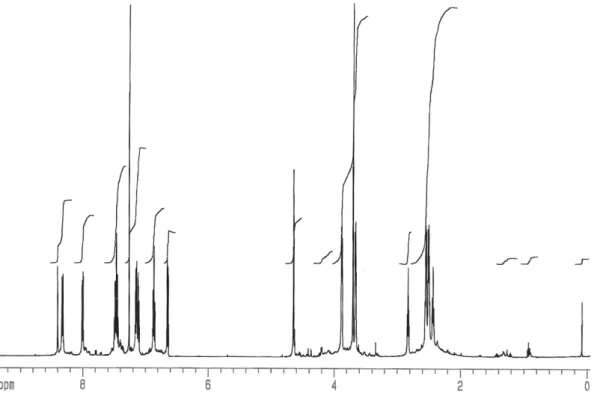
2.5.2 - Spectrophotometric and spectrofluorimetric measurements ... 44 2.5.3 - Chemical and starting materials ... 44 2.5.4 - Synthesis of macrocycle L ... 45
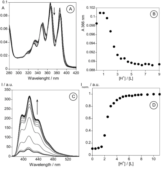
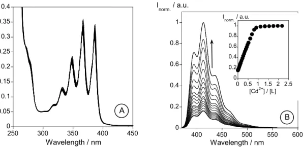
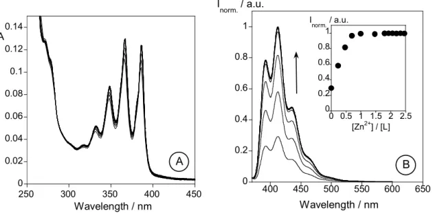
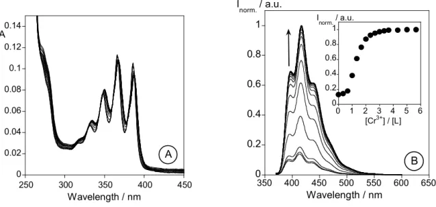
2.6 - Results and discussion... 46 2.6.1 - Synthesis and characterization of L ... 46 2.6.2 - NMR Spectra of L... 47 2.6.3 - Spectrophotometric and spectrofluorimetric studies... 47 2.6.3.1 - Studies in organic media... 47 2.6.4 - Metal Complexes... 49 2.6.4.1 - Li+, Na+, K+, Ca+ and Mg2+ titrations... 50 2.6.4.2 - Zn2+ and Cd2+ titrations... 50 2.6.4.3 - Cu2+ and Hg2+ titrations ... 52 2.6.4.4 - Trivalent metal ions: Al3+ and Cr3+... 52 2.6.5 - Studies in mixtures water-organic media ... 54
2.7 - Conclusion... 57
2.8 - Acknowledgements... 57
2.9 - Supporting Information... 58
2.10 - References... 62
Page
Chapter 3: Exploring the emissive properties of new azacrown compounds bearing aryl, furyl or thienyl moieties: a special case of Chelation
Enhancement of Fluorescence upon interaction with Ca2+, Cu2+ or Ni2+…...65 3.1 - Abstract... 69
3.2 - Resumo... 70
3.3 - Introduction... 71
3.4 - Experimental Section... 72 3.4.1 - Materials and Apparatus... 72 3.4.2 - Spectrofotometric and spectrofluorimetric measurements ... 73 3.4.3 - EPR Measurements. ... 73 3.4.4 - X-ray Crystal Structure Determinations ... 74 3.4.5 - Chemicals and starting materials... 75 3.4.6 - Synthesis of ligands ... 76 3.4.7 - Synthesis of solid complexes. General method ... 78
xix
3.6 - Crystallography data... 92
3.7 - Conclusions... 92
3.8 - Acknowledgment... 93
3.9 - Supporting Information Available... 93
3.10 - References... 94
Page
Chapter 4: Synthesis, Characterization and Metal Ion Detection of Novel
Fluoroionophores Based on Heterocyclic Substituted Alanine…………... 99
4.1 - Abstract... 103
4.2 - Resumo... 104
4.3 - Introduction... 105
4.4 - Results and Discussion... 106 4.4.1 - Synthesis ... 106 4.4.2 - Photophysical study ... 107 4.4.2.1 - Spectrofluorimetric titrations and metal sensing effect ... 108 4.4.2.2 - Protonation effects ... 108 4.4.2.3 - Deprotonation effects... 109 4.4.2.4 - Metal sensing effects ... 109
4.5 - Experimental Section... 118 4.5.1 - Synthesis general... 118 4.5.2 - Spectrofluorimetric titrations ... 123
4.6 - Acknowledgements... 124
4.7 - References... 124
Page
Chapter 5: Heteroaromatic alanine derivatives bearing (oligo)thiophene units:
synthesis and photophysical properties ...127
5.1 - Graphical Abstract... 131
5.2 - Abstract... 132
5.3 - Resumo... 133
5.4 - Introduction... 134
5.5 - Results and Discussion... 135 5.5.1 - Synthesis ... 135 5.5.2 - Photophysical study ... 138
xx
5.7 - Experimental Section... 140 5.7.1 - Synthesis general... 140
5.8 - Acknowledgments... 142
5.9 - References... 143
Page
Chapter 6: Synthesis, Characterization, Fluorescence and computational studies of new Cu2+,Ni2+ and Hg2+ complexes with emissive
oligothienylalanine ligands………..144
6.1 - Abstract... 149
6.2 - Resumo... 150
6.3. - Introduction... 151
6.4. - Results and Discussion... 152 6.4.1 - Synthesis and Characterization, Complexation studies ... 152 6.4.2 - Computational methods... 154 6.4.3 - Spectroscopyc Studies ... 157
6.5. - Experimental... 160 6.5.1 - Physical measurements ... 160 6.5.2 - Spectrophotometric and spectrofluorimetric measurements ... 160 6.5.3 - Computational Methods... 161 6.5.4 - Chemicals and starting materials... 161 6.5.5 - Synthesis of ligand L4 ... 161
6.5.6 - Synthesis of metal complexes - general procedure ... 162
6.6 - Acknowledgments... 164
6.7 - References... 164
Page
Chapter 7: Bio-inspired systems for Metal Ion Sensing: New Emissive Peptide Probes Based on Benzo[d]oxazole Derivatives and Their Gold and Silica
Nanoparticles………..………167
7.1 - Abstract... 171
7.2 - Resumo... 172
7.3 - Introduction... 173
xxi
7.4.2 - Synthesis of Gold nanoparticles ... 184 7.4.3 - Synthesis of the decorated silica nanoparticles with compounds L5 to L7. ... 185 7.4.4 - Photophysical Measurements ... 185 7.4.5 - MALDI-TOF-MS measurements ... 186 7.4.6 - Physical measurements ... 186 7.4.7 - Particles Size Distribution ... 187 7.4.8 - TEM measurements ... 187 7.4.9 - Chemicals and Starting Reagents ... 187
7.5 - Results and Discussion... 187 7.5.1 - Synthesis of peptides ... 187 7.5.2 - Photophysical Studies ... 188 7.5.3 - Spectrophotometric and spectrofluorimetric titrations and metal sensing effect ... 190 7.5.4 - MALDI-TOF-MS studies ... 194 7.5.5 - Gold nanoparticles and TEM measurements... 196 7.5.6 - Silica nanoparticles obtained by surface derivatization ... 198 7.5.7 - The use of core/shell water soluble silica nanoparticles ... 201
7.6 - Conclusions... 203
7.7 - Acknowledgements... 203
7.8 - References... 208
Page
xxiii
INDEX OF FIGURES
Figure 2.1- Absorption (full line), emission (broke line) and excitation (dotted line) spectra of
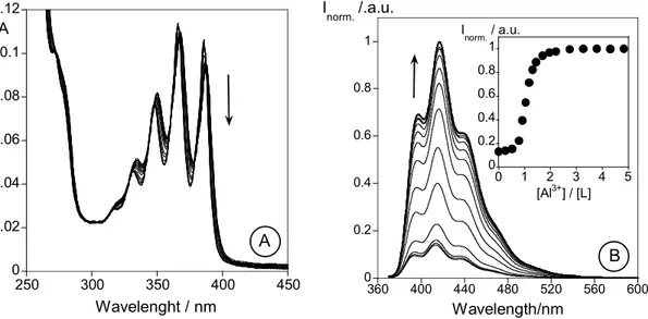
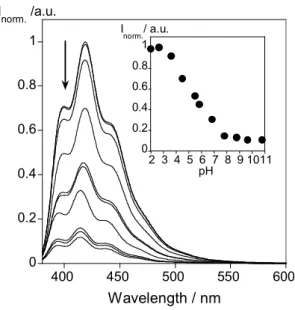
L ( exc = 367 nm; em= 413 nm, [L] =9.00 10-6 M) in methanol at room temperature...47 Figure 2.2.- Absorption (A) and emission (C) spectra of methanol solutions of L as a function of added HBF4. Figures (B) and (D) show (respectively) the absorption at 366 nm and the normalized fluorescence intensity at 416 nm. ([L] = 9.0 10-6 M, exc = 367 nm)...49 Figure 2.3.- Absorption (A) and emission (B) spectra of methanol solutions of L as a function of increasing amounts of Cd(NO3)2. The inset shows the normalized fluorescence intensity at 413 nm. ([L] = 1.35 10-5 M, exc = 367 nm)...50 Figure 2.4.- Absorption (A) and emission (B) spectra of methanol solutions of L as a function of increasing amounts of Zn(NO3)2. The inset shows the normalized fluorescence intensity at 413 nm. ([L] = 1.25 10-5 M, exc = 367 nm)...52 Figure 2.5.- Absorption (A) and emission (B) spectra of methanol solutions of L as a function of increasing amounts of Cr(NO3)3. The inset shows the normalized fluorescence intensity at 413 nm. ([L] = 1.35 10-5 M, exc = 367 nm)...53 Figure 2.6.- Absorption (A) and emission (B) spectra of methanol solutions of L as a function of increasing amounts of AlCl3. The inset shows the normalized fluorescence intensity at 413 nm. ([L] = 1.35 10-5 M, exc = 367 nm)...54 Figure 2.7.- Emission spectra of L in water/methanol solutions (50/50, v/v) as a function of pH. The inset shows the normalized fluorescence intensity at 419 nm. ([L] = 1.35 10-5 M,
xxiv
Figure 3.3.- Spectrofluorimetric titrations of compounds 1 (A), 2 (B) and 3 (C), in the presence of F-, in acetonitrile. The inset represents the emission for 1 (A) at 400 nm, for 2
(B) at 431 nm and for 3 (C) at 455 nm and 490 nm...83 Figure 3.4. – Absorption (A and C) and emission titrations (B and D) of compounds 2 and 3
with the addition of increased amount of Ca2+ in acetronitrile (2) and absolute ethanol (3) solution. The inset represents the absorption at 326 nm (A) and 325 nm (C), and the emission at 403 nm (B), and 430 nm (D) as a function of [Ca2+]/[2] or [Ca2+]/[3]. ([2] = 9.06 10-6 M, [3] = 6.34 10-6 M, [Ca(CF3COO)2] = 1.46 10-2 M, exc2 = 320 nm nm, exc3 = 325 nm, T=298K)...84 Figure 3.5 – Absorption (A and C) and emission titrations (B and D) of compounds 2 and 3
with the addition of increased amount of Ni2+ in acetronitrile. The inset represents the absorption at 320 nm, 350 nm (A) and 325 nm (C), and the emission at 423 nm (B), and 433 nm (D) as a function of [Ni2+]/[2] or [Ni2+]/[3]. ([2] = 9.06 10-6 M, [3] =8.55 10-6 M, [Ni(BF4)2] = 1.62 10-2 M, exc2 = 320 nm nm, exc3 = 325 nm, T=298K)...85 Figure 3.6 –Spectrofotometric (A) and spectrofluorimetric (B) titration of 3 in the presence of Hg2+, in an acetonitrile solution. The inset represents the absorption (A) at 325, 350 and 510 nm, and the emission (B) at 427 nm and 450 nm, as a function of [Hg2+]/[3]. ([3] = 6.66 10-6 M, [Hg (CF3COO) 2] = 1.7 10-2 M, exc3 = 325 nm, T=298K)...86 Figure 3.7 – Absorption (A, C and E) and emission titrations (B, D and F) of compounds 1
xxv
Figure 3.11.- X-Ray crystallographic structures of compound 3...92 Figure SI 3.1. - Job’s plot for compound 3 in the presence of Cu2+. ...93 Figure 4.1. - Spectrophotometric titration (A) and fluorimetric titration (B) of an ethanolic solution of 2b with a standard solution of Hg(CF3SO2)2 in absolute ethanol ([2b] = 1.30E-5 M, T = 298 K, λexc = 316 nm. Inset: normalized emission at 394 nm)...111 Figure 4.2. - Spectrophotometric titration and fluorimetric titration of an ethanolic solution of
2b with a standard solution of Cu(CF3SO2)2 in absolute ethanol. ([2b] = 1.30E-5 M, T = 298 K, λexc = 316 nm. Inset: normalized emission at 394 nm)...112 Figure 4.3. - Spectrophotometric titration (A) and fluorimetric titration (B) of an ethanolic solution of 2f with a standard solution of Hg(CF3SO2)2 in absolute ethanol. ([2f] = 1.00E-5 M, T = 298 K, λexc = 334 nm. Inset: normalized emission at 395 nm)...113 Figure 4.4.- Spectrophotometric titration and fluorimetric titration of an ethanolic solution of
2f with a standard solution of Cu(CF3SO2)2 (A) and Ni(BF4)2 (B) in absolute ethanol. ([2f] = 1.00E-5 M, T = 298 K, λexc = 334 nm. Insets: normalized emission at 396 nm in both cases).113 Figure 4.5.- Spectrophotometric titration (A) and fluorimetric titration (B) of an ethanolic solution of 2d with a standard solution of Hg(CF3SO2)2 in absolute ethanol. ([2d] = 1.00E-5 M, T = 298 K, λexc = 315 nm. Inset: normalized emission at 393 nm)...114 Figure 4.6.- Spectrophotometric titration and fluorimetric titration of an ethanolic solution of 2d with a standard solution of Cu(CF3SO2)2 (A) and Ni(BF4)2 (B) in absolute ethanol. ([2d] = 1.80E-5 M, T = 298 K, λexc = 315 nm. Insets: normalized emission at 394 nm in both cases).115 Figure 4.7. - Spectrophotometric titration and fluorimetric titration of an ethanolic solution of
2g with a standard solution of Cu(CF3SO2)2 (A) and Ni(BF4)2 (B) in absolute ethanol. ([2g] = 1.00E-5 M, T = 298 K, λexc = 336 nm. Inset: normalized emission at 397 and 398 nm, respectively)...116 Figure 5.1. - Normalized UV-visible absorption and emission spectra of compounds 4a,5a
and6a in absolute ethanol at T = 298 K (4a, λexc = 314 nm; 5a, λexc = 365 nm; 6a, λexc = 400 nm) (absorption, full line; emission, dotted line)...138 Figure 6.1. – Solid-state emission spectra of L1(A), L2(B) and L3(C) and its corresponding metal complexes with Cu2+, Ni2+ and Hg2+ ( excL1,L2 = 315 nm, excL3 = 366 nm, T=298K)...154 Figure 6.2 - DFT structures of ligand L2 in the presence of one and two equivalents of Hg(II)...156
xxvi
=1.02 10-2M, [Ni(BF4)2] =1.00 10-2M, T = 298 K, exc = 366 nm. (insets: normalized emission at 447 nm)...158
Figure 6.4. – Absorption, emission and excitation spectra of ligand L4 (T=298 K, [L4] = 1.40 10-6 M, exc = 374 nm) (A) and spectrophotometric (B) and spectrofluorimetric titration (C) of L4 with a standard solution of Hg(CF3SO3)2 in dichloromethane, ([L4] = 1.40×10⁻⁶ M, exc = 434 nm, the inset shows the normalized emission at 433 nm, and the fitting by Sigma Plot). Panel D shows the JOB plot for L4/Hg2+ interaction...159
Figure 7.1.- Absorption (bold line), normalized emission (full line) and excitation spectra (dotted line) of compound L3 (A) and L7 (B) in dichloromethane. On the right corner is represented the relative fluorescence quantum yield of L3 (A) and L7 (B) in dichloromethane...189 Figure 7.2.- Absorption and emission spectra of L2 in the presence of Cu2+ in absolute ethanol solution. The Inset shows the intensity of emission as a function of [Cu2+]/[L2] at 388 nm. (T=298K, [L2] = 3.22 10-6M, exc = 316 nm)...192 Figure 7.3. – Spectrofotometric (A) and spectrofluorimetric titration (B) of L3 in the presence of Hg2+ in abs. ethanol solution. The inset represents the absorption at 316 nm and 334 nm (A), and the emission (B) at 388 nm, as a function of [Hg2+]/[L3]. (T=298K, [L3] = 8.8 10-6M,
exc = 316 nm)...192 Figure 7.4 – Spectrophotometric (A) and spectrofluorimetric (B) titration of L7 in the presence of Hg2+ in absolute ethanol solution. The inset represents the absorption at 366 nm and 400 nm (A), and the emission (B) at 440 nm and 480 nm, as a function of [Hg2+]/[L7]. (T=298K, [L7] = 7.69 10-6M, [HgCF3COO)2]=3.76 10-3M, exc = 366 nm)...194 Figure 7.5. – (A) Absorption (full line) and emission (dotted line) spectra of L (black line) and its gold(0) nanoparticles (red line) in dichloromethane. (T=298K, [L] = [Lnanop.] 4.13
xxvii
Figure 7.8. – Water-soluble core-shell dye-doped silica nanoparticles decorated with L3 or
L5 in the presence of Ag+ and Hg2+...202 Figure S7.1. – Absorption and emission spectra of L1 in the presence of Hg2+ in abs. ethanol solution. The Inset represents the intensity of emission as a function of [Hg2+]/[L1] at 387 nm. (T=298K, [L1] = 1.26 10-5 M, exc = 316 nm)...204 Figure S7.2. – Absorption and emission spectra of L2 in the presence of Hg2+ in absolute ethanol solution. The inset shows the intensity of emission as a function of [Hg2+]/[L2] at 388 nm. (T=298K, [L2] = 3.22 10-6 M, exc = 316 nm)...205 Figure S7.3. – Absorption and emission spectra of L4 in the presence of Hg2+ in abs. ethanol solution. The inset represents the intensity of emission as a function of [Hg2+]/[L4] at 388.5 nm. (T=298K, [L4] = 1.22 10-5 M, exc = 316 nm). ...205 Figure S7.4. – Spectrophotometric and spectrofluorimetric titration in absolute ethanol of compound L7 with addition of an ethanolic solution of Ag(BF4). [L7]= 7.69 10-6 M, [Ag(BF4)] = 5.70 10-3 M, T=298 K, exc = 366 nm. The inset shows the intensity of emission as a function of [Ag+/[L7] at 440 and 460 nm...206 Figure S7.5. – Spectrofluorimetric titration of silica nanoparticles of compound L5 with the addition of Ag+ (A) and Hg2+ (B) in absolute ethanol. The inset represents the emission at 450 nm, as a function of [Ag+]/[L5] (A) and as a function of [Hg2+]/[L5]. ([L5] = 8.45 10-6 M,
xxix
INDEX OF SCHEMES
xxx
Scheme 1.17. – Schematic pathway of DDNs silica nanoparticles. A – Low concentration of fluorophore, (All molecules are totally encapsulated). B – High concentration of fluorophore, (Molecules are partially encapsulated, being necessary other silica shell to cover all the molecules). The fluorophores of outside shell interact with those present in the inside shell...25 Scheme 1.18.- Ligand L discussed in Chapter 2 and 15-crown-5-imidazo crown ethers derivates (1 to 3) discussed in Chapter 3...26 Scheme 1.19.- Alanine derivatives (2a-g) discussed in Chapter 4, and (Oligo)thienylbenzoxazolyl-alanine derivatives 4a-c to 6a-c discussed in Chapter 5...27 Scheme 1.20.- Alanine derivatives discussed in Chapter 6...27 Scheme 1.21.- Sensors discussed in Chapter 7...28 Scheme 2.1 - Several scorpionate ligands containing anthracene which have been successfully used for metal ion chelation...43 Scheme 2.2 - Synthetic pathways of ligand L...46 Scheme 2.3. - Schematic representation of the photoinduced electron and energy transfer mechanism observed in system L upon complexation with Al3+, Cr3+, Zn2+, Cd2+, Hg2+ and Cu2+...56 Scheme 3.1.- Synthesis of 2,4,5-Tri(hetero)aryl-imidazo-crown Ether Ligands 1-3...79 Scheme 4.1. - Synthesis of tyrosine derivatives 1a-e and alanine derivatives 2a-g.
Reagents and conditions: a) (Boc)2O, NaOH 1 M aq solution, rt, 2 days; b) 1,4-cyclohexadiene, Pd/C, MeOH, reflux, 24h; c) 2-formylthiophene, EtOH, rt, 5 days; d) 2,4,5-trimethoxybenzaldehyde, EtOH, rt, 3 days; e) 1d or 1e, LTA, DMSO, rt, 3 days; f) NaOH 1 M
aq solution, dioxane, rt, 3h; g) trifluoacetic acid/dichloromethane, 1:1, rt, 2h...106 Scheme 4.2. - Schematic representation of the complexation mechanism proposed for alanine 2d upon complexation with Cu2+, Ni2+ and Hg2+. Fluorescence spectra of 2d in the presence of one and two equivalents of Cu2+ in absolute ethanol...117 Scheme 5.1. - Synthesis of fully protected (oligo)thienylbenzoxazolyl-alanine derivatives 4a to 6a...136 Scheme 5.2. - Synthesis of N- and C-terminal deprotected (oligo)thienylbenzoxazolyl-alanine derivatives 4a-c to 6a-c...137 Scheme 6.1 – Structure of benzoxazolyl-alanine derivatives studied...152
xxxi
xxxiii
INDEX OF TABLES
Table 2.1. Quantum yields in methanol at 298 K ...51 Table SI2.1. 1H NMR data at 500 MHz of L in CDCl3...59 Table 3.1.- Crystal data and structure refinement for ligand 2 and 3...75 Table 3.2. – Optical data of compounds 1 to 3 in protic and aprotic solvents...81 Table 3.3.- Stability Constants with Compounds 1-3 by Hypsec Program...83 Table 3.4 – Luminescence quantum yield of compound 3 in the presence of Ca2+ and Cu2+...90 Table 4.1. - Synthesis data of tyrosine derivatives 1d-e and alanine derivatives 2a-g...107 Table 4.2. - UV-vis and fluorescence data for alanine derivatives 2a-g...108 Table 4.3. - Complexation constants for alanine derivatives 2b-d and 2f-g with Cu2+, Ni2+ and Hg2+ in absolute ethanol...110 Table 5.1. - Yields, UV-visible absorption and emission data for (oligo)thienylbenzoxazolyl-alanines 4-6 in absolute ethanol...137 Table 6.1. - DFT (CAM-B3LYP/def2-svp (PCM, ethanol)) electronic and free energies in kcal/mol for the mercury-L2 complexes studied. The relative values have been calculated with respect to Hg(H2O)22+, the free ligand L2, and H2O...156 Table 6.2.- Complexation constants for benzoxazolyl-alanine ligands L1 to L4 with Cu2+, Ni2+ and Hg2+ in absolute ethanol calculated with Hypspec program. (logK)...158
Chapter 1
Introduction
“If I have seen further it is by standing on the shoulders of giants”
3
Index
1.1 - Supramolecular Chemistry: Fluorescent Chemosensors... 5
1.2 - Mechanisms of detection... 7
1.3 - Chemosensors based on macrocycle detection receptors.... 10
1.4 - Chemosensors containing bio-inspired units. From single amino acids to peptide chains as receptors.... 12
1.5 - Some aspects on the biological applications on emissive fluorophores... 16
1.6 - The importance of metal ions. Recognition and Quantification.... 18
1.7 - On nanoparticle science. Gold or Silica nanoparticles.... 20 1.7.1 - Gold nanoparticles ... 20 1.7.2 - Silica nanoparticles ... 23
1.8 - Aim of thesis………..………25
5
1.1 - Supramolecular Chemistry: Fluorescent Chemosensors.
In the middle of the 19th century G. Stokes reported that the fluorescence emission spectrum appears at a longer wavelength than the excitation spectra1, and with this physical observation known as “Stokes shift” started the key conceptual basis for the fluorimetric analysis.
The development of new chemosensors changed dramatically since the pioneering work of Czarnik2, and these modifications altered substantially the analytical applications in chemical analysis.3
One of the first papers reporting the uses of luminescence as an analytical tool appears later in 1923. Harvey reported in Science the minimum of concentration required of the molecule luciferin, 1, a light-emitting biological dye found in some organisms capable of bioluminescence, to detect visible light.4 Later Sousa and Larson described the concept of a fluorescence chemosensor using some functionalized naphthalene azacrown-ether ligands,
2, for alkaline metal ion detection (Scheme 1.1).5
Scheme 1.1 – Structure of ligands 1 and 2.
Since that, a fluorescent chemosensor can be defined as “a compound of abiotic origin that
complexes to an analyte reversibly with a concomitant fluorescent signal transduction”.6,7
This classical definition cannot be confused with the more extended definition given by the
IUPAC for a chemical sensor, “as a device that transform chemical information, ranging from
the concentration of a specific sampler to total composition analysis into analytically useful
signals”8
As was reported by A.P. de Silva and co-workers, a classical fluorescent chemosensor is
constituted by three basic units: i) a receptor (responsible for molecule recognition), ii) a
fluorophore (responsible for signaling the recognition) and iii) a spacer (a chemical brigde
that links the receptor and the fluorophore controlling their separation and geometric N
S
N S
OH
O HO
N
N
H n=1 to 4
6
arrangement). 9,10 Sometimes, one part of the molecule can act by performing two or more
aforementioned functions. From a structurally point of view fluorescent chemosensors can be
classified into two general classes: intrinsic11chemosensors in which both functions (binding
and signaling) are performed by the fluorophore (A; Scheme 1.2) and conjugated
chemosensors in which binding/recognizing and signaling parts are separated by a spacer
(FSR) (B; Scheme 1.2).
In all cases mentioned, a good fluorescent chemosensor must have a strong affinity and
selectivity for the analyte, be photostable, and the environmental interferences should not
disturb in the fluorescence signal.12 The recognition can be occurs in four different ways: i) a
chelation enhancement of the fluorescence emission (CHEF), ii) a chelation enhancement of
the quenching emission (CHEQ), iii) formation of an exciplex or excimer probes (C; Scheme
1.2), and iv) as a chemodosimeters (D; Scheme 1.2).13,14 The three first mechanism are
reversible and the last one is irreversible.14
Scheme 1.2 – Schematic representation of the different approaches between the
chemosensor and the analyte. (A) – Intrinsic Fluorescence probes, (B) –
Fluorophore-spacer-receptor systems (Conjugated), (C) - exciplex or excimer probes, (D) -
Chemodosimeters14
Fluorophore = Receptor
Analyte
Analyte
A
Spacer
Fluorophore Receptor
Analyte
Spacer
Fluorophore Receptor
Analyte
Analyte
B
Spacer Spacer Spacer
Fluorophore Receptor Fluorophore
Analyte
C
Sp acer
Spacer
Spa ce
r Analyte
Fluorophore weak emissive
Analyte
7
1.2 - Mechanisms of detection
Fluorescence is an important analytical tool to sense relevant species, for example metal ions and anions. This application can be useful for analytical purposes in vitro and in vivo
studies.3
It is important to mention that different processes can occur: ones involving intramolecular and others intermolecular processes.
In an excited molecule radiative and non-radiative transition between electronic states can occur, for example, photon absorption, fluorescence, internal conversion, intersystem crossing, phosphorescence, delay fluorescence and triplet-triplet transitions (see Scheme 1.3).
Concerning the interaction with other molecules several photophysical processes can happen as for example, electron transfer, proton transfer, energy transfer, excimer or exciplex formation.15
In more detail, fluorescence is a radiative transition where an emission of a photon from the excited state to the ground state ocurrs. Based on Stoke´s rule mentioned below and the energy loss in the excited state due to the vibrational relaxation, the wavelength of the emission is always located at higher values.15
An internal conversion is a non-radiative transition that occurs between two electronic states with the same spin multiplicity, being described as a vibrational relaxation from the lowest vibrational level to the final electronic state.
8
Delay fluorescence is contrary to the intersystem crossing, when the triplet state T1 has a long lifetime, and the energy difference between S1 and T1 is small, the transition from T1 to S1 can occur. A rise in temperature increases the probability of the process occurence.15 Triplet-triplet transitions occur when a molecule in the excited state absorb another photon at different wavelength.15
In Scheme 1.3 are represented schematically the processes mentioned above.
Scheme 1.3 – Photophysical processes, radiative and non-radiative transitions between electronic states of a molecule in the excited state in solution.15
There are many processes that can quench the fluorescence emission, like, double-bond torsion, low energy n * levels, heavy metals, weak bonds, photoinduced electron transfer (PET) or electronic energy transfer (EET).9
When the fluorophore and the receptor (FSR) are electronically decoupled, any signaling mechanism has to occur by electronic transfer (ET) mechanisms.
A PET process happens when the receptor has nitrogen or other atoms, with a free lone pair of electrons, which can quench the luminescence by photoinduced transfer processes. On the other hand, this mechanims can be prevented by complexation or protonation of the lone pair located in the donor atoms.
S
0S
1T
1T
2Intersystem crossing
delayed fluorescence
fl
u
o
re
sce
n
ce
in
te
rn
a
l
conve
rsi
on
triplet-triplet absorption
p
h
o
s
phor
e
9
In more details, in the PET-FSR, the HOMO of the receptor is energetically located between the HOMO and LUMO of the fluorophore. So, when promotion of an electron occurs from the HOMO to the LUMO of the fluorophore, happens a fast electron transfer, eT, from the receptor’s HOMO to the HOMO of the fluorophore, promoting thus a quenching in the emission fluorescence.14
However, when the lone electron pair is engaged, the HOMO energy decreases, and the eT from the HOMO receptor to the HOMO fluorophore is switched off, resulting an emissive fluorescence (see Scheme 1.4). 14,14
Scheme 1.4 – Schematic representation of an excited-state photoinduced electron transfer (PET) process and its inhibition. 14,15
Finally the formation of excimers and exciplexes can occur when two or more fluorophores with a long lifetime, for example, pyrene and naphthalene, are present.16,17,18
The intramolecular/intermolecular excimer is formed in the excited state, by collision of an excited molecule/fluorophore that interacts with other identical molecule/fluorophore in the ground state.15 The fluorescence band corresponding to an excimer is located at highest wavelengths than the monomer and normally is none vibrationally resolved. The monomer/excimer relationship is highly temperature dependent, with higher temperatures increasing the monomer formation.15
The exciplex formation occurs by the interaction of one molecule in the excited state with other different molecule or lone pair of electrons in the ground state, forming an excited complex. 15 The exciplex band changes with the solvent polarityas solvent polarity increases the band is red-shifted.
HOMO LUMO
HOMO
Excited Fluorophore
HOMO HOMO
LUMO
Excited Fluorophore
Free Receptor Free Receptor
light
ene
rg
y
PET process Inhibition of PET process
en
er
10
On the other hand, colorimetric molecular devices have attracted much scientific attention due to the so called “naked-eye” detection. This strategy reduces the use of expensive and complicated equipment to metal-ion measurements.3
1.3 - Chemosensors based on macrocycle detection receptors.
Cyclic polydentate ligands are defined as macrocycles if they feature nine or more atoms (including heteroatoms) and at least three donor atoms.19 As an example, three representative macrocyclic systems, a crown-ether (3), a polyamine ligand (4) and a poly thia-aza system (5) are shown in scheme 1.5.
As the definition of macrocycle is very broad, the number of possible systems that can be designed is unlimited. For example just by changing the donor atoms the synthetic possibilities are infinites. The donor atoms can be oxygen, nitrogen, phosphorous, sulphur, selenium, or arsenium. Depending of the homo or hetero type of atoms used crown-ethers (O), polyamine (N)20,21,22, polythioethers (SO)23,24, polythia-aza (SN)25 can be designed.
Scheme 1.5 – Macrocyclic systems, 3 – crown ether macrocycle, 4 - Polyamine macrocycle,
5 - Polythiaza macrocycle
Apart from the type of donor atoms other parameters such as its cavity size, shape, conformation, topology and rigidity are important because they influence the thermodynamic and kinetics properties of the corresponding metal complexes.19
The uses of macrocyclic ligands are important in chemistry due to the higher thermodynamic and kinectic stability shown in comparison with the acyclic ligands, called the chelate and macrocycle effects.26,27
Polyamine macrocycle receptors provides versatility to the chemosensor applications, since its properties can be modulated in huge extension by the pH, the interaction with metal ions,
N N
S S
S O
O O O
O
O
HN NH
HN
HN HN HN
5
4
11
anions etc. For example, the protonation of the polyamine unit at acidic pH values renders an anion sensor while at basic pH the unprotonated forms are ideal to form stable metal complexes modulating the properties to sense protons and/or metal ions.
In systems containing of nitrogen donor-atoms, the electron transfer process from the lone pair of the aliphatic amino groups to the excited luminophore occurs when the lone pair is free, and this process is blocked after protonation or complexation. This phenomenon was observed in organic solvents and in water.
When in the skeleton of the receptor unit an aromatic heterocycle is included, such as a pyridine, bipyridine, terpyridine, antracene, naphthalene or phenanthroline unit, for example as in compound 6, (see Scheme 1.6), their protonation leads to a photoinduced electron transfer from the - * excited state of the hydrocarbon fluorophore to the protonated heterocycle. In some special cases, when a hydrocarbon fluorophore (anthracene, naphthalene, etc) is connected to a nitrogen containing aromatic heterocycles through a polyamine chain, both quenching effects can occur, defining a pH window where the fluorescence emission appears.28
Scheme 1.6 – Structure of compound 6.
Also, the use of crown ether as metal ion chemosensors, artificial membranes29, smart material30, actives components of molecular machines31 and biomedical application32 as potencial antitumor agents was reported by several authors.33,34.
Replacement of the oxygen by nitrogen atoms, can changes their properties in metal sensing. For example, the aza-crowns (7 and 8)presented at Scheme 1.7 , were immobilized on an amino cellulose fibre substrate, for the detection of Na+, K+ levels in blood.35,36
NH N HN
12
Scheme 1.7 – Structures of compounds 7 and 8.
15-Crown-5 systems are usually used for the interaction with Na+,37 whereas 15-crown-5 monoazacrown ethers show better results for Ca2+.38 In this case the size of the metal ion influenced notably the answer of the chemosensor.
In general, the addition of metal ions or protonation of the aza-crown ethers, “switches ON” the fluorescence emission, due the protection of the lone pair of electrons present in the nitrogen, preventing thus the PET process.
1.4 - Chemosensors containing bio-inspired units. From single amino acids to
peptide chains as receptors.
A bio-inspired sensor has a similar structure of a classical chemosensor, but in this case, the receptor is formed by an amino acid (natural or synthetic) or by a peptide chain.3
The insertion of amino acids in the backbone of synthetic polymers can lead us to macromolecules containing biomimetic characteristics, with a specific structure and biological properties.
Their properties, as luminescence, conducting ability, higher thermal stability and metal ions or other analyte recognition can be modified with synthetic manipulation at the amino acids side chain.
Amino acids and peptides contain sites available for metal binding and recognition, making them good biosensors for metal detection in solution and in solid state. 39
13
An amino acid is formed by amine, and carboxylic terminals and a R-group at the side chain. Depending of the R-group, peptides can adopt different conformations, where a peptide can interact with other peptide by covalent linkage, and also by non-covalent linkage, via ionic, hydrophobic, hydrogen bonding and - stacking interactions. The 20 amino acids, due to different characteristics of the R-groups, can be divided in hydrophobic, hydrophilic, charged or “other” (see Scheme 1.8).
H2N OH
O H2N
OH O
H2N OH O
H2N OH O
Alanine Leucine Valine Isoleucine
H2N OH O
S
Methionine
H2N OH O
H2N OH O
N H
H2N OH O
OH H2N
OH
O
N NH
Phenylalanine Tryptophan Tyrosine Histidine
H2N
OH O N H NH NH2 Arginine
H2N OH O
NH2
Lysine
H2N OH O
HO O
H2N
OH O
O OH
Aspartic acid Glutamic acid
H2N OH O
OH
H2N OH O
O NH2
H2N OH O
OH
H2N OH O
O NH
2
H2N OH O SH
H2N OH O OH O H N
Serine Asparagine Threonine Glutamine Cysteine Glycine Proline = Hydrophobic =Negatively charged = Positively charged = Hydrophilic = Others
Scheme 1.8.- Amino acids structures.
In the hydrophobic aminoacids are included the aliphatic alanine, isoleucine, leucine, methionine and valine, the aromatic aminoacids are phenylalanine, tryptophan and tyrosine.
Aromatic residues can be very important in the interaction with proteins and peptides folding due to their - stacking characteristics. The hydrophilic residues are formed by serine, threonine, asparagine, and glutamine, and they can be involved in the hydrogen bonding interactions.
14
Taking into account, the peptide skeleton presented in this PhD dissertation, we are going to focus only on the properties of alanine, tryptophane and cysteine.
Alanine is a non-essencial amino acids, which means that the human body can produce it, being not necessary its inclusion through diet. It is present in food, particularly in meat, and Grosser et al. described their important anti-oxidant properties. 41
Cysteine represents a unique chemical reactive, where due to the properties of its side chain, can be used as target for chemical modifications and interpeptide crosslinking. The presence of a sulfur atoms, gives it the ability for linkage at gold surfaces. 40
Tryptophan is an essential amino acid in the human diet. This L- form amino acid is used in structural or enzyme proteins. The distinguishing structural characteristic of tryptophan is that it contains an indole functional group. It is an essential amino acid as defined by its growth effects on rats.42
Tryptophan can be found in health food stores, as a dietary supplement; and shows some effectiveness for the treatment of a variety of conditions associated with low serotonin levels in the brain, being a considerable promise as an antidepressant.43,44,45
The oxygen, nitrogen and sulfur atoms present in the alanine, tryptophan and cysteine amino acids, makes these compounds good candidates as molecular probes for metal detection. Many publications containing alanine derivatives for metal detection have been previously described. 46
The synthesis of metal complexes containing amino acids and transition-metal ions are knowing a huge development due to their importance in biology, pharmacy and industry.47 Properties, such as antibacterial, antitumour, and anticancer activities has made them the target of many studies.48
In biological processes, there are many classes of enzymes with a metal ion as cofactor, and with the purpose of understanding these biological process, as reactivity, the interaction of metal complexes containing alanine amino acids has been tested49.
Transition metals could also be used in the future as cleavage reagent for peptides and proteins in Peptide Mass Mapping (Proteomics Approach). 50
15
Scheme 1.9. – Cleavage mechanism of palladium (II) complex proposed by Anbalagan51. Concerning other metal ions studied with these bio-inspired probes, copper(II) can be found in some important biological process, such as oxidation transport and electron transfer, and also complexed with proteins containing hydrophobic sides52. For example Marino et al.,
have published some interesting theoretical studies of the interaction of metal ions as Cu+ and Cu2+ with alanine amino acids, where Cu2+ ions, are preferentially bound to the C-terminal carboxylate group of alanine amino acids53.
Mercury (II) as a soft metal ion has a strong affinity for sulfur-donor ligands, so the use of cysteine derivatives is very important for Hg2+ removal from waste waters. The complexation of mercury (II) with inorganic or natural organic ligands can change the speciation of Hg2+, such as, its transport, transformation and bioavailability in natural water. There are many papers published on Hg2+, demonstrating that it is normally linked to acid units, like, carboxylic acids, phenols, alcohols and thiols. 54,55,56
Scheme 1.10.- MeHg complexes with seleno amino acids.55
H3C N
H O
O H N
N- N
O
N H
O OH
(Alanine) Pd2+
H3C N H O O N OH NH N N -O OH
Pd2+(Alanine)
O N
O N
N-Pd2+ N
H H3C
O
(Alanine)
H2N O OH HO O NH2 Se Methylmercury-l-selenocysteinate HO O NH2 SeHgCH3 Methylmercury-d,l--selenopenicillaminate HO O
NH3+ Se+
CH3 HgCH3
HgCH3
Methylmercury-l-selenomethionate (via Hg-Se bonding, low pH)
O O
NH2HgCH3 Se
CH3
Methylmercury-l-selenomethionate (via Hg-N bonding, high pH)
16
Finally, Belcastro et al. published the interaction of cysteine with Zn2+, Cd2+ and Hg2+, soft atoms coordination57, and through theoretical studies they could conclude that these metals had different coordination sites in the amino acids. Zn2+ and Cd2+ link preferably to carbonyl oxygen, nitrogen and sulfur atoms, whereas, Hg2+ links at the sulphur atom of one the zwitterions forms of the amino acid residue.58
Scheme 1.11.- Theoretical structures of the complexes of cysteine with M2+ (= Cu2+, Zn2+, Cd2+ and Hg2+). Depending on the metal ions, the distance in the molecule:metal ions changes.58
1.5
-
Some aspects on the biological applications of emissive fluorophores
Peptides and proteins do not have fluorescent properties strong enough to be useful as intrinsic fluorescence chemosensor for sensing in the environment. So, their conjugation with emissive fluorophores can enhance their properties, for sensing and developing fluorescence peptide sensors.
Joshi et al. synthesized a fluorescent peptide containing a dansyl group as a fluorophore, with an amino acid sequence, and studied their interaction with metal ions. They showed that the peptide probe successfully exhibited a turn on and a ratiometric response for several metal ions, as Cd2+, Pb2+, Zn2+ and Ag+. 59 (See Figure 1.12.)
1 2 3 4 5
17 N S O O H
N Ala AlaCys Ala Ala His Cys Trp AlaGluNH2
SH HS
N H
Cys
Ala Ala His Cys
SH HS
Ala
Trp
Ala Glu NH2
S O O N HN Hg2+ Hg2+ hv (CHEF)
Scheme 1.12.- Possible mechanism for binding of Hg2+ metal ions in a fluorescent peptide containing dansyl as a fluorophore.59
Fluorescent molecular sensors based on conjugated aminoacids for in vivo applications, have to be soluble in aqueous media, penetrate in cell membranes, exhibit fluorescent changes with the pH variation and also for metal detection, the excitation wavelength must be located in the visible or near infrared, because UV radiation can damage cells and tissues.60
However, the majority of sensors developed until now have short-wavelength fluorophores, such as, coumarin, benzoxazol, anthracene or pyrene, which are suitable for abiotic analysis, but for in vivo applications they do not have optimal spectral conditions.
To improve the point, common fluorophores used for biological applications are fluorescein or rhodamine. They have long excitation wavelength (ca. 500 nm), high fluorescence quantum yields and extinction coefficients.61,62
Other fluorophores, such as, BODIPY dyes, squaraines and cyanine are organic fluorescent emitting between 500 nm and at ca. 900 nm. Compounds containing Nile Red, Nile Blue are interesting for biological sensors, they are more photochemical stable than cyanine, but due to their structure, their solubility in aqueous solution is very limited. 63
In our case, due to its biological properties, 2-Benzoxazol derivatives have been the target of many research and developments. They have high lipophilicity, a broad biological activity and they can act as antifungal, antimicrobial and anticancer agents. They also present good optical properties, as high molar coefficients and fluorescent quantum yields, and can be applied as fluorescent /colorimetric probes for metal or anionic detection.39,64
18
their stability (oligo)thiophenes could be used for cellular imaging and fluorescence resonance energy transfer experiments.63
1.6 - The importance of metal ions. Recognition and Quantification.
As is well known, metal ions are present in nature, being essential to plants and animal life.60 They can be grouped in different families, depending of their properties. In this dissertation we will discuss the following families, the alkaline, alkaline-earth and transition metal ions.
Alkaline Na+, K+ and alkaline earth, Mg2+, Ca2+ are present in large quantities in the human body, contrary to the transition metal ions Cu2+, Ni2+ and Pb2+; and post-transition metals Zn2+, Cd2+ and Hg2+, (d-block elements) that are present in small quantities.3
Pearson’s theory of hard and soft acids and bases states that “Hard Acids prefer to bind with hard bases, and soft acids to soft bases”.57 In this way, a compound containing oxygen atoms is considered hard; sulfur is soft and nitrogen is an intermediate, because it can link to soft or hard metal ions.
Alkaline, Li+, Na+, K+, and alkaline earth, Mg2+, Ca2+, Sr2+ and Ba2+; Cu2+, Ni2+, Zn2+, Pb2+ under Pearson theory are considered hard metal ions; whereas Ag+, Cu+, Cd2+, Hg2+ are considered soft metal ions.57
Metal ions have a very important role, such as, the stabilization and reactivity of proteins. However, for human and environmental welfare, they must exist in optimal quantities. Contrary, they can promote metabolic disorders, being easily absorbed and accumulated from the environment causing toxicity and diseases.60
Zinc (II) is the second most abundant metal ion in the human body, is an important structural or catalytic cofactor of many proteins (for example carbonic anhydrase and zinc finger proteins) and can chelate on brain, pancreas and spermatozoa.65
Copper (II) is the third in abundance in human bodies, is highly toxic for some bacteria and viruses, and is suspect of causing infant liver damage. The alteration of its level in cellular environmental can causes neurodegenerative diseases such as Alzheimer’s disease.66
![Figure 2.1- Absorption (full line), emission (broke line) and excitation (dotted line) spectra of L ( exc = 367 nm; em = 413 nm, [L] =9.00 10 -6 M) in methanol at room temperature](https://thumb-eu.123doks.com/thumbv2/123dok_br/16483474.732623/87.892.284.599.707.1021/figure-absorption-emission-excitation-dotted-spectra-methanol-temperature.webp)

![Figure SI2.3. Relative fluorescence intensity at 413 nm of L in methanol to 1 equiv. of metal ions ([L] = 1.25 10 -5 M, exc = 367 nm)](https://thumb-eu.123doks.com/thumbv2/123dok_br/16483474.732623/101.892.259.675.232.532/figure-relative-fluorescence-intensity-methanol-equiv-metal-ions.webp)
