João Pedro Fernandes Andrade
Licenciatura Biologia
Pleistocene biostratigraphic markers of
the North Pacific, IODP Site U1340:
Proboscia barboi
,
Proboscia
curvirostris
and
Thalassiosira jouseae
Dissertação para obtenção do Grau de Mestre em Paleontologia
i
João Pedro Fernandes Andrade
Licenciatura Biologia
Pleistocene biostratigraphic markers of
the North Pacific, IODP Site U1340:
Proboscia barboi
,
Proboscia
curvirostris
and
Thalassiosira jouseae
Dissertação para obtenção do Grau de Mestre em Paleontologia
Orientadora: Zuzanna Stroynowski, Investigadora, IPMA
Co-orientador: Paulo Legoinha, Professor auxiliar, FCT
Júri:
Presidente: Fernando Reboredo Arguente: Fátima Abrantes Vogal: Zuzanna Stroynowski
iii
v
Foreword and acknowledgements
The original aim of this thesis was to study Miocene diatoms from the Belverde Borehole (Península de Setúbal) in order to: 1) establish a diatom-based biostratigraphy and complement the well-established biostratigraphy based on foraminifera and nannoplankton (Legoinha and Flores, 2014), and 2) characterize diatom paleoecological associations and thereby better understand the paleogeographic and paleoenvironmental evolution of the Lower Tagus Basin and complement previous studies based on other microfossil groups. Unfortunately, the Belverde Borehole proved to be barren of any siliceous microfossils and only one outcrop sample yielded diatom frustules. Therefore the subject had to be reformulated, having my advisor Dr. Zuzia Stroynowski conveniently suggested the study of Plio-Pleistocene diatom species at IODP Site U1340 in the Bering Sea. The study of the Belverde Borehole diatoms is presented in the Annex.
I should express my gratitude to my advisor Dr. Zuzia Stroynowski, for the help and counseling throughout the process of accomplishing this dissertation, my co-advisor Dr. Paulo Legoinha for his assistance and accessibility, Dr. Fátima Abrantes, who gave me the opportunity to work on my master’s degree dissertation at the Instituto Português do Mar e da Atmosfera (IPMA) by indication of Dr. Paulo Legoinha, and Dr. Brian Ottway for helping identify the diatoms from the Belverde Borehole study. I am also thankful to my colleagues at IPMA who were always willing to help and support me.
vii
Abstract
Diatoms are a large and widespread group of phytoplankton with an important role in ecosystems as primary producers. They are of great use in biostratigraphic and paleoclimatic studies, namely in the Bering Sea, where they are abundantly preserved in sediments. Proboscia barboi, Proboscia curvirostris and Thalassiosira jouseae are Plio-Pleistocene centric diatoms of mid to high latitudes of the North Pacific and North Atlantic Oceans, and are important biostratigraphic markers and datums in these regions. In this study, the biostratigraphy of these species at IODP Site U1340 (Bowers Ridge, Bering Sea) is refined and their abundance record interpreted in light of the paleoclimatic context of the North Pacific during the Plio-Pleistocene, using environmental information from the diatom assemblage in order to better understand the ecology of these extinct species. On a morphological approach, T. jouseae and its close related species Thalassiosira nidulus are described based on specimens of Site U1340 and their differences discussed. In addition, evidence for the evolutionary link between P. barboi and P. curvirostris is provided and discussed.
Keywords: diatoms, Bering Sea, Site U1340, Proboscia curvirostris, Proboscia barboi,
ix
Resumo
As diatomáceas são um grande grupo de fitoplâncton com uma distribuição global, que desempenha um papel importante nos ecossistemas como produtores primários. Têm uma aplicação fundamental em estudos de biostratigrafia e paleoclimatologia, nomeadamente no Mar de Bering, onde se encontram abundantemente preservados nos sedimentos. Proboscia barboi, Proboscia curvirostris e Thalassiosira jouseae são diatomáceas cêntricas de latitudes
média-altas do Pacífico Norte e Atlântico Norte, e importantes marcadores e datums biostratigráficos nestas regiões. Neste estudo, a biostratigrafia destas espécies na sondagem do IODP U1340A (Bowers Ridge, Mar de Bering) é refinada e o seu registo de abundância interpretado no contexto paleoclimático do Pacífico Norte durante o Plio-Plistocénico e recorrendo a espécies de diatomáceas indicadoras de ambiente, de maneira a estudar sua ecologia. De uma perspectiva morfológica, T. jouseae e uma espécie taxonomicamente próxima
Thalassiosira nidulus, são descritas com base em exemplares da sondagem U1340A e as suas diferenças discutidas. Adicionalmente, a relação evolutiva entre P. barboi e P. curvirostris é evidenciada.
Termos chave: diatomáceas, Mar de Bering, Sondagem U1340A, Proboscia curvirostris,
xi
List of Contents
Foreword and acknowledgements ... v
Abstract ... vii
Resumo ... ix
List of Figures ... xiii
List of Tables ... xv
Acronyms ... xvii
1 Introduction ... 1
1.1 Diatoms ... 1
1.2 The frustule ... 2
1.3 Reproduction ... 3
1.4 Resting spores ... 4
1.5 Application in biostratigraphy and paleoclimatology ... 4
2 Regional Setting ... 9
2.1 Bering Sea Oceanography ... 9
2.2 Surface Water Currents ... 10
2.3 Ecosystem productivity ... 11
2.4 Site U1340 ... 11
3 Fossil diatoms of the Bering Sea... 13
3.1 Proboscia ... 13
3.1.1 Introduction... 13
3.1.2 Morphology of Proboscia ... 13
3.1.3 Ecology ... 15
3.1.4 Proboscia barboi and Proboscia curvirostris: phylogenetic relation ... 16
3.2 Proboscia curvirostris (Jousé) Jordan and Priddle, 1991 ... 16
3.2.1 Geographic distribution and stratigraphic range ... 16
3.2.2 Description ... 17
3.3 Proboscia barboi (Brun) Jordan and Priddle, 1991 ... 18
3.3.1 Geographic distribution and biostratigraphy ... 18
3.3.2 Description ... 19
3.4 Thalassiosira ... 20
3.4.1 Introduction... 20
3.4.2 Thalassiosira jouseae Akiba, 1986 ... 21
xii
4.1 Samples ... 25
4.2 Method ... 26
4.3 Proboscia curvirostris and Proboscia barboi ... 28
4.3.1 Identification ... 28
4.3.2 Measurements ... 28
4.4 Thalassiosira jouseae ... 29
4.5 Paleoecology – used counts and data ... 29
4.6 Environmental proxies ... 30
5 Results and Discussion... 31
5.1 Biostratigraphy ... 31
5.2 Paleoecology ... 32
5.2.1 Proboscia barboi ... 32
5.2.2 Proboscia curvirostris ... 34
5.2.3 Thalassiosira jouseae ... 37
5.3 Morphology ... 41
5.3.1 Intermediate specimens of Proboscia ... 41
5.3.2 Description of Thalassiosira jouseae and Thalassiosira nidulus ... 42
6 Conclusions ... 45
7 References ... 46
8 Plates ... 51
xiii
List of Figures
Figure 1.1 - Gross morphology of the frustule in cross section view. (Hasle et al., 1996). ... 3 Figure 1.2 - Correlation between high latitude North Pacific diatom zonations. Dotted lines represent indirect correlation between magnetostratigraphy and diatom biohorizons. Adapted from Yanagisawa and Akiba (1998). ... 6 Figure 1.3 - Correlation of the diatom zonation of Schrader (1973) with Barron (1980) and Koizumi (1975b). Adapted from Barron (1980). ... 7 Figure 1.4 - Location of the main Drilling Sites in the North Pacific. Red – IODP Exp. 323; Black – DSDP Leg 19 (Koizumi, 1973); Blue – ODP Leg 145 (Barron and Gladenkov, 1995); Green – Akiba, 1986, Akiba and Yanagisawa, 1986 and Yanagisawa and Akiba, 1998 (DSDP Leg 87 and 57); Yellow – Schrader, 1973 (Leg 18 DSDP). Map generated by Ocean Data View 4.0. ... 8 Figure 1.5 - Location of mentioned ODP Sites in the North Atlantic. Map generated by Ocean Data View 4.0. ... 8 Figure 2.1 - Map of the Bering Sea with the location of Site U1340 (red star) and other sites of the IODP Expedition 323 (red dots). Surface water currents are illustrated by the black arrows. On the right is a scale for the water depth. Map generated by Ocean Data View 4.0. ... 9 Figure 2.2 - Volume transport in the corresponding main Aleutian Passes and in the Bering Strait. (Takahashi, 2005). ... 10 Figure 3.1 - General morphology of Rhizosolenia spp. (Hasle et al., 1996). ... 14
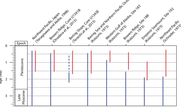
Figure 3.2 – SEM images of P. alata. Scale bar: a, b - 10 µm; c – 1 µm. Adapted from Takahashi et al. (1994) ... 15 Figure 3.3 – SEM images of Proboscia curvirostris (a) and its distal end (b). Arrow indicates longitudinal slit. Scale bar: a - 10 µm; b - 5 µm (Akiba and Yanagisawa, 1986). ... 18 Figure 3.4 – Stratigraphic ranges of P. barboi and P. curviostris in several sites of the North Pacific. P. barboi either disappears near the LO or FO of P. curvirostris, varying with site. Dashed
line indicates scarce occurrences. Blue –P. barboi; Red –P. curvirostris. ... 19 Figure 3.5 – SEM images of Proboscia barboi (a) and its distal end (b). Arrow indicates longitudinal slit. Scale bar: a – 5 µm; b – 1 µm (Akiba and Yanagisawa, 1986). ... 20 Figure 3.6 - Schematics of Thalassiosira jouseae (valve view). Legend: 1 – valve face; 2 – sub-marginal spine; 3 – base of sub-marginal spines; 4 – marginal rim; 5 – marginal ribs; 6 – margin. ... 21 Figure 3.7 - Holotypes of T. nidulus (Tempère and Brun) Jousé (a) and T. jouseae Akiba (b). Magnification 1500x. (Akiba, 1986). ... 22 Figure 3.8 - Stratigraphic ranges of T. jouseae, T. nidulus and variations in North Pacific Sub-arctic, and T. nidulus (Koç and Scherer, 1996) in North Atlantic Sub-arctic. Note: T. nidulus var.
xiv
Figure 4.4 - Measurement of the tube’s curvature. ... 29 Figure 5.1 - a) Productivity of Proboscia barboi, P. curvirostris and Thalassiosira jouseae plotted
with depth (blue, this work) superimposed with the productivity of Stroynowski, 2015; Values higher than 107 valves/g were clipped off. b) Productivity and biostratigraphic events of the same species (this work). Red asterisk: peaks not considered. ... 33 Figure 5.2 - RA of P. barboi, P. curvirostris and T. jouseae in cores U1341B and U1343E. Green arrow denotes the increase in RA of P. curvirostris. Plots constructed with the supplementary data of Onodera et al. (2013) and Teraishi et al. (2013); Chaetoceros considered. ... 35
Figure 5.3 - RA of P. barboi and P. curvirostris compared with RA of Rhizosolenia spp. Red area
denotes the rise of Rhizosolenia spp. (mainly R. hebetata). ... 36 Figure 5.4 - Average RA of P. barboi, P. curvirostris and T. jouseae at Sites U1341, U1340 and U1343; Chaetoceros considered. ... 38 Figure 5.5 –T. jouseae compared with the sea ice species and Chaetoceros sp. records in RA (a) and productivity (b). Values above 107 were clipped off. ... 39 Figure 5.6 - RA of P. barboi, P. curvirostris and T. jouseae juxtaposed with selected environmental
xv
List of Tables
Table 4.1 - Area (mm2) observed for each sample. Note: counts of T. jouseae on samples 24H-5 and 25H-5 were performed on 40.66 mm2 and 39.23 mm2 respectively. ... 27 Table 4.2 - List of the environmental proxies according to Sancetta (1982) and von Quillfeldt (2000, 2001). ... 30 Table 5.1 - Datums of P. curvirotris, P. barboi and T. jouseae of core U1340A (this work). ... 31 Table 5.2 - Mean percentages and range of RA of P. barboi, P. curvirostris and T. jouseae of
xvii
Acronyms
APC – Advanced piston corer AS – Alaskan Stream
BSC – Bering Slope Current DSDP – Deep Sea Drilling Project DSF – Drilling depth below seafloor FCO – First common occurrence FO – First occurrence
FOV – Fields of view
IODP – International Ocean Drilling Project LM – Light microscope
LCO – Last common occurrence LO – Last occurrence
MFSF – Mat-forming shade flora species MPT – Mid-Pleistocene Transition NHG – Northern Hemisphere Glaciation NPD – North Pacific Diatom (zonation/zone) RA – Relative abundance
1
1
Introduction
1.1
Diatoms
Diatoms are a large group of unicellular phototrophic algae with sizes ranging from 10 to 200 µm, which inhabit a wide range of aquatic and semi-aquatic environments. The most distinguishing feature of diatoms is the silica shell or exoskeleton - frustule - which consist of a highly differentiated cell wall heavily impregnated with silica. The frustule is characterized for its diverse shapes and ornamentations and has been since the first microscopic observations in the XVIII century, the main source of taxonomic characters. Diatoms often establish colonies by a variety of means e.g. producing mucus threads, and thereby forming chains of several millimetres.
Being photosynthetic microorganisms, diatoms are restricted to aquatic and semi-aquatic environments with sufficient light exposure. They commonly live as part of the phytoplankton community in the surface water (down to 200 m) or have a benthic lifestyle, and as such have an important role in ecosystems as primary producers. In fact, they are the dominant marine primary producers, being responsible for 40-45% of ocean’s primary production (Mann, 1999). Also, production of biogenic silica in the ocean is mainly attributed to diatoms which therefore play an important role in the ocean’s silica cycle, in addition to the carbon cycle (Tréguer and De La Rocha, 2013). In coastal, equatorial and high latitude upwelling regions, diatoms dominate the phytoplankton communities and typically exhibit bloom-and-bust cycles where their numbers exponentially increase when nutrients and light become available, and upon depletion of nutrients (e.g. Si), sinking rates increase, spores may form and eventually fall to the sediment. In some cases in the past, such massive quantities of diatoms were deposited on the sea floor that through diagenetic processes, the deposits turned into diatomite, a rock entirely formed of frustules that today is of great commercial interest. Thus diatoms play a key role as producers and regulators of the ocean’s both silica and carbon cycles.
Diatoms belong to Division Chrysophyta, class Bacillariophyceae and are organized in two orders based on symmetry (Abrantes and Gil, 2007):
2
Pennate diatoms have bilateral symmetry and mostly live in freshwater, soil or attached to a substrate (e.g. sand grains, plants). Many pennate diatoms bear an unsilicified groove along the apical axis of the valve, called the raphe, which is involved in the secretion of mucus that aids locomotion. The raphe is exclusive to pennate diatoms. Centric diatoms have radial symmetry and often live in marine waters as phytoplankton.
The oldest diatom fossils date back to the Early Jurassic and consist of centric forms, while pennate diatoms are a more recent group, only appearing in the Late Cretaceous from a radiation of centric diatoms. However, genomic studies suggest an earlier origin for diatoms, during the Mesozoic (Armstrong and Brasier, 2013; Sims et al., 2006).
1.2
The frustule
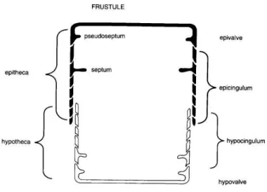
The frustule is the silica exoskeleton of the diatom cell and during its lifetime, is covered by a thin organic coat. Although characterized for its diverse morphology, the frustule has a basic composition, organization and morphologic features common to all diatoms.
The frustule is a multipartite structure composed of two valves, the epivalve and the hypovalve, which are connected by a series of thinner linking structures called the girdle elements. These surround the region in between the two valves and are collectively named girdle or cincture. The girdle elements associated with the epivalve are termed epicingulum, which together with the epivalve form the epitheca, while the hypovalve together with the hypocingulum forms the hypotheca. The organization of the frustule resembles the two halves of a petri-dish in the sense that the hypotheca underlies the edge of the epitheca (fig. 1.1). All components of the frustule fit together very closely and enclose the cytoplasm, allowing communication with the exterior, mainly via pores and slits in the wall components.
3
Figure 1.1 - Gross morphology of the frustule in cross section view. (Hasle et al., 1996).
The valve is regularly perforated by areolae or pores, whose arrangement forms a pattern (i.e. areolation) which is an important taxonomic feature. Rows of areolae form striae and the non-perforated areas of the valve surface between striae are called interstriae or costae. The valve mantle is the marginal area or edge of the valve (Round et al., 1990).
1.3
Reproduction
Diatoms reproduce vegetatively by binary fission, forming two individuals within the frustule of the parent cell. As a consequence of this type of cell division, with the formation of new siliceous components inside the parent cell, the cell size of each generation progressively diminishes. Maximal cell size is restored by auxospore formation, a process usually linked to sexual reproduction. It is also a size dependent process which usually occurs when a cell reaches about a third of its maximal size and cannot take place below this threshold. Small cells unable to develop auxospores keep dividing until division is no longer viable. This in turn, means that certain species are not found beyond a certain size range, and again, are a diagnostic feature used in taxonomic identification.
4
expansion is nearly spherical whereas in pennate taxa expansion is usually bipolar. Once this process finishes, the initial thecae are synthesized. The initial valves may have a modified, more rounded shape than the valves of vegetative cells due to the constraints inside the auxospore and sometimes have a simpler morphology, lacking structures such as spines as in Melosira and
Stephanodiscus (Hasle et al., 1996; Round et al., 1990)
1.4
Resting spores
Diatoms, like some other microorganisms have the capacity to enter a dormant phase in their life cycle when unfavourable environmental conditions arise. Some freshwater planktonic species enter a resting stage, which does not differ morphologically from the vegetative cell except for the thicker cell walls and cytoplasmic content. In most cases however, dormancy is associated with the formation of morphologically distinct cells, called resting spores. Formation of resting spores is a common occurrence in centric diatoms and occurs mostly in species with a distribution in coastal waters and upwelling regions but also at ice fronts (Hargraves, 1986).
Although usually the morphology of the resting spore fairly resembles that of the vegetative cell (e.g. Coscinodiscus furcatus; Syvertsen, 1985), some resting spores differ so drastically that they could be classified as a different genus, family or order (e.g. Chaetoceros
spp.; Hargraves, 1986). Resting spores are generally characterized for their heavily silicified cell walls which usually results in alteration or loss of wall perforations, often coarser areolation and sometimes loss of cingulum bands. There are three main types of resting spores based on the relationship of the mature resting spore to the parent cell i.e. whether the spore is enclosed by the vegetative cell: exogenous, semi-endogenous and endogenous. The same species may produce all types of resting spore as observed in clonal cultures of Thalassiosira nordenskioeldii
Cleve (Hasle et al., 1996).
Resting spores also occur in fossil species and typically can account for the majority of the fossil assemblage, due to a greater resistance to dissolution (Barron, 1985; Tsukazaki et al., 2013). The resting or vegetative spores of extinct species are often difficult to associate with their initial valve, unless examples of resting cell division are found (e.g. Suto, 2004).
1.5
Application in biostratigraphy and paleoclimatology
5
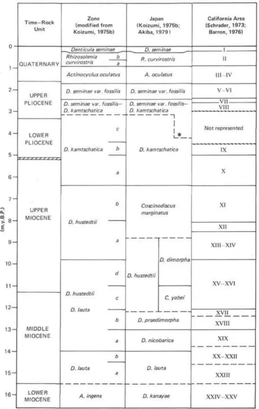
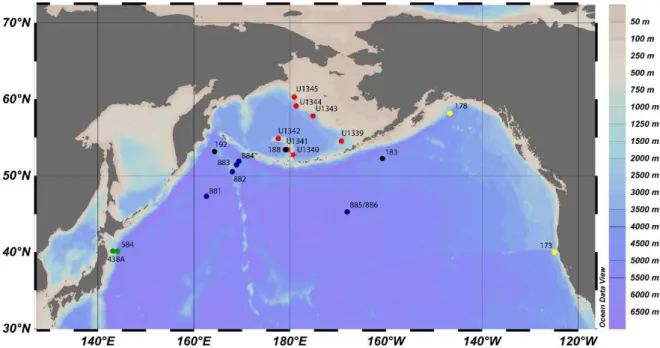
associations. These zonations are generally applicable to a broad geographical range but may differ according to latitude and global region. For instance, a diatom zonation for the North Pacific high latitudes (e.g. Yanagisawa and Akiba, 1998) may not be very suitable in the North Atlantic. As in the following pages diatom zones are frequently mentioned, the main zonations of the North Pacific are presented below (figs. 1.2 and 1.3; Akiba, 1986; Barron, 1980; Barron and Gladenkov, 1995; Koizumi, 1973; Schrader, 1973; Yanagisawa and Akiba, 1998). The location of the main drill hole sites considered in this study are also presented below (figs. 1.4 and 1.5).
Diatoms can also be used to better understand the environment and ecological parameters such as sea surface temperature during a given geological time. By knowing the ecology of a given species or group of close related species, this can be used to reconstruct past environments and as a proxy for a given environmental parameter (Sancetta, 1982; von Quillfeldt, 2000, 2001).
6
7
8
Figure 1.4 - Location of the main Drilling Sites in the North Pacific. Red – IODP Exp. 323; Black – DSDP Leg 19 (Koizumi, 1973); Blue – ODP Leg 145 (Barron and Gladenkov, 1995); Green – Akiba, 1986, Akiba and Yanagisawa, 1986 and Yanagisawa and Akiba, 1998 (DSDP Leg 87 and 57); Yellow – Schrader, 1973 (Leg 18 DSDP). Map generated by Ocean Data View 4.0.
9
2
Regional Setting
2.1
Bering Sea Oceanography
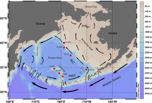
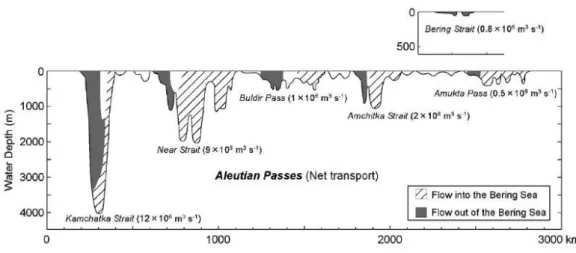
The Bering Sea is a marginal sea in the North Pacific with 2.29x10⁶ km2 of surface area, making it the third largest marginal sea in the world. It is bounded by Siberia and Alaska, semi-enclosed by the Aleutian Islands in the South, and connected to the Arctic Ocean through the Bering Strait in the North. The Bering Sea is characterized by its extensive eastern continental shelf, which covers roughly half of its area from northwest to southeast, creating a vast neritic area (<200 m; fig. 2.1). The central and southern area consists of the Aleutian basin (3500 m) with two main structural highs: the Bowers Ridge (which extends from the Aleutian Island arc into the Aleutian Basin), and the Shirshov Ridge (extending from Kamchatka, Siberia). Three major rivers discharge into the Bering Sea waters: Koskokwin and Yukon draining Central Alaska and Anadyr River draining Siberia (Takahashi, 2005).
10
2.2
Surface Water Currents
The Alaskan Stream (AS) flows westward along the Aleutian Islands and is the main source of water input to the Bering Sea. When combined with part of the Subarctic current joining the northward flow to the Bering Sea, the AS results in a total of 11x106 m3/s (Ohtani, 1973; Takahashi, 2005). The warm waters of the AS enter the Bering Sea through various Aleutian passes and flow eastward to become the Aleutian North Slope Current (ANSC), then flowing north-westward along the shelf break of the eastern continental shelf as the Bering Slope Current (BSC; fig. 2.1). AS waters enter the Bering Sea mostly through the Amchitka Strait, Near Strait and the Kamchatka Strait while other shallower passes only allow a less significant water exchange (fig. 2.2). The water input is balanced by the outflow, mainly through the Kamchatka Strait. A limited amount of water also flows out unidirectionally through the Bering Strait into the Arctic Ocean. The surface water circulation of the Bering Sea basin follows a large-scale anti-clockwise motion, being thereby commonly described as a cyclonic gyre, except in the Bowers basin where surface water circulation is clockwise. On the eastern Bering Sea shelf water circulation is generally northwestward and despite being important to the Arctic Ocean and global water circulation, it has virtually no influence on the circulation in the Bering Sea basin (Stabeno et al., 1999).
Figure 2.2 - Volume transport in the corresponding main Aleutian Passes and in the Bering Strait. (Takahashi, 2005).
11
and closed, cutting the connection to the Arctic Ocean and affecting global water circulation, heat and salt balance (Takahashi, 2005).
2.3
Ecosystem productivity
The Bering Sea is a very productive and rich ecosystem from the basal producers up to the higher trophic levels of the food web. The edge of the Eastern continental shelf is commonly called the Green Belt for being a highly productive area of the Bering Sea, due to upwelling along the Bering Slope, among other physical processes (Springer et al., 1996).
Sea ice is also a fundamental element to ecosystem dynamics and productivity of the Bering Sea. Melting at the ice edge changes the seawater’s physical and chemical properties, such as increasing water column stratification and stability, altering water salinity and temperature. These changes in the water column near the ice-edge may enhance and prolong spring blooms, and therefore significantly contribute to the annual primary productivity in the eastern Bering Sea (Alexander and Niebauer, 1981). Sea ice forms seasonally, first in northern Bering Sea in November, moving southward and covering hundreds of kilometres of the Eastern continental shelf, whereas it is rarely present in the southwestern areas (Niebauer et al., 1999). Sea ice is part of the ecology of many diatom species. Epontic diatoms live attached to the underside of sea ice cover or within brine channels in the ice and bloom in the spring, when enough sunlight penetrates the ice. Another type of bloom, named marginal ice zone bloom occurs when sea ice begins to break up on the Bering Sea shelf, releasing nutrients and freshwater from the ice and thereby promoting the bloom (Caisse, 2012).
2.4
Site U1340
12
13
3
Fossil diatoms of the Bering Sea
3.1
Proboscia
Family Probosciaceae Jordan and Ligowski, 2004 Genus Proboscia Sundström, 1986
3.1.1
Introduction
Proboscia is a recent genus of rhizosolenioid diatoms which includes a number of species transferred from Rhizosolenia, following the taxonomic reviews of this genus (Glezer et al., 1988; Round et al., 1990; Simonsen, 1979; Sundström, 1986; Takahashi et al., 1994). Rhizosolenia alata
Brightwell was the first species moved to Proboscia and was set as the holotype species (Sundström, 1986). Since then several other extinct and extant species of Rhizosolenia, some of
them sub-taxa of Rhizosolenia alata were moved to Proboscia (Takahashi et al., 1994).
Rhizosolenioid is a general term used for diatoms of the family Rhizosoleniaceae and historically related taxa such as the recent family Probosciaceae. Generally, rhizosolenioid diatoms are adapted to stratified waters with a strong nutricline and thermocline, as they are able to vertically migrate between the nutrient rich deeper layers of the water column and the euphotic zone. Some taxa within the Rhizosolenia genus, have been found to host an endosymbiotic Nitrogen-fixing cyanobacteria. Species with this ecology are commonly called shade flora and include other species such as: Thalassiothrix spp., Coscinodiscus spp.,
Stephanopyxis palmeriana and Proboscia alata. The cumulative production by shade flora
diatoms during periods of stratified waters, generally during summer, and subsequent sinking and sedimentation in autumn/winter (i.e. “Fall dump”), when mixing breaks down the nutricline, accounts for a significant proportion of export production, comparable or even superior to that yielded by spring blooms (Kemp et al., 2000). The sedimentation of shade flora diatoms may form laminae in the sediments and thus may also be referred to as mat-forming species (Kemp et al., 2000; Sukhanova et al., 2006).
3.1.2
Morphology of
Proboscia
14
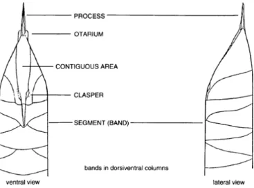
often curved, distinct from the external process, and is diagnostic of Proboscia (Hasle et al., 1996; Medlin and Priddle, 1990).
Figure 3.1 - General morphology of Rhizosolenia spp. (Hasle et al., 1996).
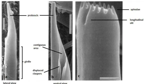
The frustule of Proboscia is cylindrical and the valves are subconical. The ends of the valves lack the external process and terminate into the tubular structure known as proboscis (fig. 3.2). The distal end of the proboscis usually bears spinulae (i.e. short spines; e.g. P. alata) and/or spines (P. curvirostris and P. barboi). Many species form chains of individual cells by
attaching the terminal end of the proboscis to the groove formed by clasper-like structures of the host cell and thereby binding to its ventral side (i.e. the contiguous area). Species such as P. subarctica (= Rhizosolenia alata Brightwell f. curvirostris Gran, 1900), not bearing these morphologic features, characteristic of chain-forming species, are either solitary or form chains through other mechanisms (Hasle et al., 1996; Takahashi et al., 1994). In fossil species, the basal portion of the valve is missing; therefore it is not possible to know whether they formed chains as do extant species, although alternative mechanisms may have been present. Auxospores are terminal, in contrast to the lateral position of auxospores of Rhizosolenia. The proboscis usually bears a longitudinal slit on the dorsal side at the distal end which, in extant species is longer and closer to the tip in comparison to fossil species.
15
Figure 3.2 – SEM images of P. alata. Scale bar: a, b - 10 µm; c – 1 µm. Adapted from Takahashi
et al. (1994)
3.1.3
Ecology
Extant Proboscia have a wide distribution, with species distributed from the subarctic (e.g. P. subarctica) and antarctic (e.g. P. truncata and P. inermis) to tropical environments (e.g.
P. indica; Hernández-Becerril, 1995; Jordan and Priddle, 1991; Sunesen and Sar, 2007). P. alata
is common in polar waters but also found in tropical and subtropical waters (Hernández-Becerril, 1995; Jordan and Ligowski, 2004) although this widespread distribution may be due to P. alata
actually being a complex of cryptic species (Jordan and Ligowski, 2004; Sundström, 1986). P. alatahas been considered a key “fall dump” species in the Gulf of Alaska, and in the Walvis Ridge, South Atlantic (Kemp et al., 2000; Takahashi et al., 1994; Treppke et al., 1996). In the Bering Sea shelf, P. alata is one of the dominant contributors to phytoplankton biomass and abundance from late May to early September, with the highest peak in August, during a period typified by stratified waters. However, in subtropical/tropical regions such the Arabian Sea and off the Somalian coast, P. alata along with Rhiziosolenia spp. is a dominant species prior to or
early in the upwelling season (Gordon and Seckbach, 2012; Kohning et al., 2001; Smith, 2001). The ability to adjust their buoyancy allows these species to migrate to deeper layers bellow the euphotic zone and reach the nutrients during the onset of upwelling events, before other species (Koning et al., 2001; Villareal, 1988).
The seasonal life cycle of P. subarctia on the other hand differs from that of P. alata. Its
16
towards winter, indicating that this species proliferates under high nutrient and low light conditions (Takahashi et al., 1994), and so does not appear to fit in the fall dump annual cycle.
3.1.4
Proboscia barboi
and
Proboscia curvirostris
: phylogenetic relation
Proboscia extends back from Late Cretaceous to the present day. Taxa with long proboscis, particularly the winter forms, bear the closest resemblance to the fossil taxa.
P. barboi and P. curvirostris were two former Cenozoic Rhizosolenia extinct species. Akiba and Yanagisawa (1986) assumed from the strong morphologic similarities, that P. barboi
which proceeded from P. praebarboi, evolved into P. curvirostris, thereby forming a continuous evolutionary lineage. However, Jordan and Priddle (1991) remark that the existence of previous species with curved probosces and bearing terminal spines argue against the hypothesis of that evolutionary sequence. Furthermore, Hajós (1976) suggests that P. interposita is an intermediate species between P. cretacea and P. curvirostrisand shows “close affinity” with the
latter species. Therefore, the evolutionary hypothesis presented by Akiba and Yanagisawa (1986) is not consensual and needs more evidence.
It has also been remarked in the literature that P. subarctica, whose morphology is quite
different from other extant species namely due to the absence of contiguous area and claspers, bears a striking similarity to P. barboi and P. curvirostris (Donahue, 1970; Jordan and Priddle, 1991; Takahashi et al., 1994).
3.2
Proboscia curvirostris
(Jousé) Jordan and Priddle, 1991
Original description: Jousé, 1968, p. 19, pl. 3, fig. 2.Synonymy: Rhizosolenia curvirostris, Akiba, 1986; Akiba and Yanagisawa, 1986
3.2.1
Geographic distribution and stratigraphic range
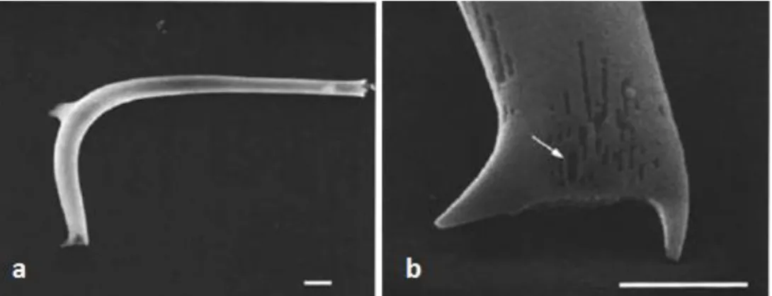
P. curvirostris is an extinct species found in Pleistocene sediments of both the North Atlantic and North Pacific with a mid to high latitude distribution. Only the proboscis of the valve is preserved in the sediments. Being a dissolution-resistant species and having a short stratigraphic span makes it a good Pleistocene biostratigraphic marker.
17
The LO of P. curvirostris defines the top of the Proboscia curvirostris Zone (NPD11) and varies between 0.26 and 0.35 in the North Pacific although it reveals some diachroneity according to location (Barron and Gladenkov, 1995; Koizumi and Tanimura, 1985; Yanagisawa and Akiba, 1998; Onodera et al., 2013; Teraishi et al., 2013). In Northernmost Emperor Seamount it is dated at 0.26 Ma (Katsuki and Takahashi, unpublished data, in Takahashi et al., 2011b) and in the Bowers Ridge at 0.28 Ma +-0.02 (IODP Site U1341; Onodera et al., 2013; fig. 3.4). The extinction of P. curvirostris and the subsequent replacement by the cold water species
R. hebetata Bailey in the North Pacific is related to the Mid-Brunhes event and a transition to more extreme glacial conditions (Jansen et al., 1986; Sancetta and Silvestri, 1984).
In the North Atlantic, its extinction is latitudinally diachronous through MIS 9-8. It first disappeared in the northern areas which are more sensitive to climatic forcing at 0.31 Ma (Irminger Basin, ODP 919) and survived in the mid latitudes, approximately 40°N until 0.26 Ma. The overlap in age between the North Atlantic and North Pacific strongly suggests that the LO of P. curvirostris is relatively synchronous in both oceans (Koç et al., 2001).
3.2.2
Description
18
Figure 3.3 – SEM images of Proboscia curvirostris (a) and its distal end (b). Arrow indicates
longitudinal slit. Scale bar: a - 10 µm; b - 5 µm (Akiba and Yanagisawa, 1986).
3.3
Proboscia barboi
(Brun) Jordan and Priddle, 1991
Original description: Brun, 1894, p. 87, pl.5, figs. 16-17, 23 as Pyxilla (Rhizosolenia?) barboi Brun. Synonymy: Rhizosolenia curvirostris var. inermis Jousé, 1971
3.3.1
Geographic distribution and biostratigraphy
P. barboi is an extinct species found in Miocene-Pleistocene sediments of both the North
Atlantic and North Pacific with a high- to middle-latitude distribution. Like P. curvirostris, this species is known from the valves’ probosces that are preserved on the sediments
.
In the North Pacific, the FO of P. barboi goes back to the Upper Middle Miocene (NPD5B; Akiba and Yanagisawa, 1986). Contrary to P. curvirostris, the LO of P. barboi is more variable, depending on location (fig. 3.4). The LO of P. barboi is divided into two main ages: close to 0.3 Ma, at the same time P. curvirostris disappears or close to the FO P. curvirostris (1-2 Ma). In
many locations such as the Bowers Ridge (IODP Site U1341), P. barboi disappears a little earlier than P. curvirostris, at 0.42-0.47 Ma (Onodera et al., 2013), at IODP Site U1343 (Teraishi et al., 2013) the LO is roughly at 0.6 Ma (age corresponding to 140 m depth; fig. 4.2), and in the Northwest Pacific, off Japan’s east coast, it occurs at the same age as P. curvirostris (0.3 Ma;
19
curvirostris zone, ~0.3 Ma). Nevertheless, on the South side of the Aleutian Islands Arc (Sites 183 and 192) and on the Western flank of Bowers Ridge (Site 188), the LO of P. barboi approximately
matches the FO of P. curvirostris near the top, ~1 Ma; bottom, ~2 Ma; and middle, ~1.5 Ma of
Actinocyclus oculatus zone, correspondingly.It is odd that while sites 188 and U1341 are both located in the same region (Western flank of the Bowers Ridge), they have such a large age discrepancy on the LO of P. barboi.
In the Atlantic Ocean on the Iceland Plateau (Site 907), P. barboi disappears at 3.3 Ma, approximately 3 Ma earlier than the extinction of P. curvirostris (Koç and Scherer, 1996).
Figure 3.4 – Stratigraphic ranges of P. barboi and P. curviostris in several sites of the North
Pacific. P. barboi either disappears near the LO or FO of P. curvirostris, varying with site. Dashed line indicates scarce occurrences. Blue –P. barboi; Red –P. curvirostris.
3.3.2
Description
20
Figure 3.5 – SEM images of Proboscia barboi (a) and its distal end (b). Arrow indicates longitudinal slit. Scale bar: a – 5 µm; b – 1 µm (Akiba and Yanagisawa, 1986).
3.4
Thalassiosira
Family Thalassiosiraceae (Lebour) Hasle, 1973 Genus Thalassiosira (Cleve) Hasle, 1973
3.4.1
Introduction
Thalassiosira is a large genus of marine diatoms, with more than 100 species. Cells are
discoid or cylindrical and form chains by connecting organic threads. Thalassiosira species are found in a wide variety of environmental settings including sea ice, upwelling zones, highly stratified waters, cold, and warm waters. The type species is Thalassiosira nordenskioeldii Cleve, a species part of the spring bloom in the marginal ice zone (Caisse, 2012; von Quillfeldt et al., 2003).
The classification of the genus and its species is mostly based on the strutted, occluded and labiate processes present on the valve face, that are either isolated or in ring formations, and are best observed by electron microscopy. These structures are openings in the valve face consisting of tubes that differ in their internal structure. The first two mentioned structures are diagnostic of Thalassiosiraceae. The number, arrangement and position of these structures are important characters of the genus’ taxonomy, while the valve size and areolae density are not as taxonomically diagnostic as they vary considerably within the species rank. The strutted processes are involved in colony formation by extruding threads that link frustules together (Hasle et al., 1996; Makarova, 1980). Resting spores occur in Thalassiosira and have been fairly
21
3.4.2
Thalassiosira jouseae
Akiba, 1986
Description: Akiba, 1986, p. 440, pl. 6, figs. 8-10Synonym: Thalassiosira nidulus (Tempère and Brun) Jousé, 1961, p. 63, pl. 3, figs. 4-5;
Thalassiosira nidulus (Tempère and Brun) Jousé var. nidulus (nomen nudum), Barron, 1980a, p. 673, pl. 6, fig. 5.
3.4.2.1 Geographic distribution and biostratigraphy
Thalassiosira jouseae is a species with a mid to high latitude distribution in the Pacific and Atlantic Oceans. It appears in the uppermost Miocene (Yanagisawa and Akiba, 1998), and becomes extinct approximately at 0.3 Ma in the North Pacific (fig. 3.8), the same age as P. curvirostris (Onodera et al., 2013; Teraishi et al., 2013; Yanagisawa and Akiba, 1998). Its LO is a secondary biohorizon defining the top of zone NPD11 (Akiba, 1986), and is synchronous in both North Pacific and North Atlantic Oceans (Koç et al., 2001).
3.4.2.2 Description
T. jouseae (fig. 3.6, 3.7b and 3.9) has a circular valve with 9-29 µm in diameter. The valve is sparsely areolated in the central part of the valve face. Areolae are quadrangular, more or less isolated from each other, 8-10 areolae in 10 µm. The most distinguishing feature of the valve is the conspicuous sub-marginal tapering spines that form a crown-like structure by uniting their basal parts (Akiba, 1986). These structures correspond to the marginal strutted processes.
22
Figure 3.7 - Holotypes of T. nidulus (Tempère and Brun) Jousé (a) and T. jouseae Akiba (b).
Magnification 1500x. (Akiba, 1986).
3.4.2.3 Thalassiosira nidulus: taxonomy and biostratigraphy
Akiba (1986) discovered and described T. jouseae based on specimens that had been assigned to Thalassiosira nidulus by Jousé (1961). The latter species has an older taxonomic
history. It was originally described as Stephanopyxis nidulus Tempère and Brun in Brun and Tempère (1889), and then transferred to the genus Thalassiosira by Jousé (1961). Akiba (1986) places T. nidulus (Tempère and Brun) Jousé 1961, as a synonym of T. jouseae. However, the author is only referring to T. nidulus of plate 3, figs. 4-5 of Jousé (1961) and not the other specimens (plate 1, figs. 3-4). Therefore, the taxon T. nidulus represents a real species and
should not be used interchangeably with T. jouseae.
T. nidulus (fig. 3.7a and 3.10) appears in the Upper Miocene (DSDP Hole 438A; Akiba, 1986) and in North Atlantic in the Lower Pliocene (ODP Site 907; Koç and Scherer, 1996). The LO of T. nidulus is synchronous with that of T. jouseae although from the end of the Miocene epoch onwards its occurrences become rare or sporadic (DSDP Hole 584; Akiba, 1986). In the North Atlantic, T. nidulus has a shorter stratigraphic range, within the lower-middle Pliocene (ODP Site 907; Koç and Scherer, 1996).
Barron (1980) actually seems to precede Akiba (1986) by distinguishing a variation “nidulus” of T. nidulus (Tempère and Brun) Jousé from the Pacific subarctic, although not
accompanied with a description. Akiba (1986) listed said variation as a synonym of T. jouseae. Indeed T. nidulus var. nidulus Barron looks identical to T. jouseae and both LOs match very closely, defining the top of Zone Rhizosolenia/Proboscia curvirostris (Barron, 1980; Yanagisawa and Akiba, 1998). Barron (1980) also considers T. nidulus of Schrader (1973, pl. 11, figs. 1-7) as
T. nidulus var. nidulus which would imply that T. nidulus (Schrader, 1973) are actually T. jouseae.
23
Figure 3.8 - Stratigraphic ranges of T. jouseae, T. nidulus and variations in North Pacific Sub-arctic, and T. nidulus (Koç and Scherer, 1996) in North Atlantic Sub-arctic. Note: T. nidulus var.
nidulus = T. jouseae Akiba; T. nidulus var. delicata = Thalassiosira delicata Akiba. Dashed line
indicates rare to sporadic occurrences.
The LO of T. nidulus (Schrader, 1973) was established in lower NPD Zone III (0.92-1.3 Ma) somewhat earlier than the LO of Rhizosolenia curvirostris which defines the top of NPD Zone II (Schrader, 1973), whereas in the literature the LO of T. jouseae occurs later (~0.3) and is coincident with the LO of P. curvirostris (fig. 3.8).
3.4.3.3 Distinguishing Thalassiosira jouseae from Thalassiosira nidulus
The original description of T. nidulus (Jousé, 1961) is not accessible by most means, which complicates its recognition and contributes to the confusion with T. jouseae. Nevertheless, Akiba (1986) remarks that T. jouseae is distinguished from T. nidulus by the “united basal parts of sub-marginal spines, smaller valve, and sparser areolae on valve face” (fig.3.7b; Akiba, 1986).
24
notice significant differences between them. The former images show a T. jouseae specimen (fig. 3.9; 10 µm valve diameter) with a convex valve face and areolation restricted to the central area, leaving the periphery hyaline. The striated margin displays the ribs connecting to the marginal rim. On the other hand, in the T. nidulus specimen (fig. 3.10; 17 µm valve diameter) the valve face is flat and the areolation covers all of its area till the base of the sub-marginal spines. The areolae are also arranged closer to each other. The marginal rim seems to be missing, leaving only the ribs. The sub-marginal spines of T. nidulus do not seem to have united basal parts and thus can be said to have separate spines, whereas the spines of T. jouseae have united basal parts as noted by Akiba (1986).
In the TEM images of Akiba (1986), the holotype of T. nidulus is densely areolated, with contiguous irregularly sized areolae covering all of the valve face and arranged in (imperfect) concentric rows (fig. 3.7a).
Although T. jouseae and T. nidulus are fairly distinguishable on these SEM images, the identification of T. jouseae on LM is often ambiguous. In the morphology chapter, T. jouseae and
T. nidulus are described based on specimens from U1340A samples of the Pleistocene.
Figure 3.9 - SEM image of T. jouseae in valve view (Koç et al., 1999).
25
4
Methodology
4.1
Samples
The studied core samples were obtained from International Drilling Program (IODP) repository from selected intervals from Core U1340A. The Site is located at 53°24.0008’ N, 179°31.2973’ W, on the eastern flank of the southern part of Bowers Ridge in the Bering (fig. 2.1). Four holes were drilled during IODP Expedition 323 in 2009, and cored with the advanced piston (APC) and extended core barrel (XCB) coring systems at a water depth of 1294.7 m. All analysed samples are from Hole U1340A, which was cored to 604.6 m drilling depth below seafloor (DSF) using both APC and XCB coring systems. Diatom biostratigraphy at Site U1340 was based on core catcher samples from Hole A and a biostratigraphic zonation was constructed until the lowermost Pliocene, ca. 5 Mya (Subzone NPD 7Bb; Yanagisawa and Akiba, 1998; Takahashi et al., 2011b). The age models of Site U1340 (hole A) and other IODP Exp. 323 Sites are presented bellow (fig. 4.1 and 4.2).
26
Figure 4.2 - Age-depth plot for Sites U1339-U1345 (Takahashi et al., 2011a).
4.2
Method
In order to study the biostratigraphy and morphology of P. barboi, P. curvirostris and T. jouseae, a total of 43 samples were used (3H-CC, 23.12 m to 28H-3, 238.55 m; table 4.1), covering an age interval from 0.18 to 1.63 Ma, roughly encompassing the stratigraphic span of
P. curvirostris. The observed samples and slides are the same as the ones previously used for the
biostratigraphy of core U1340A (Stroynowski et al., 2015). Counts of the three mentioned species were performed on light microscope (LM; Nikon eclipse 80i) under x1000 (or under x600, when suitable) magnification, by running transects on the first slide of each sample.
The productivity of each species was calculated by applying the following formula (Abrantes et al., 2005):
No. valves/gram = ((N*(S/s))*(V/v))/W N - number of valves counted
S - area of the evaporation tray (mm2) S - observed area of the slide (mm2) V - volume of solution in the beaker (ml)
V - aliquot or volume of solution put into the evaporation tray (ml) W - weight of raw sample (g)
Radius (slide) = 10 mm
27
The area observed on each slide was calculated considering the shape of each transect as approximately that of a rectangle. Thus the observed area is the sum of the areas of all transects run on the slide. The height of one transect is equal to the diameter of the field of view (FOV), which depends of the microscope used. The FOV diameters (D) of each magnification are: D1000x = 0.22 mm; D600x = 0.40 mm; D400x = 0.61 mm
Table 4.1 - Area (mm2) observed for each sample. Note: counts of T. jouseae on samples 24H-5 and 25H-5 were performed on 40.66 mm2 and 39.23 mm2 respectively.
Core U1340A sample # Mid-depth (cm) Depth (Mid CSF m) Area observed (mm2)
3H-CC 0.5 23.12 314.16
4H-3 135.5 25.75 314.16
4H-5 135.5 28.75 314.16
4H-CC 0.5 32.56 628.32
5H-CC 0.5 42.13 314.16
6H-CC 0.5 51.68 314.16
7H-CC 0.5 61.2 314.16
8H-3 135.5 65.25 314.16
8H-5 135.5 68.25 314.16
8-CC 0.5 70.69 314.16
9H-3 135.5 75.95 314.16
9H-5 135.5 77.75 314.16
9H-CC 0.5 80.11 40.46
10H-3 135.5 84.26 314.16
10H-5 135.5 87.25 40.06
10H-CC 0.5 89.63 157.08
11H-5 135.5 96.75 34.21
11H-CC 0.5 99.17 38.72
12H-CC 0.5 108.61 37.22
13H-5 135.5 115.75 37.55
13H-CC 0.5 118.06 36.28
14H-3 105.5 121.95 40.94
14H-6 135.5 126.75 38.28
15H-5 150 134.89 37.60
16H-5 150 144.39 41.14
17H-3 150 150.89 36.76
17H-5 150 153.89 36.26
18H-3 150 160.39 39.25
18H-5 150 163.39 39.78
19H-3 136 169.75 35.42
19H-5 137 172.76 28.09
20H-3 136 179.25 33.64
20H-5 136 182.25 33.84
23H-3 136 191.04 34.67
23H-5 136 194.04 37.69
24H-3 136 200.54 36.87
24H-5 136 203.54 90.40
25H-3 136 210.04 157.08
25H-5 136 213.04 93.61
26H-3 136 219.55 45.63
26H-5 136 222.55 314.16
27H-3 137 229.06 314.16
28
4.3
Proboscia curvirostris
and
Proboscia barboi
4.3.1
Identification
Identification of these species was based on the presence or absence of the dorsal fin. Heavily dissolved frustules but with distinguishable remains of the dorsal fin and intermediate specimens between P. barboi and P. curvirostris with a dorsal fin were counted as P. curviostris.
4.3.2
Measurements
In order to study the intermediate specimens of P. barboi and P. curvirostris, 13 specimens from sample 11H-CC and 14H-3 respectively, were measured as well as 13 specimens considered to be intermediate, from sample 24H-5. The samples were selected for containing fairly well preserved and morphologically average specimens of P. barboi and P. curvirostris,
suitable for the measurements. Although a larger sampling would be necessary to make a rigorous morphometric study, this number of specimens allows to have a reference point to which the intermediate specimens can be compared, which was the main purpose.
Apart from the dorsal fin, two main features were used to distinguish the P. curvirostris
and P. barboi: the width and curvature of the tube. The width (µm) was measured at the region between the maximum curvature and the tip of the tube (fig. 4.3), closer to the former, as the tube in some specimens appears to become narrower towards the apex. The curvature of the tube was measured on the ventral side. The employed method consisted in drawing two straight lines (on the ventral side), one parallel to the direction of the tube’s distal end and another parallel to its more proximal part and afterwards measuring the angle of the two lines (fig. 4.4).
29 Figure 4.4 - Measurement of the tube’s curvature.
4.4
Thalassiosira jouseae
4.4.1.1
Identification
Identification was based on Akiba (1986) and the identification of T. nidulus relied on Akiba (1986) and SEM imaging of Koç and Scherer (1996). A few T. nidulus valves were included under T. jouseae during the counting process, although not in a sufficient number that would undermine the counts.
4.4.1.2
Description
The description of T. jouseae was made based on a sizeable number of specimens more than a hundred, from samples U1340A-4H-CC (32.56 Mid CSF m) to 40H-1 (343.28 Mid CSF m) which corresponds to a 2.13-0.26 Ma time interval in the Pleistocene Epoch. It is merely a qualitative description of T. jouseae with the purpose of complementing the original description
by Akiba (1986). Likewise, T. nidulus was also described based on a few specimens from samples U1340A-20H-3, 19H-5, 9H-5, 9H-3 and 8H-3, where this species was identified.
4.5
Paleoecology
–
used counts and data
30
4.6
Environmental proxies
The environmental proxies used in this study are summarized in the following table.
Table 4.2 - List of the environmental proxies according to Sancetta (1982) and von Quillfeldt (2000, 2001).
Proxy group Species Environmental
factor
Neodenticula spp.
Neodenticula kamtschatika, Neodenticula koizumii and
Neodenticula seminae Alaskan Stream
Sea ice species
Thalassiosira gravida, Thalassiosira antiqua, Thalassiosira nordenskioeldii, Thalassiosira hyalina, Stellarima microtrias, Porosira glacialis, Paralia sol, Paralia sulcata, Nitzschia sp., Fossula arctica, Fragilariopsis curta, Fragilariopsis cylindrus
Presence or influence of sea
ice (e.g. iceberg transportation)
Chaetoceros
spp. - Productivity
Mat-forming shade flora
species
Rhizosolenia hebetata f. hebetata, Rhizosolenia hebetata f. hiemalis, Rhizosolenia hebetata f. seminspina, Rhizosolenia stylisformis, Coscinodiscus marginatus
water stratification
Rhizosolenia spp.
Rhizosolenia hebetata f. hebetata, Rhizosolenia hebetata f. hiemalis, Rhizosolenia hebetata f. seminspina, Rhizosolenia stylisformis.
31
5
Results and Discussion
5.1
Biostratigraphy
At U1340, the LO of P. curvirostris, T. jouseae and P. barboi occur at 0.33-0.26 Ma (sample U1340A-5H-CC), at 0.26-0.23 Ma (sample U1340A-4H-CC) and at 1.39-1.37 Ma sample 24H-5, respectively (table 5.1; fig. 5.1b). In samples up-core from the observed LO of P. barboi
at sample U1340A-24H-5, no P. barboi valves were found except for samples 23H-3 (191.04 m;
one valve), 11H-CC (99.17 m; 21 valves) and 4H-CC (32.56 m; 1 valve). Specimens of P. barboi in sample 11H-CC were well preserved, while P. curvirostris is absent which may indicate that these events in the same sample are not a mere coincidence and not a result of reworking. Hence, an alternative LO for P. barboi could be set at 0.67-0.65 Ma (sample U1340A-11H-CC, 99.17 m),
which is also approximately the same age when this species disappears at Site U1343. However, the LO of P. barboi was set at sample 24H-5 due to the great scarcity of occurrences after this sample. On the other hand, in Takahashi et al. (2011b) the LOs of these species both occur at 0.3 Ma (sample U1340A-5H-CC). The previously established LOs of these species in core U1340A are all set at 0.3 Ma (Takahashi et al., 2011b) differing on the LOs of T. jouseae and P. barboi.
Table 5.1 - Datums of P. curvirotris, P. barboi and T. jouseae of core U1340A (this work).
Age (Ma) Sample Depth (mid CSF m) #valves counted
P. curvirostris
LO 0.33-0.26 U1340A-5H-CC 42.13 94
FO 1.52-1.56 U1340A-26H-5 222.55 1
FCO 1.44-1.39 U1340A-24H-5 203.54 60
P. barboi LO 1.39-1.37 U1340A-24H-5 203.54 4
LCO 1.44-1.39 U1340A-25H-3 210.04 30
T. jouseae LO 0.26-0.23 U1340A-4H-CC 32.56 4
LCO 0.38-0.33 U1340A-6H-CC 51.68 51
An important observation in core U1340A is that the occurrences of P. barboi and P. curvirostris do not overlap and are mutually exclusive, that is, both species virtually never occur in the same sample. In sample 11H-CC, when there is an abundance spike of P. barboi, P. curvirostris becomes abruptly absent. In the few samples where both species co-occur (samples
23H-3 and 24H-5) the quantity of one of the species is negligible and the specimens found are dubious.
The counts performed in this study match well those of Stroynowski et al. (2015) despite the lower values of productivity which might be due to the higher area observed (fig. 5.1). Nevertheless, at approximately 160 m depth (mid CSF), a few valves of P. barboi were found
32
213 m depth mid CSF (sample U1340A-25H-5; Stroynowski et al., 2015) which were not observed during the counts of this study.
5.2
Paleoecology
Note: the following discussion of the paleoecology of the studied species is made in reference to fig. 5.6 unless otherwise indicated.
5.2.1
Proboscia barboi
P. barboi appears at 2.7 Ma, when the first signs of sea ice diatoms subtly increase at Bowers Ridge (Site U1340) and also coinciding with the inception of Northern Hemisphere Glaciation (NHG; Takahashi et al., 2011; Maslin et al., 1996). At this time in the Bering Sea, diatom biodiversity and productivity increased with a marked change in the diatom assemblage, indicating enhanced seasonality and stratification (Stroynowski et al., 2015). Fluctuations of relative abundances (RA) of P. barboi show an increasing trend starting from 2.1 Ma, coinciding with a long term progressive increase of sea ice assemblages in Site U1340, up to 10%-20% of the total respective assemblage (Takahashi et al., 2011). From 2 to 1.2 Ma, the Bering Sea is characterized by stratified and nutrient-depleted summer waters as indicated by the absence and reduced presence of the high-productivity indicators Chaetoceros (resting and vegetative spores) and Thalassiothrix longissima (Stroynowski et al., 2015). The authors suggest that the most likely cause of stratification is by ice melt. Rhizosolenia spp. (R. hebetata f. hebetata; R. hebetata f. hemialis; R. hebetata f. semispina and R. styliformis), which are shade flora species, also began to appear with more frequency after 2.0 Ma (Stroynowski et al., 2015), and their RA fluctuations show good correlation with P. barboi (fig. 5.3). The fairly coincident rise and positive correlation of Rhizosolenia spp. and P. barboi, together with the positive responses to sea ice influence and resultant water stratification, support the notion of P. barboi being a shade flora species that proliferated colder waters.
P. barboi disappears from Site U1340 geological record at 1.4 Ma, being replaced at the
same time by P. curvirostris, despite the curious isolated abundance spike of P. barboi
33
34
As discussed above, the LO’s of P. barboi vary depending on location and may be divided into two groups: sites where both the LO of P. barboi and P. curvirostris co-occur, and sites where
the LO of P. barboi occur around the same time as the FO of P. curvirostris. At Site U1340, the LO of P. barboi belongs to the latter group as P. barboi disappears at 1.4 Ma where the FO of P. curvirostris is established. Whereas at Site U1341, P. barboi disappeared at 0.42-0.47 Ma (Onodera et al., 2013), although its RA somewhat decreases after the FO of P. curvirostris. Hence, at two close locations, one on the western flank and the other on the south-eastern part of the Bowers Ridge, P. barboi disappears at different ages, which suggest differences in the environmental conditions affecting P. barboi. However, at Site 188 (Koizumi, 1973) which is in the vicinity of Site U1341 (fig. 1.4), the LO of P. barboi does not match that of the latter Site, as it occurs near the FO of P. curvirostris and therefore the stratigraphic range of P. barboi in this
region should be further investigated.
5.2.2
Proboscia curvirostris
P. curvirostris appearance around 1.4 Ma is marked by a large peak of 14% RA and coincides with a peak of AS marker species Neodenticula spp. (56 %) and a drop in sea ice species (1%), which indicates that its appearance occurred in a relatively warm period under relatively high influence of AS waters. It is also not a particularly nutrient-abundant period with frequent upwelling as can be observed by the low abundance of Chaetoceros spp. Furthermore, the FCO of P. curvirostris is the largest RA peak which indicates that the environmental conditions referred above were favourable or at least tolerable when P. curvirostris appeared.
The abundance record shows a general tendency for a progressive decline in the amplitude of peaks of P. curvirostris which may be a reflection of the progressive intensification of the NHG. However, in neighbouring Site U1341, an opposite trend, with an increase of the RA peaks is observed (fig. 5.2). The first peak occurs at 150 m depth with 8 % RA, followed by another peak of 10 % at 97 m depth, and lastly a peak of 16 % at 70 m depth. As discussed above, during the past 270 kyr, the western and eastern sides of the Bowers Ridge faced different influence of AS waters and consequently sea ice as the eastern Aleutian passes (e.g. Amchitka Pass), were more restricted during glacial periods (Katsuki and Takahashi, 2005). Extending this reasoning to an earlier time interval, it is possible that the opposite trends in RA of P. curvirostris
35
Figure 5.2 - RA of P. barboi, P. curvirostris and T. jouseae in cores U1341B and U1343E. Green arrow denotes the increase in RA of P. curvirostris. Plots constructed with the supplementary data of Onodera et al. (2013) and Teraishi et al. (2013); Chaetoceros considered.
The sudden absence of P. curvirostris at ~0.7 coincides well with a big 40% drop of N. seminae (from 50% to 7% RA), a large peak of sea ice species (31%) and a peak of Chaetoceros spp. (17%)which indicate a cold period with enhanced sea ice influence and somewhat nutrient rich waters. The RA of other mat-forming shade flora (MFSF) species also drops. The combination of these environmental factors, especially the cold/sea ice factor may well be the cause for the short absence of P. curvirostris. Curiously at the same time, P. barboi unexpectedly resurfaces. The absence of P. curvirostris was registered in 2 consecutive samples (U1340A-12H-CC, 108.61 mid CSF m; 11H-(U1340A-12H-CC, 99.17 Mid CSF m) while P. barboi re-appears in a single sample (11H-CC).
The LO of P. curvirostris occurs at 0.3 Ma, in agreement with the other North Pacific sites. Close to the extinction of P. curvirostris, the Rhizosolenia MFSF group (mostly comprised of R. hebetata f. hebetata) shows a significant increase up to 27% (fig. 5.3), equivalent to its rise in the Subarctic, where Sancetta and Silvestri (1984) suggested that R. hebetata“ecologically replaces” P. curvirostris and suggested that the intensification of glacial conditions wiped out P. curvirostris and created a new or expanded niche which allowed R. hebetata to flourish.
36
Mean values of RAs of P. barboi and P. curvirostris at Site U1340 are about 4 times higher than at Site U1343 (fig. 5.4; table 5.2). The low abundances of P. curvirostris and P. barboi at Site
U1343 suggest that they do not have a particular preference for the upwelling regimes or sea ice influence of the Bering Slope and fits into the likely shade flora ecology of P. curvirostris and
P. barboi, since Proboscia in high latitudes are generally not associated with upwelling or part of the sea ice community (Takahashi et al., 1994).
At the Southern Bering Sea sites as well as the North Subarctic Pacific where the Alaskan Current is the dominant water current (Leg 19 DSDP), RA of both Proboscia species vary between 0.5% and 2.5% and less often between 3% and 9.5% in a 200 total valve count (Chaetoceros not counted; Koizumi, 1973). Their low abundance in the Alaskan Stream domain requires further investigation, however it does suggest an ecological preference for colder, less saline waters.
37
5.2.3
Thalassiosira jouseae
Subtle occurrences of T. jouseae started at around 2.5 Ma, and can be considered part of the diatom assemblage reorganization and enhanced productivity that began circa. 2.7 Ma, coinciding with the onset of NHG (Stroynowski et al., 2015). With the exception of the sudden abundance spike at 0.65 Ma (24.3% at sample 10H-CC), the RA of T. jouseae remained low and at relatively stable fluctuations below 4% with no apparent long term trend which shows that T. jouseae over the long term, did not respond to the progressive trend of seasonally stratified waters (Stroynowski et al., 2015), nor the development of glacial/interglacial cycles. Hence, water stratification does not seem to be a very important part of the ecology of T. jouseae. Strong negative correlation with Neodenticula spp.also supports a preference for colder waters and positive response to cold periods.
38
Figure 5.4 - Average RA of P. barboi, P. curvirostris and T. jouseae at Sites U1341, U1340 and U1343; Chaetoceros considered.
Table 5.2 - Mean percentages and range of RA of P. barboi, P. curvirostris and T. jouseae of coresU1341B, U1340A and U1343E. The depth interval considered for the calculations is also shown. Chaetoceros considered.
0 2 4 6 8 10 12 Site U1341B (Onodera et al., 2013)
Site U1340A (Stroynowski et al., 2015)
Site U1343E (Teraishi et al., 2013)
Av
erage
RA
(%
)
Abundance according to Bering Sea sites
%P. barboi %P. curvirostris %T. jouseae
Core U1341B (Onodera et al.,
2013)
Core U1340A (Stroynowski et
al., 2015)
Core U1343E (Teraishi et al.,
2013)
%P. barboi
Average 1.9 1.6 0.4
Range (min. – max.) 0-12.3 0 - 15.1 0 - 8.0
depth interval (m) 51.15-532.65 99.17-489.27 139.73-777.34
%P. curvirostris
Average 4.3 3 0.8
Range (min. – max.) [0 - 18.0] [0 - 14.6] 0 - 4.8 depth interval (m) 41.64-157.99 42.13-213.04 81.45-505.79
%T. jouseae
Average 0.6 1 5.5
Range (min. – max.) 0 - 7.0 0 -24.3 0-27.0
39