McArdle’s disease: an underestimated or underdiagnosed
myopathy in rheumatologic practice? Cases series and
literature review
Pablo Arturo Olivo PalloI, André Macedo Serafim da SilvaII, Edmar ZanoteliII, Samuel ShinjoI
DOI: 10.5935/MedicalExpress.2018.mo.008
I Universidade de São Paulo, Faculdade de Medicina, Division of Rheumatology, Hospital das Clínicas HCFMUSP, São Paulo, Brazil (BR). II Universidade de São Paulo, Faculdade de Medicina, Department of Neurology, Hospital das Clínicas HCFMUSP, São Paulo, Brazil (BR).
OBJECTIVE: McArdle’s disease is a metabolic myopathy that manifests with varied clinical conditions and is often confounded with other diagnoses. Herein, the authors report a case series and carry out a literature review.
METHODS: A cross-sectional single-center study evaluating 12 patients with McArdle’s disease was conducted.
RESULTS: Mean age at onset of symptoms was 28.0±17.4 years, while age at disease diagnosis was 39.0±14.8 years. History of intolerance to physical exercises was observed in 10 cases; muscle weakness in 9, second wind phenomenon in only 1 case. The presence of cramps, fatigue and myalgia was observed in 12, 11 and 9 of the cases respectively. Median creatine phosphokinase level was 5951U/L. Most of the patients (83.3%) were initially diagnosed with another condition (polymyositis, inclusion body myositis, fibromyalgia and/or muscular dystrophy), and approximately half had received glucocorticoids and/or immunosuppressants prior to definitive diagnosis. All patients underwent muscular biopsy, which revealed the presence of subsarcolemmal vacuoles characterized by glycogen deposits, and negative histochemical reaction for the myophosphorylase enzyme.
CONCLUSION: The present study reinforces the presence of clinical variability among patients and shows that McArdle’s disease should be considered one of the differential diagnoses of inflammatory myopathies and other rheumatic diseases.
KEYWORDS: Fibromyalgia; glycogen storage disease; myopathies; myophosphorylase; myositis.
Olivo Pallo PA, Silva AMS, Zanoteli E, Shinjo S. McArdle’s disease: an underestimated or underdiagnosed myopathy in rheumatologic practice? Cases series and literature review. MedicalExpress (São Paulo, online). 2018;5:mo180008
Received for Publication on April 17, 2018; First review on May 18, 2018; Accepted for publication on June 10, 2018; Online on August 20,2018
E-mail: pablolivo@yahoo.es
■
INTRODUCTIONMcArdle’s disease or glycogen storage disease
type V is a metabolic myopathy caused by the deficiency
of myophosphorylase, an enzyme encoded by the PYGM gene, which catalyzes the degradation of glycogen into glucose in skeletal muscle.1-4
There is scant epidemiological data about McArdle’s disease. The disease has an estimated prevalence of approximately 1 per 100000 - 167000
population,5,6 and affects individuals of both sexes at a
mean age of 44 years.6
The symptoms of McArdle’s disease usually commence during adolescence or young adulthood.7 The
disease is characterized by exercise intolerance, myalgias and/or muscle cramps.8 The “second-wind” phenomenon
is very characteristic of the disease, in which muscle pain may dissipate after a brief rest period and allow the patient to resume exercise at the previous or a slightly reduced level.5 Half of patients have myoglobinuria and
maximum) values were calculated for continuous variables that did not present a normal distribution. All analyzes were performed using SPSS 15.0 software (Chicago, USA).
■
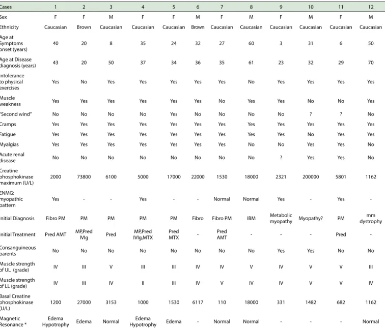
RESULTSThe general characteristics of the 12 patients with McArdle’s disease are shown in Table 1. There was a predominance of females and Caucasians. The mean age at the onset of symptoms was 28.0 ± 17.4 years, while the mean age at diagnosis of the disease was 39.0 ± 14.8 years.
A history of intolerance to physical exercise was observed in 10 of the cases, while muscular weakness occurred in 9, and “second wind” phenomenon was described in only 1 case. In addition, the presence of cramps, fatigue and myalgia was observed in 12, 11 and 9 of the cases, respectively. A history of acute renal failure was reported in 2 cases. At admission of the patients to our service, with the exception of 2 patients, all had some degree of muscle weakness of the upper and/or lower limbs. Regarding family history, 2 patients had a history of consanguineous parents.
The mean peak CPK level was 5951 (1162 - 200000) U/L, while the mean baseline CPK serum level was 1341 (110 - 27000) U/L. Only 1 patient had a normal baseline CPK level. Electroneuromyography, performed in half of the cases, revealed a myopathic pattern in about two thirds of the cases. Magnetic resonance imaging of the thigh muscles had been performed in 8 of the 12 patients, four of whom had edema of the muscle compartments.
Ten of the 12 patients were initially treated with the hypothesis of polymyositis, inclusion body myositis,
fibromyalgia and/or muscular dystrophy. Half of the
patients had received glucocorticoid, immunosuppressants and/or human intravenous immunoglobulin. One patient had a myopathy hypothesis to clarify, and only 1 case was admitted with the initial suspicion of metabolic myopathy.
All of these patients underwent muscle biopsy, which showed the presence of subsarcolemmal vacuoles characterized by glycogen deposits (Figure 1).
■
DISCUSSIONIn the present study, the demographic, clinical and
laboratory profiles of 12 patients with histologically-confirmed McArdle’s disease were described; most of the
cases were initially treated as other rheumatic diseases. Although no molecular analysis was performed to investigate possible mutation in the PYGM gene, the patients included in the study were carefully selected according
to the histological findings and with demonstrated deficiency of the myophosphorylase enzyme activity. All
had histological analysis of muscle biopsy, the presence exacerbation.8,9 Muscle biopsy reveals the presence of
subsarcolemmal vacuoles, characterized by deposits of glycogen.11-13 In addition, histochemistry shows a deficiency
in myophosphorylase enzyme activity.11-13
However, the variability of the clinical manifestations and lack of knowledge about the disease can lead to delayed and erroneous diagnosis, such as polymyositis,
inclusion myositis, fibromyalgia, among others. Therefore,
recognition of McArdle’s disease is of great relevance in rheumatologic practice, especially as one of the differential
diagnoses of inflammatory myopathies. Thus, we present
a case series of patients with McArdle’s disease, followed by a review of the literature.
■
MATERIALS AND METHODSAn inception single-center cohort study describing 12 consecutive patients with McArdle’s disease14 in the
period from 2010 to 2017 was carried out. The study was approved by the Local Ethics Committee (case # 0039/10).
Inclusion criteria: patients presenting with objective skeletal muscle weakness, physical exercise intolerance
and/or the presence of a “second wind” phenomenon; elevated serum CPK, without apparent cause; muscle biopsy
with evidence of subsarcolemmal vacuoles characterized by glycogen deposits, and negative histochemical reaction for the myophosphorylase enzyme.
Definition of variables: demographic, clinical and
laboratory data were obtained through a systematic review of medical records. The following parameters were
analyzed: sex; ethnicity; age at onset of muscle symptoms; age at diagnosis of the disease; history of intolerance to
exercise, myalgias, fatigue, cramps, objective muscular weakness, presence of the second wind phenomenon, acute renal failure, family history, serum CPK level (basal
and maximum); electromyography; findings on magnetic resonance imaging of skeletal muscle; initial diagnosis and
type of treatment initially received.
The following parameters refer to patient admission to our service: magnetic resonance imaging of the thighs, biopsy of the vastus lateralis muscle or brachial biceps, serum CPK (automated kinetic method), evaluation of muscular strength
of the limbs - grade 0: absence of muscular contraction; grade 1: mild contractility deficit, grade 2:normal amplitude
movements but does not overcome the action of gravity; grade 3: normal amplitude movements against the action of gravity;
grade 4: integral mobility against action of gravity and some
degree of resistance; grade 5: complete mobility against severe
resistance and against the action of gravity).15
Cases 1 2 3 4 5 6 7 8 9 10 11 12
Sex F F M F F M F M F M F M
Ethnicity Caucasian Brown Caucasian Caucasian Caucasian Brown Caucasian Caucasian Caucasian Caucasian Caucasian Caucasian
Age at Symptoms onset (years)
40 20 8 35 24 32 27 60 3 31 6 50
Age at Disease
diagnosis (years) 43 20 50 37 34 36 35 61 23 32 29 70
Intolerance to physical exercises
Yes No Yes Yes Yes Yes Yes No Yes Yes Yes Yes
Muscle
weakness Yes Yes Yes Yes Yes Yes No Yes Yes No No Yes
“Second wind” No No No No Yes No No No No ? ? No
Cramps Yes Yes Yes Yes Yes Yes Yes Yes Yes Yes Yes Yes
Fatigue Yes Yes Yes Yes Yes Yes Yes Yes Yes No Yes Yes
Myalgias Yes Yes Yes Yes Yes Yes Yes No No Yes Yes No
Acute renal
disease No No No No No No No No ? Yes Yes No
Creatine phosphokinase maximum (U/L)
2000 73800 6100 5000 17000 22000 1530 18000 2321 200000 5801 1162
ENMG: myopathic pattern
Yes - - Yes - - Normal Normal Yes - Yes
-Initial Diagnosis Fibro PM PM PM PM PM Fibro Fibro PM IBM Metabolic
myopathy Myopathy? PM
mm dystrophy
Initial Treatment Pred AMT MP,Pred
IVIg Pred
MP,Pred IVIg,MTX
Pred
MTX
-Pred
AMT - - - Pred
-Consanguineous
parents No No No No No No No No Yes Yes No No
Muscle strength
of UL (grade) IV III V III III IV IV V IV V V III
Muscle strength
of LL (grade) IV III IV II III IV V IV IV V V IV
Basal Creatine phosphokinase (U/L)
1200 27000 3153 1000 1530 6117 110 18000 331 1482 682 1162
Magnetic Resonance *
Edema
Hypotrophy Edema Normal
Edema
Hypotrophy Edema - Normal Normal - - - Normal
AMT: amitriptyline; ENMG: electroneuromyography; F: female; Fibro: fibromyalgia; IS: immunosuppressant; IVIg: human intravenous immunoglobulin; M: male; IBM: inclusion body myositis; LL: lower limbs; UL: upper limbs; mm: muscular; MP: methylprednisolone; MTX: methotrexate; PM: polymyositis; Pred: prednisone; cc: cause to clarify. *: At middle third of the thighs.
Table 1. General characteristics of the 12 patients with McArdle's disease.
of subsarcolemmal vacuoles due to the accumulation of glycogen.
The time of onset of symptoms in McArdle’s disease is variable, ranging from early childhood to adulthood, with disease predominance in females.13,16-18 In the present
study, this characteristic was confirmed, with the majority of patients presenting the first symptoms in adulthood. In
addition, although the onset of symptoms is rare after age 50, there was one case (#8) with initial symptoms at the age of 60.
According to the classification of Vieitz et al.19 there
are three clinically distinguishable groups of patients
with McArdle’s disease: 1) with exercise intolerance;
2) with permanent muscular weakness and 3) oligo or asymptomatic. Exercise intolerance is the most common clinical form reported in most studies,16-18,20 as observed
in our cohort.
In patients with exercise intolerance, the “second wind” phenomenon, described as an improvement in exercise tolerance after a short period of sustained effort (about 12 minutes) is a classic clinical manifestation.21-23
However, in our cohort, the “second wind” phenomenon was observed in only one case.
The most common initial symptoms identified in
Figure 1. Muscle biopsy of a patient with McArdle's disease. A) Subsarcolemmal and intracytoplasmic vacuoles in the muscle fiber (Hematoxylin & Eosin) (arrows). B) Schiff's periodic acid (PAS) showing subsarcolemmal accumulation of glycogen (arrow head). C) Absence of reaction staining for myophosphorylase (negative myophosphorylase reaction). D) myophosphorylase normal reaction in a patient without McArdle's disease. Black bar = 100 um.
information.16,17 Fatigue was also observed in most patients
and is usually associated with a hyperkinetic circulation response to exercise.21,24
Although McArdle’s disease is not a potentially fatal condition, rhabdomyolysis can occur and lead to acute renal failure, described in up to 50% of cases.17,21,25 In our
series, about one-fifth of cases evolved with rhabdomyolysis
without the need for hemodialysis.
The presence of muscle weakness ranges from 16 to 33%14,17 and is generally of proximal predominance. In the
present study, the complaint of muscle weakness was observed in 10 of the 12 patients, affecting both upper and lower limbs. Serum levels of muscle enzymes are also variable between the rest period and physical activity.16,17,21 Even during rest periods,
our patients had elevated levels of CPK.
With regard to electroneuromyography findings these can be normal, nonspecific, neuropathic or, in most cases, myopathic findings.18,26 In our series, two thirds of the
The confirmatory diagnosis of McArdle’s disease
can be established by evidence of mutation in the PYGM
gene or by muscle biopsy disclosing a deficiency of
myophosphorylase activity.14 Over 100 types of mutations
in the PYGM gene have been described to date, the most common being the p.R50X variant (previously known as p.R49X).18,28 However, access to genetic testing in Brazil is
limited and therefore the diagnosis is typically confirmed
by muscle biopsy, usually indicated before the suspicion of myopathy under investigation.
The diagnosis of McArdle’s disease can pose a challenge in clinical practice, since its clinical manifestations can be extremely variable, as shown in the present study, and depends on a high level of suspicion. Patients are often initially diagnosed as having depression, Parkinson’s,
chronic fatigue syndrome, fibromyalgia, muscular dystrophy or inflammatory myopathies.16-18
Currently, there is no curative treatment for
McArdle’s disease, with no evidence of significant benefit from any specific nutritional or pharmacological treatment,
such as ramipril, verapamil, oral ribose, branched chain amino acids, or dantrolene.29-35 Similarly, there was no
benefit from treatment of the disease with pyridoxine
over placebo, except in one study in which one patient was supplemented with vitamin B6 and showed enhanced myophosphorylase activity.36,37 Use of low dose creatine
produced a slight benefit,38 but a high dose of creatine
caused myalgia39 in patients with McArdle’s disease.
In general, recommendation for patients with exercise intolerance is primarily the indication of a diet high in carbohydrates (65%) and low in fat (20%), which
has been shown to be a beneficial intervention that relieves
intolerance and protects against rhabdomyolysis.40
In addition, supervised aerobic training and self-awareness about the “second wind” phenomenon are considered fundamental in the treatment of McArdle’s disease. Patients may learn to recognize the point at which exercise becomes better tolerated, when glycolytic pathway deviation to lipid beta-oxidation occurs metabolically. This approach has an impact on quality of life and promotes improvement in exercise intolerance.
The limitations of this study were the small number of patients and the lack of genetic analysis, not routinely performed in Brazil.
■
SUMMARYMcArdle’s disease has broad clinical variability, often being misdiagnosed as other diseases, leading to costly and unnecessary treatments. Knowing the characteristics of this condition and assuming a high level of suspicion in clinical practice can help establish a faster and more accurate diagnosis.
■
AUTHOR CONTRIBUTIONP A Olivo Pallo: planning, reviewing literature, executing and writing the present article.
E Zanoteli: reviewing literature and writing the present article.
A M S Silva: reviewing literature and writing the present article.
S K Shinjo: planning, reviewing literature, executing and writing the present article.
■
CONFLICT OF INTERESTAll authors declare no conflict of interest.
■
ACKNOWLEDGEMENTSThis work was supported by: Federico Foundation, Faculdade de Medicina and Fundação Faculdade de Medicina to S.K.S.
DOENÇA DE MCARDLE: UMA MIOPATIAS SUBESTIMADA E SUBDIAGNOSTICADA NA PRATICA REUMATOLÓGICA? SÉRIE CLÍNICA E REVISÃO DE LITERATURA
OBJETIVO: A doença de McArdle é uma miopatia
metabólica que se manifesta com condições clínicas variadas e muitas vezes é confundida com outros diagnósticos. Os autores relatam uma série de casos e realizam uma revisão de literatura.
MÉTODOS: Estudo transversal de um único centro
em que foram avaliados 12 pacientes com doença de McArdle.
RESULTADOS: A média de idade no início dos
sintomas foi de 28,0±17,4 anos, enquanto a idade no diagnóstico da doença foi de 39,0±14,8 anos. História de
intolerância ao exercício físico foi observada em 10 dos casos; fraqueza muscular em 9; fenômeno do “second
wind” em apenas 1 caso. A presença de câimbras, fadiga e mialgia foi observada, respectivamente, em 12, 11 e 9 dos casos. O nível mediano de creatinafosfoquinase foi de 5951U/L. Oito pacientes foram inicialmente diagnosticados com outra condição (polimiosite, miosite de corpos
de inclusão, fibromialgia e/ou distrofia muscular), e
aproximadamente metade havia recebido glicocorticoides
e/ou imunossupressores antes do diagnóstico definitivo.
CONCLUSÕES: O presente estudo reforça a presença de variabilidade clínica entre pacientes e mostra que a doença de McArdle deve ser considerada um dos
diagnósticos diferenciais de miopatias inflamatórias e
outras doenças reumáticas.
PALAVRAS-CHAVE: Fibromialgia, moléstias de
armazenamento de glicogênio, miopatias, monofosforilase, miosite.
■
REFERENCES1. McArdle B. Myopathy due to a defect in muscle glycogen breakdown.
Clin Sci. 1951;10:13-35.
2. Haller RG. Treatment of McArdle disease. Arch Neurol. 2000;57:923-4.
DOI:10.1001/archneur.57.7.923
3. DiMauro S, Bruno C. Glycogen storage diseases of muscle. Curr Opin
Neurol. 1998;11:477-84. DOI:10.1097/00019052-199810000-00010
4. Kitaoka Y. McArdle disease and exercise physiology. Biology.
2014;3:157-66. DOI:10.3390/biology3010157
5. Tobon A. Metabolic Myopathies. Continuum (Minneap Minn).
2013;19:1571-97. DOI:10.1212/01.CON.0000440660.41675.06.
6. Lucia A, Ruiz JR, Santalla A, Nogales-Gadea G, Rubio JC, García--Consuegra I, et al. Genotypic and phenotypic features of McArdle disease: insights from the Spanish national registry. J Neurol Neurosurg
Psychiatry. 2012;83:322-8. DOI:10.1136/jnnp-2011-301593.
7. Haller RG, Vissing J. Spontaneous ‘second wind’ and glucose-induced second ‘second wind’ in McArdle disease: oxidative mechanisms. Arch
Neurol. 2002;59:1395-402. DOI:10.1001/archneur.59.9.1395 8. Mauro S Di. Muscle glycogenoses: an overview. Acta Myol. 2007;26:35-41.
9. Lopez A, Banos I, Garcia-Estan J, Garcia B, Perez J, Salmeron P.
Enfer-medad de McArdle: descripción de cuatro hermanos con déficit de miofosforilasa. An Med Interna. 2001;18:136-8.
10. Morrondo CD, Zarza LP, Tejadob BSM. McArdle Disease: 2 Case Reports.
Reumatol Clin. 2016;12:161-3. DOI:10.1016/j.reumae.2015.06.004
11. Sanjurjo E, Laguno M, Bedini JL, Miró O, Grau JM. Forearm ischemic
exercise test. Standardization and diagnostic value in the identification of McArdle disease. Med Clin. 2004;122):761-6.
DOI:10.1016/S0025-7753(04)74380-8
12. De Kerviler E, Leroy-Willig A, Duboc D, Eymard B, Syrota A. MR
quanti-fication of muscle fatty replacement in McArdle’s disease. Magn Reson Imaging. 1996;14):1137-41. DOI:10.1016/S0730-725X(96)00236-6
13. Krishnamoorthy N, Santosh V, Yasha TC, Mahadevan A, Shankar SK, Jethwani D, et al. Glycogen storage disease type V (Mc Ardle’s
disease): a report on three cases. Neurol India. 2011;59:884-6.
DOI:10.4103/0028-3886.91370
14. Lucia A, Nogales-Gadea G, Pérez M, Martín MA, Andreu AL, Arenas J. McArdle disease: what do neurologists need to know? Nature Clin
Pract Neurol. 2008;4:568-77. DOI:10.1038/ncpneuro0913
15. Medical Research Council. Aids to the examination of the peripheral
nervous system, Memorandum no. 45. Her Majesty’s Stationery Office,
London, 1981.
16. Martín MA, Rubio JC, Buchbinder J, Fernández-Hojas R, del Hoyo P,
Teijeira S, et al. Molecular heterogeneity of myophosphorylase defi
-ciency (McArdle’s disease): a genotype-phenotype correlation study.
Ann Neurol. 2001;50:574-81. DOI:10.1002/ana.1225
17. Quinlivan R, Buckley J, James M, Twist A, Ball S, Duno M, et al. McArdle disease: a clinical review. J Neurol Neurosurg Psychiatry.
2010;81:1182-8. DOI:10.1136/jnnp.2009.195040
18. Gurgel-Giannetti J, Nogales-Gadea G, van der Linden Jr H, Giannetti AV, de Castro Concentino EL, et al. Clinical and molecular characterization of
McArdle’s disease in Brazilian patients. Neuromol Med. 2013;15:470-5.
DOI:10.1007/s12017-013-8233-2 DOI:10.1007/s12017-013-8233-2 19. Vieitz I, Teijeira S, Fernandez JM, San Millan B, Miranda S, Ortolano S, et
al. Molecular and clinical study of McArdle’s disease in a cohort of 123
European patients. Identification of 20 novel mutations. Neuromuscul
20. Gordon N. Glycogenosis type V or McArdle’s disease. Dev Med Child
Neurol. 2003;45:640-4. DOI:10.1017/S0012162203001178
21. Santalla A, Nogales-Gadea G, Ortenblad N, Brull A, de Luna N, Pinós T, et al. McArdle disease: a unique study model in sports medicine. Sports
Med. 2014;44:1531-4. DOI:10.1007/s40279-014-0223-5
22. Vissing J, Haller RG. A diagnostic cycle test for McArdle’s disease. Ann
Neurol. 2003;54:539-42. DOI:10.1002/ana.10725
23. Braakhekke JP, de Bruin MI, Stegeman DF, Wevers RA, Binkhorst RA, Joosten EM. The second wind phenomenon in McArdle’s disease. Brain.
1986;109:1087-101. DOI:10.1093/brain/109.6.1087
24. Lewis SF, Haller RG. The pathophysiology of McArdle’s disease: clues
to regulation in exercise and fatigue. J Appl Physiol. 1986;61:391-401.
DOI:10.1152/jappl.1986.61.2.391
25. Pillarisetti J, Ahmed A. McArdle disease presenting as acute
renal failure. South Med J. 2007;100:313-6. DOI:10.1097/01.
smj.0000242355.27078.f3
26. Andreu AL, Nogales-Gadea G, Cassandrini D, Arenas J, Bruno C. McArdle
disease: molecular genetic update. Acta Myol. 2007;26:53-7.
27. Coleman RA, Stajich JM, Pact VW, Pericak-Vance MA. The ische-mic exercise test in normal adults and in patients with
weak-ness and cramps. Muscle Nerve. 1986;9:216-21. DOI:10.1002/
mus.880090305
28. Tsujino S, Shankse S, Di Mauro S. Molecular genetic heterogeneity
of myophosphorylase deficiency (McArdle’s disease). N Engl J Med. 1993;329:241-5. DOI:10.1056/NEJM199307223290404
29. Quinlivan R, Martinuzzi A, Schoser B. Pharmacological and nu-tritional treatment for McArdle disease (Glycogen Storage
Di-sease type V). Cochrane Database Syst Rev. 2014;12:CD003458.
DOI:10.1002/14651858.CD003458.pub5.
30. Martinuzzi A, Sartori E, Fanin M, Nascimbeni A, Valente L, Angelini
C, et al. Phenotype modulators in myophosphorylase deficiency. Ann Neurol. 2003;53:497-502. DOI:10.1002/ana.10499
31. Lane RJ, Turnbull DM, Welch JL, Walton J. A double-blind, placebo--controlled, crossover study of verapamil in exertional muscle pain.
Muscle Nerve. 1986;9:635-41. DOI:10.1002/mus.880090710
32. Steele IC, Patterson VH, Nicholls DP. A double blind, placebo con-trolled, crossover trial of D-ribose in McArdle’s disease. J Neurol Sci.
1996;136:174-7. DOI:10.1016/0022-510X(95)00320-2
33. MacLean D, Vissing J, Vissing SF, Vissing SF, Haller RG. Oral branched-chain amino acids do not improve exercise capacity
in McArdle disease. Neurology. 1998;51:1456-9. DOI:10.1212/
WNL.51.5.1456
34. Poels PJ, Braakhekke JP, Joosten EM, Stegeman DF. Dantrolene
so-dium does influence the second-wind phenomenon in McArdle’s
disease. Electrophysiological evidence during exercise in a double--blind placebo-controlled, cross-over study in 5 patients. J Neurol Sci.
1990;100:108-12. DOI:10.1016/0022-510X(90)90020-N
35. Day TJ, Mastaglia FL. Depot-glucagon in the treatment of McArdle’s
disease. Aust N Z J Med. 1985;15:748-50.
36. Phoenix J, Hopkins P, Bartram C, Beynon RJ, Quinlivan RC, Edwards RH. Effect of vitamin B6 supplementation in McArdle’s disease: a strategic
case study. Neuromuscul Disord. 1998;8:210-2.
DOI:10.1016/S0960-8966(98)00004-2
37. Sato S, Ohi T, Nishino I, Sugie H. Confirmation of the efficacy of vitamin
B6 supplementation for McArdle disease by follow-up muscle biopsy.
Muscle Nerve. 2012;45:436-40. DOI:10.1002/mus.22290.
38. Vorgerd M, Grehl T, Jager M, Muller K, Freitag G, Patzold T, et al.
Cre-atine therapy in myophosphorylase deficiency (McArdle disease): a placebo-controlled crossover trial. Arch Neurol. 2000;57:956-63.
DOI:10.1001/archneur.57.7.956
39. Vorgerd M, Zange J, Kley R, Grehl T, Husing A, Jager M, et al. Effect of high-dose creatine therapy on symptoms of exercise intolerance in McArdle disease: double-blind, placebo-controlled crossover study.
Arch Neurol. 2002;59:97-101. DOI:10.1001/archneur.59.1.97
40. Andersen ST, Vissing J. Carbohydrate- and protein-rich diets in McArdle disease: effects on exercise capacity [published