Arq. Bras. Med. Vet. Zootec., v.69, n.3, p.513-522, 2017
Involvement of mast cells, CD68+ and VEGF+ expressions in response to Himatanthus drasticus commercial latex in mice wound healing model
[
Envolvimento de mastócitos, expressão de CD68+ e VEGF+ em resposta ao látex comercial de Himatanthus drasticus em modelo de cicatrização em camundongos]
G.J.L. Santos, T.C. Ferreira, A.L.M. Rodrigues, J.C.C. Freitas, S.M. Morais, V.C.C. Girão, D.C.S. Nunes-Pinheiro*
Universidade Estadual do Ceará – UECE – Fortaleza, CE
ABSTRACT
This study aimed to evaluate Himatanthus drasticus latex in a mice wound healing experimental model. Animals were divided into four groups (n=7) according to the treatments: GI - saline 0.9% (control), GII - mineral oil (vehicle), GIII - H. drasticus commercial latex (HdCL) and GIV - H. drasticus mixed isolated fraction (MIF, 1 mg/mL). The treatments were applied topically once daily, 50 µL for 14 consecutive days. Macroscopic lesions were evaluated, considering parameters such as swelling, redness, granulation tissue and reepithelialization. VEGF+, CD68+ expressions and mast cells (Toluidin blue stain) were evaluated. HdCL induced higher contraction and exuberant granulation tissue (P > 0.05). HdCL showed a mild inflammatory process while MIF induced intense infiltrate inflammatory predominantly by lymphocytes, vascular congestion, bleeding and did not presented full reepithelialization. Reorganization of collagen fibers (red picrosirius stain) was observed. CD68+ expression and mast cells were presented as moderate, intense and mild in GI, GIII and GIV, respectively. Neovascularization occurred in all groups, while VEGF+ expression was intense in MIF in relation to HdCL. We concluded that HdCL presents wound healing potential, through modulation of mast cells, CD68+ and VEGF+ expressions that can be associated to triterpenes presence according MIF isolated from HdCL.
Keywords:latex, Apocinaceae, wound healing, angiogenesis, inflammatory infiltrate
RESUMO
Objetivou-se avaliar o látex de Himatanthus drasticus em feridas induzidas experimentalmente em
camundongos. Os animais foram divididos em quatro grupos (n=7): GI – salina 0,9% (controle), GII -
óleo mineral (veículo), GIII - látex comercial de H. drasticus (HdCL) e GIV - fração isolada mista de H.
drasticus (MIF, 1mg/mL). Os tratamentos foram aplicados topicamente uma vez ao dia (50µL), durante 14 dias consecutivos. Lesões macroscópicas, as expressões de VEGF+, CD68+ e a participação dos mastócitos (coloração azul de toluidina) foram avaliadas. HdCL induziu maior contração e tecido de granulação exuberante (P >0,05). HdCL induziu leve processo inflamatório enquanto MIF promoveu intenso infiltrado inflamatório predominantemente linfocítico, congestão vascular, hemorragia e reepitelização parcial. Observou-se reorganização das fibras colágenas (coloração picrosírius). A expressão de CD68+ e os mastócitos apresentaram-se moderados, intensos e leves em GI, GIII e GIV, respectivamente. A neovascularização foi observada em todos os grupos, enquanto a expressão de VEGF+ foi mais intensa em MIF em relação a HdCL. Conclui-se que HdCL apresenta potencial de cicatrização por meio da modulação dos mastócitos e das expressões de CD68+ e VEGF+, o que pode estar associado à presença de triterpenos de acordo com MIF isolada de HdCL.
Palavras-chave: látex, Apocinaceae, cicatrização de ferimento, angiogênese, infiltrado inflamatório
Recebido em 3 de outubro de 2016 Aceito em 16 de novembro de 2016
INTRODUCTION
Himatanthus is a midsize plant of Apocynaceae
family distributed in 14 species spread throughout South America (Mousinho et al., 2011). Among the species of this genus, H.
drasticus and H. bracteatus are restricted to
Brazil (Lucetti et al., 2010). Of these, the most used and which has greater economic importance is H. drasticus, known in Brazil for janaguba, tiborna, jasmim-manga, raivosa, pau-de-leite and joanaguba (Baldauf and Santos, 2013).
Caatinga is the only exclusively Brazilian biome, comprising a wide variety of both herbaceous and arborescent vegetation (Pinheiro et al., 2013). In Ceará, H. drasticus is found more often in Araripe, where locals remove the latex for commercialization purposes and to use in the treatment of gastritis, hemorrhoids, anemia, cancer, inflammation and wound healing (Baldauf and Santos, 2013). Although H.
drasticus latex is commonly used, it is not
described among the recognized phytotherapic products (Oliveira et al., 2012).
The lack of supporting data about the therapeutic potential of this compound justifies the need for studies to elucidate the pharmacological properties of these plants and their by-products (Okoli and Akah, 2004). Thus, this study aimed to evaluate H. drasticus latex in a mice wound healing experimental model.
MATERIALS AND METHODS
H. drasticus (janaguba) commercial latex
(HdCL) was extracted from FLONA-Araripe (Chapada do Araripe National Forest), Crato-CE/Brazil by a registered professional. The latex was dispensed into a bottle containing equal parts of water (1:1; v:v), packed and labeled in the same manner used in popular market. The obtained comercial latex was placed at 4°C until the experiments. A voucher specimen was deposited in the Herbarium Caririense Dardano de Andrade Lima, with number 11593.
Commercial latex phytochemicals assays were conducted in Natural Products Chemistry Laboratory/UECE.
HdCL was lyophilized (1 liter of latex = 6.6882g) at Research and Development
Laboratory of Technological Development Park (PADETEC) of the Federal University of Ceará (UFC). The identification of substances found in the sample was held at Northeastern Center Application and Use of Nuclear Magnetic Resonance (CENAUREMN) of UFC. Chemical tests of the lyophilized sample were performed in LQPN/UECE.
HdCL lyophilized (4 g sample) was subjected to extractions with ethyl acetate (500 mL). Then, the solvent from extract was removed on rotoevaporator at 77ºC for sample concentration, obtaining a white solid (1.5498 g, η = 38.7%). Subsequently, this was subjected to thin layer chromatographic analysis (TLC). The chemical structures of the compounds were determined by spectroscopic analysis of nuclear magnetic resonance (NMR - Bruker Avance DRX-500), named mixed isolated fraction (MIF). For use, it was dissolved in mineral oil (1 mg/mL) in ultrasonic bath for 30 min.
Microbiological analyzes on HdCL were carried out for bacteria and fungi. Therefore, HdCL aliquots were plated with platinum loop aid in chocolate agar plates, MacConkey agar, Blood chromogen agar for Escherichia coli and
Candida spp. and tubes containing Sabouraud
agar. The plates and tubes were incubated at 37°C. Growth evaluations were carried out at 24 and 48 h.
Swiss mice, female, 25-30g and 60 days old were divided into four groups (n = 7 per group) and housed at the Laboratory of Immunology and Biochemistry Animal of State University of Ceará (UECE) under controlled humidity (40-45%) and temperature (23-25ºC). They received water and commercial food ad libitum and underwent 12 hours clear/dark cycle, in accordance with the ethical principles of animal experimentation. Experimental protocol was approved by the Ethics Committee for the Use of Animals (CEUA/UECE), protocol number 12769794-2.
according to treatment: GI - saline 0.9% (control), GII - mineral oil (vehicle), GIII - H.
drasticus commercial latex (HdCL) and GIV - H.
drasticus mixed isolated fraction (MIF, 1
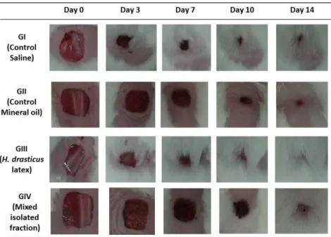
mg/mL). The treatments were applied topically once daily, 50 µL for 14 consecutive days. Mice bearing skin lesions were placed in an apparatus that allowed image capture at a fixed height of 22 cm for morphometric analysis using the Image J® software (Garros et al., 2006). Macroscopic evaluation was performed daily, considering parameters such as swelling, redness, granulation tissue and reepithelialization (Oliveira et al., 2010).
Skin samples were collected from animals on days 0, 3, 7, 10 and 14 and were subjected to histology (H&E) to evaluate the parameters as reepithelization, ulceration, necrosis; congestion, edema, fibroblast proliferation, mononuclear and polymorphonuclear cells and neovascularization that were graded as absent, mild, moderate or intense for dermal or epidermal remodeling. All sections were blindly assessed by the same investigator (Akkol et al., 2009) under a light microscope (Nikon Eclipse E200®). To evaluate the extracellular collagen matrix, tissue sections were stained with red picrosirius and analyzed by polarized light microscopy (Rich and Whittaker, 2005).
The immunohistochemical evaluations were conducted in paraffin embedded skin sections for VEGF+ (clone VG1; Dako®) and CD68+ (SC-59103 clone, Santa Cruz Biotechnology®). For this, 5 µm sections were mounted on silanized glass slides and subjected to antigen retrieval process (Dako® EnVision TMFLEX Target Retrieval Solution High pH Code DM828) or low pH (Code DM829) for 20 min at 97ºC using the Dako pre-treatment (PT) link module (Dako®, Glostrup, Denmark). The endogenous peroxidase activity was inhibited by peroxidase block (Dako®) for 5 min, and slides received the anti-human CD68+ murine monoclonal antibody diluted 1:100; and anti-human VEGF+ murine monoclonal antibody diluted 1:100 and incubated for 1 h at room temperature. Then, slides were washed three times in phosphate buffered saline (PBS, pH 7.2), and then incubated with the reagent polymer (EnVision TMþ Dual LinkSystem/HRP; Dako®) for 30 min at room temperature and finally diaminobenzidine (DAB, Dako®) for 10 min.
The sections were counterstained with Mayer's hematoxylin and observed by polarized light microscopy using the scores absent, mild, moderate or intense. In order to obtain the scores, all slides were analyzed by the same observer and compared to the control group.
In order to evaluate mast cells participation in wound healing process, mice paws were subjected to conventional histological processing, stained with toluidine blue according the scores absent, mild, moderate and intense (Farahani et al., 2010).
Data were presented as mean ± standard deviation. Data were previously subjected to Grubbs test for outliers exclusion. Then, the Kolmogorov-Smirnov test and ANOVA for homoscedasticity and homogeneity evaluation were used. For wound contraction analysis, Kruskal-Wallis test followed by Dunn (P<0.05) were employed.
RESULTS
HdCL showed pinkness supernatant, milky appearance and whitish sedimentation. In phytochemical qualitative analysis, it was detected the presence of the following compounds: phenols, flavanonois, flavonols, flavanones and free steroids. No bacterial or fungi agents that could compromise the healthiness of the compounds used were identified.
series. The cinnamoyl moiety was confirmed by the following chemical shifts in δ128.2 (C-2’ and C-6’), δ129.0 (C-3’and C-5’), δ130.3 (C-4’) and δ134.8 (C-1’) of an aromatic ring and in δ119.1, δ144.4 e δ167.0 from the unit (CH=CH-COO). Then, the compound isolated from Janaguba
latex analyzed by 13C- and 1H NMR spectroscopy showed to be a mixture of three cinnamoyl derivatives of lupeol (1), α-amyrin (2) and β-amyrin (3) (Figure 1C). All the assignments of 13C and 1H NMR spectra of three compounds are shown in Table 1.
Table 1. 1H and 13C NMR spectra assignments of the mixture of three cinnamoyl derivatives of lupeol (1), α-amyrin (2) and β-amyrin (3) obtained from H. drasticus latex
Carbon cinnamate Lupeol cinnamate α-Amyrin β-Amyrin cinnamate δ1H
(300 MHz) δ
13C
(75 MHz) δ
1H
(300 MHz) δ
13C
(75 MHz) δ1H (300 MHz) δ13C (75 MHz)
1 38.6 38.5 38.7
2 23.6 23.4 23.7
3 81.2 4.66 (m, J= 6.1;
15.0) 81.2 4.66 (m, J= 6.1; 15.0) 81.2
4 37.6 37.9 38.3
5 55.5 0.79 (d, J=13.5) 55.6 0.79 (d, J=13.5) 55.8
6 17.7 18.5 18.2
7 34.4 32.8 32.7
8 41.7 40.2 40.0
9 50.6 47.8 47.9
10 37.1 37.3 37.6
11 21.2 17.1 17.7
12 26.8 5.18 (dt, J=3.9) 124.5 5.18 (dt, J=3.9) 121.9
13 38.1 139.8 145.4
14 41.9 42.3 43.5
15 27.6 1.94 (td, J= 3.3;
10.6) 28.6 1.94 (td, J= 3.3; 10.6) 28.3
16 35.8 1.72 (dd, J=5.0;
11.0) 26.3 1.72 (dd, J=5.0; 11.0) 26.1
17 47.0 33.6 33.5
18 48.2 59.3 59.3
19 47.8 39.8 39.8
20 15.1 39.8 39.8
21 29.9 31.2 31.4
22 39.8 1.82 (dt, J=3.0;
8.0) 41.7 1.81 m 41.9
23 28.2 0.94 s 28.3 0. 93 s 28.9
24 15.9 0.77 s 16.9 0.90 s 16.9
25 16.2 0.81 s 15.7 0.77 s 15.9
26 16.4 0.86 s 16.9 0.94 s 15.8
27 14.7 1.01 s 23.6 1.17 s 23.9
28 17.7 0.94 s 28.3 1.06 s 28.6
29 4.59 s; Hα
4.67 s; Hβ 109.6 0.83 s 23.7 0.86 s 23.4
30 21.6 0.77 s 21.5 0.81 s 21.5
1’ 134.8 134.8 134.8
2’ 128.2 128.2 128.2
3’ 129.0 129.0 129.0
4’ 130.3 130.3 130.3
5’ 129.9 129.9 129.9
6’ 128.2 128.2 128.2
7’ 7.71 (d, J=16.0) 144.4 7.66 (d, J=16.0) 144.4 7.66 (d, J=16.0) 144.4 8’ 6.49 (d, J=16.0) 119.1 6.44 (d, J=16.0) 119.1 6.44 (d, J=16.0) 119.1
Figure 1. (A) 1H NMR spectra of the mixture of three cinnamoyl derivatives of lupeol (1), α-amyrin (2) and β-amyrin (3) (CDCl3, 300 MHz). (B) 13C NMR spectra of a mixture of three cinnamoyl derivatives of lupeol (1), α-amyrin (2) and β-amyrin (3) (CDCl3, 75 MHz). (C) Structural representation of compounds: cinnamoyl derivatives of lupeol (1), α-amyrin (2) and β-amyrin (3), obtained from H. drasticus latex.
Wound contraction results are shown in Tab. 2. HdCL induced higher contraction on days 3, 7 and 10, in relation to MIF, but did not differ from negative control. Wound healing macroscopical analysis on different groups was shown in Figure 2. The granulation tissue formation was more exuberant in GII and GIV on days 3, 7 and 10;
presented it discretely in GI and GIII on days 3 and 7, where the fur growth showed up at an accelerated rate. GIV present granulation tissue formation more exuberante, while GIII presented a more accelerated re-epithelialization than other groups.
(A)
(B)
(C)
1
2
Table 2. Effect of HdCL and MIF on the wound contraction in excision model Contraction (%) – Treatments
Day Saline 0.9% Mineral Oil HdCL MIF
0 0 0 0 0
3 35.79 ± 12.44b 4.73 ± 10.15a 37.83 ± 23.30b 3.79 ± 6.84a 7 55.72 ± 13.11b 21.24 ± 5.68a 69.24 ± 18.12b 14.56 ± 10.51a 10 76.97 ± 14.46a 60.35 ± 20.59ab 81.56 ± 15.77a 45.32 ± 26.89b 14 99.11 ± 1.08a 95.68 ± 5.11a 97.07 ± 7.76a 97.32 ± 2.53a
Different letters in the same row mean significant difference among treatments (P < 0.05).
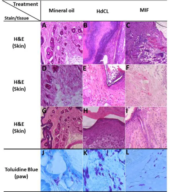
To evaluate the microscopic findings, conventional histological processing were performed, where the main findings are presented in Fig. 3. GIII showed a mild inflammatory process (Fig. 3B). On day 14, the inflammation was already solved in GIII, with reepithelialization (Fig. 3B) and return to tissue integrity. HdCL induced even complete epithelialization on day 14, as can be seen in Fig. 3E. There was intense inflammation in GIV characterized predominantly by lymphocytes, vascular congestion and the red blood cells presence (Fig. 3C). Treatment with MIF delayed the healing process, which, although it was resolved on day 14, did not have full reepithelialization (Fig. 3I). Neovascularization was observed in MIF (Fig. 3F).
Mast cells analysis is presented in Fig. 3. Treatments induced mast cells influx to lesion
tissue in different cellularity degrees. GI was described as absent (data not shown), GII as mild (Fig. 3J), GIII as intense (Fig. 3L) and GIV as moderate (Fig. 3K).
VEGF+ and CD68+ expressions are shown in Fig. 4 and were assessed on day 7. CD68+ expression is presented as moderate, intense and mild intensity in control, HdCL and MIF groups, respectively (Fig. 4A, B and C). Treatment with MIF induced more intense VEGF+ expression when compared to treatment with HdCL, presented as moderate (Fig. 4D, E and F).
Collagen analysis is shown in Fig. 4. On day 7, the animals that underwent treatments showed disorganized collagen fibers (Fig. 4G and H). On day 14, reorganization of collagen fibers and tissue remodeling were observed in all groups (Fig. 4J and K).
DISCUSSION
Plants are excellent sources of compounds that present many properties such as antibiotics, hemostatics, anti-oxidants, anti-inflammatory and healing potential (Bhagyashri et al., 2015, Parwani et al., 2012). Angiosperms produce an exudate known as latex that is present at Euphorbiaceae, Moraceae, Asclepiadaceae, Apocynaceae, and some members from the Compositae family (Rajesh et al., 2007).
HdCL used in our study can appear whitish, yellowish or pink colored, and phytochemical compounds identified are consistent with other studies that detected proteins, alkaloids, tannins, terpenes, sugar, starch, oils, resins, enzymes, rubber, terpenoids, phenolic compounds and proteases (Rajesh et al., 2007; Badgujar, 2014). The highly diversified molecular profile of plant latex allows it to modulate inflammatory agents (Arya and Kumar, 2005; Fernandes et al., 2015; Kumar et al., 2015).
Figure 4. Effect of HdCL and MIF on CD68+ (A, B, C) and VEGF+ (D, E, F) expressions. Staining intensity was scored as mild (C, D, E), moderate (A, F) and intense (B). Extracellular matrix organization (G, H, I) and 14 (J, K, L), disorganization (G, H, I) and reorganization (J, K, L) collagen fibers. 100x magnification. Images were assessed on day 7 (4A to 4H) and on day 14 (4J, 4K).
In our study, the wound healing process induced by topical application of HdCL increased the production of granulation tissue when compared with topical application of MIF, although both induced inflammatory cells recruitment (Fig. 2 and 3). It is noteworthy that even though the wound contraction process was delayed until day 10, data suggests that there was no negative interference in the healing process resolution, which was already resolved by day 14. We must emphasize that for wound contraction it was taken into account not only the regression of the
area lesion, but macroscopic, histologic and cellular aspects and reorganization of collagen fibers, as shown is figures 2, 3 and 4. the sum of all factors corroborate the inference of acceleration of wound healing, interpreted by the production of scab and granulation tissue, angiogenesis and modeling of all epithelial layers.
predominant in MIF. These data suggest recruitment and activation of sentinel cells that express CD68+, which are primarily macrophages, dendritic cells (Vinish et al., 2016) and mast cells. The increase of CD68+expression found in our study can be attributed not only to the recruitment of inflammatory cells to injured site but also to activation of these cells, although the mechanisms are unknow. Analougosly, C.
procera latex increased macrophage influx
(Seddek et al., 2009). Unfortunately, there is a lack of studies that correlates use of plant latex with CD68+ expression, but the anti-inflammatory properties of several plant latex suggest that there is inhibition of CD68+ cells associated to their use. E. lactea (Euphorbiaceae) potently inhibted macrophage activation (Fernandez-Arche et al., 2010), while Hancornia
speciosa (Apocynaceae) inhibited cell migration,
including neutrophils and macrophages (Marinho
et al., 2011), which were atributed to triterpenes
isolated from latex. The possible difference between the results found in many latex products suggests that the presence of biomolecules in the latex composition can give different outcomes.
Angiogenesis is a very important parameter on wound healing. MIF stimulate VEGF+ expression (Fig. 4F). In different stages of healing process, VEGF is produced by several cells, such as polymorphonuclear, mononuclear and endothelial cells (Bao et al., 2009). In the present work, wound contraction was delayed, which may be related to the observed cell type in the inflammatory infiltrate (Fig. 3B and 3C). This process is initially influenced by cytokines and growth factors, such as VEGF+, culminating with the advent of new blood vessels, which transport nutrients and inflammatory cells, accelerating the recovery of damaged tissue (Brown et al., 2002). Thus, we can then infer that the latex pro-inflammatory properties have a direct impact on their ability to modulate the scarring process.
CONCLUSION
Based on our results, H. drasticus commercial latex presents wound healing potential. It can be inferred that HdCL positively modulates the wound healing parameters by the participation of mast cells, CD68+ and VEGF+ expressions that can be associated to presence of lupeol, α-amyrin and β-amyrin contained in MIF. For its
application in veterinary medicine, it is necessary studies in the skin tissue of different species, in order to justify the use of commercially available phytomedicines and the easy access to the population and veterinary clinical staff.
REFERENCES
AKKOL, E.K.; KOCA, U.; PESIN, I. et al. Exploring thewound healing activity of Arnebia
densiflora (Nordm.) Ledeb by in vivo models. J.
Ethnopharmacol., v.124, p.137-141, 2009.
ARYA, S.; KUMAR, V.L. Antiinflammatory efficacy of extracts of latex of Calotropis
procera against different mediators of
inflammation. Mediat. Inflamm., v.4, p.228-232, 2005.
BADGUJAR, S.B. Evaluation of hemostatic activity of latex from three Euphorbiaceae species. J. Ethnopharmacol., v.151, p.733-739, 2014.
BALDAUF, C.; SANTOS, F.A.M. Ethnobotany, Traditional knowledge, and diachronic changes in non–timber forest products management: a case study of Himatanthus drasticus (Apocynaceae) in the Brazilian Savanna. Econ. Bot., v.67, p.110-120, 2013.
BAO, P.; KODRA, A.; TOMIC-CANIC, M.B.A. et al. The role of vascular endothelial growth factor in wound healing. J. Surg. Res., v.153, p.347-358, 2009.
BHAGYASHRI, C.; JOGENDRA, H.; AVINASH, P. Plant latex: an inherent spring of pharmaceuticals. World J. Pharm. Sci., v.4, p.1781-1796, 2015.
BROWN, N.J.; SMYTH, E.A.E.; CROSS, S.S. et al. Angiogenesis induction and regression in human surgical wounds. Wound Repair Regen., v.10, p.245-251, 2002.
FARAHANI, S.S.; NAVABAZAM, A.; ASHKEVARI; F.S. Comparison of mast cells count in oral reactive lesions. Pathol. Res. Pract., v.206, p.151-155, 2010.
FERNANDEZ-ARCHE, A.; SAENZ, M.T.; ARROYO, M. et al. Topical anti-inflammatory effect of tirucallol, a triterpene isolated from
Euphorbia lactea latex. Phytomedicine, v.17,
FERNANDES, H.B.; MACHADO, D.L.; DIAS, J.M. et al. Laticifer proteins from Plumeria
pudica inhibit the inflammatory and nociceptive
responses by decreasing the action of inflammatory mediators and pro-inflammatory cytokines. Rev. Bras. Farmacogn, v.25, p.269-277, 2015.
GARROS, I.C.; CAMPOS, A.C.L.; TÂMBARA, E.M. et al. Extrato de Passiflora edulis na cicatrização de feridas cutâneas abertas em ratos: estudo morfológico e histológico. Acta Bras. Cir., v.21, p.55-65, 2006.
KUMAR, V.L.; GURUPRASAD, B.; CHAUDHARY, P. et al. Protective effect of proteins derived from Calotropis procera latex against acute inflammation in rat. Auton
Autacoid Pharmacol., v.35, p.1-8, 2015.
LUCETTI, L.D.; LUCETTI, E.C.P.; BANDEIRA, M.A.M. et al. Anti-inflammatory effects and possible mechanism of action of lupeol acetate isolated from Himatanthus drasticus (Mart.) Plumel. J. Inflamm., v.7, p.60-71, 2010.
MARINHO, D.G.; ALVIANO, D.S.; MATHEUS, M.E. et al. The latex obtained from
Hancornia speciosa Gomes possesses
anti-inflammatory activity. J. Ethnopharmacol., v.135, p.530-537, 2011.
MOUSINHO, K.C.; OLIVEIRA, C.C.; FERREIRA, J.R. et al. Antitumor effect of laticifer proteins of Himatanthus drasticus (Mart.) Plumel – Apocynaceae. J.
Ethnopharmacol., v.137, p.421-426, 2011.
OKOLI, C.O.; AKAH, P.A. Mechanisms of the anti-inflammatory activity of the leaf extracts of
Culcasia scandens P. Beauv (Araceae).
Pharmacol. Biochem. Behav., v.79, p.473-481,
2004.
OLIVEIRA, A.P.; SILVA, L.R.; FERRERES, F.
et al. Chemical assessment and in vitro
antioxidant capacity of Ficus carica latex. J.
Agric. Food Chem., v.58, p.3393-3398, 2010.
OLIVEIRA, S.G.D.; MOURA, R.R.F.; DEMARCO, F.F. et al. An ethnomedicinal survey on phytotherapy with professionals and patients from Basic Care Units in the Brazilian Unified Health System. J. Ethnopharmacol., v.140, p.428-437, 2012.
PARWANI, L.; BHATNAGAR, M.; BHATNAGAR, A. et al. Reactive oxygen species control by plant biopolymers intended to be used in wound dressings. Int. J. Pharm.
Pharm. Sci., v.4, p.506-510, 2012.
PINHEIRO, E.A.R.; COSTA, C.A.G.; ARAÚJO, J.C. Effective root depth of the Caatinga biome. J. Arid Environ., v.89, p.1-4, 2013.
RAJESH, R.; SHIVAPRASAD, H.V.; RAGHAVENDRA, C.D. et al. Comparative study on plant latex proteasis and their involvement in hemostasis: a special emphasis on clot inducing and dissolving properties.
Planta Med., v.73, p.1061-1067, 2007.
RICH, L.; WHITTAKER, P. Collagen and picrosirius red staining: a polarized light assessment of fibrillar hue and spatial distribution. Braz. J. Morphol. Sci., v.22, p.97-104, 2005.
SEDDEK, A.L.S.; MAHMOUD, M.E.; SHIINA, T. et al. Extract from Calotropis procera latex activates murine macrophages. J. Nat. Med., v.6, p.297-303, 2009.