Expression of enterovirus 71 capsid protein VP1 in
Escherichia coli
and its clinical
application
Mei Shi
1, Yaping Zhou
2, Limin Cao
2, Cuijun Ding
3,Yun Ji
1, Qinbo Jiang
1, Xiping Liu
1,
Xiang Li
1, Xueling Hou
1, Hongjun Peng
1, Weifeng Shi
11
Department of Clinical Laboratory, The Third Affiliated Hospital of Suzhou University, Changzhou, Jiangsu, P.R. China.
2
Changzhou 21st Century Biotech Research Institute, Changzhou, Jiangsu, P.R. China. 3
Department of Clinical Laboratory, Changzhou Children’s Hospital, Changzhou, Jiangsu, P.R. China.
Submitted: April 27, 2012; Approved: April 04, 2013.
Abstract
The VPl gene of enterovirus 71 (EV71) was synthesized, construct a recombinant plasmid pET15b/VP1 and expressed inE. coliBL21. The recombinant VP1 protein could specifically react with EV71-infected patient sera without the cross-reaction with serum antibodies of coxsackievirus A16 (CA16), A4, A5, B3 and B5 as well as echovirus 6. In acute and convalescent phases, IgM and IgG antibodies of 182 serum samples were detected by ELISA with recombinant VP1 protein as a coated antigen. The results showed that the sensitivity, specificity, positive predictive value (PPV) and negative predictive value (NPV) of IgM antibodies in serum samples for the diagnosis of EV71 infection were 90.1, 98.4, 98.8 and 88.7%, respectively; similarly, those of IgG antibodies in serum samples were 82.4, 89.1, 91.5 and 78.1%, respectively. Five of 80 samples (6.25%) from CA16-infected patients were detected positive by ELISA with recombinant VP1 protein in which indicated the cross reactions and 0 of 5 samples from patients infected with other enteroviruses including CA4, CA5, CB3, CB5 and echovirus 6. Therefore, the recombinant VP1 protein of EV7l may provide a the-oretical reference for establishing an effective antibody screening of IgM for EV71-infected patients with clinically suspected hand, foot, and mouth disease (HFMD).
Key words:Enterovirus 71, Gene cloning, Recombinant VPl protein, ELISA.
Introduction
EV71 is one of the most important pathogens in the family ofPicornaviridaethat can cause severe complica-tions, from mild HFMD to severe neurological syndromes, such as encephalitis, pulmonary edema, and even death. Outbreaks of EV71 infection have been reported around the world since 1969.(Melnick et al., 1974; Schmidt et al., 1974; Herreroet al., 2003; Wuet al., 2010; Zhanget al., 2010) It often causes a mild illness, and most patients usu-ally recover quickly. However, in the past decade, a signifi-cant increase in EV71 epidemics is observed, and it has emerged as a serious threat to public health throughout the Asia-Pacific region, such as Taiwan, Malaysia and Singa-pore as well as Guangdong, Hunan, Jiangsu and Fuyang in
China.(Yanet al., 2000; Chanet al., 2003; Herrero,2003; Maoet al., 2010; Zhang,2010; Zhuet al., 2010) EV71 and CA16 infections mainly occur in children under 5 years old. However, patients infected with EV71 are more liable to develop severe complications, including encephalitis, asep-tic meningitis, pulmonary edema or haemorrhage, and acute flaccid paralysis. (Iwaiet al., 2009; Ooiet al., 2010; Solomonet al., 2010)
EV71 is a small genus of human enterovirus RNA vi-rus family A, and closely related to CA16. EV71 possesses a single-stranded RNA genome of approximately 7400 bp, consisting of a single open reading frame (ORF) flanked by 5’-untranslated regions (5’-UTR) and 3’-untranslated re-gions (3’-UTR).(Chuaet al., 2008) The ORF is expressed as a large polyprotein that can be cleaved into P1, P2, and
Send correspondence to W. Shi. Department of Clinical Laboratory, Third Affiliated Hospital of Suzhou University, No. 185 Juqian Road, Changzhou 213003, P.R. China. E-mail: [email protected].
P3 regions. The P1 region encodes four structural proteins including VP1, VP2, VP3, and VP4.(Lalet al., 2006) The P2 and P3 regions encode nonstructural proteins, such as proteases 2A, 2B and 3CD, responsible for virus replication and virulence. Variation of capsid proteins, except VP4, is responsible for the antigenic diversity among entero-viruses, but neutralizing epitopes reside mainly on VP1. (Fooet al., 2008)
Traditional detection of EV71 infection is primarily dependent on virus cultivation, serodiagnosis and real-time PCR assays.(Liet al., 2002; Singhet al., 2002; Solo-mon,2010) However, virus culture and real-time PCR as-says are time-consuming and need special facilities (Rigo-nanet al., 1998; Zhanget al., 2009; Chenet al., 2011). Early diagnosis can be helpful for the adminstration of ap-propriate treatments which may limit the spread of this vi-rus and reduce the mortality of patients. In this study, the entire VP1 gene of EV71 was synthesized and expressed inE. coliBL21 (DE3). The capsid VP1, as a natural pro-tein with molecular mass of 36 kDa evaluated by SDS-PAGE, had the desired immunogenicity against EV71 an-tibody. The aim of this study is to obtain a recombinant VP1 antigen for establishing a rapid serological test for the diagnosis and epidemiological investigation of EV71 infection.
Materials and Methods
Specimen collection
From March to September of 2009, a total of 176 rec-tal and 176 throat swabs were collected from 176 patients with HFMD under the age of 5 years old enrolled in Changzhou Hospital in China. During acute (0-5 days) and convalescent (14-30 days) phases, 182 serum samples were harvested in duplicates for the detection of IgM and IgG an-tibodies. 64 control serum samples were collected from healthy children with the mean age of 2.5±1.3 years old. These children showed no disease symptoms and didn’t present with a previous history of EV71 and CA16 infec-tion at the time of sample harvesting. In addiinfec-tion, 80 serum samples from CA16-infected patients (0-5 days) with the mean age of 2.5±1.3 years old. The harvested serum sam-ples were stored at -80 °C for future use. CA16 serum (horse) was provided by American Type Culture Collection (ATCC). Five serum samples collected from patients in-fected by coxsackievirus A4 (CA4), coxsackievirus A5 (CA5), coxsackievirus B3 (CB3), coxsackievirus B5 (CB5) and echovirus 6 were gifts from Changzhou Center for Dis-ease Control and Prevention, and Nanjing Medical Univer-sity. This study was approved by the local ethics committee and all parents/guardians of children were provided with a description of the study and were asked to give infromed consents.
Detection of rectal and throat swabs by PCR fluorescence probing assay
Fecal specimens were mixed thoroughly with 5 to 10 volumes of phosphate-buffered saline (PBS) (pH 7.4) to generate homogeneous suspensions. The mixtures were clarified by centrifugation at 13, 000 g for 5 min. Viral RNA was extracted from supernatants of fecal suspensions and throat swabs using QIAamp viral RNA Mini kit (QIAGEN, Germany) according to the manufacturer’s in-structions. The RNA was eluted from the QIAspin column in a final volume of 100mL of elution buffer and kept at -80 °C until further analysis. The PCR fluorescence prob-ing assay reagent (DaAn Gene, China) for EV71 and CA16 were commercially available. The cDNA was generated in a 20mL of reaction volume for 30 min at 40 °C using ran-dom primers and SuperScript II reverse transcriptase (Invi-trogen, USA) according to the instructions. The EV71 cycling conditions were composed of 5 min at 94 °C, fol-lowed by 40 cycles with 93 °C for 15 s, 55 °C for 45 s and 72 °C for 1 min, and a final extension cycle at 72 °C for 10 min. The primers of EV71/VP1 were designed according to the complete gene sequences (2800-2930 bp) published in GenBank (accession No. AY465356). Forward primer EV71-F: 5’-AAA GGT GGA GCT GTT CAC CTA CAT GCG CTT TGA C-3’, reverse primer EV71-R: 5’-AAT CTG GCT TGG GGG CCC CAG GTG GTA CAA-3’, and oligonucleotide probe EV71-P: 5’-CCC ACC GGG GAA GTT GTC CCA CAA TTG CTC C-3’. The CA16 cycling conditions were composed of 3 min at 94 °C, followed by 40 cycles with 93 °C for 15 s, 55 °C for 45 s and 72 °C for 1 min, and a final extension cycle at 72 °C for 10 min. The primers of CA16 were designed according to the complete gene sequences (1909-1970 bp) published in GenBank (ac-cession No. EU262658). Forward primer CA16-F1: 5’-CAT GCA GCG CTT GTG CTT-3’, CA16-F2: 5’- CAT GCA ACG ACT GTG CTT TC-3’. Reverse primer CA16-R1: 5’-CAC ACA ATT CCC CCG TCT TAC -3’, and CA16-R2: 5’-CAT AAT TCG CCC GTT TTG CT-3’.
Virus isolation and identification
Clinical specimens including rectal swabs and throat swabs were inoculated into rhabdomyosarcoma (RD) cells and human laryngeal carcinoma (Hep-2) cells (Chinese Academy of Sciences Cell Bank of Type Culture Collec-tion, CBTCCCAS) for the isolation of EV71. Cytopathic effects (CPE) were examined under an inverted microscope after 2 to 7 days. Enterovirus strains were identified by immunofluorescence test using EV71 monoclonal antibody (Chemicon International Inc).
Construction of expression vector
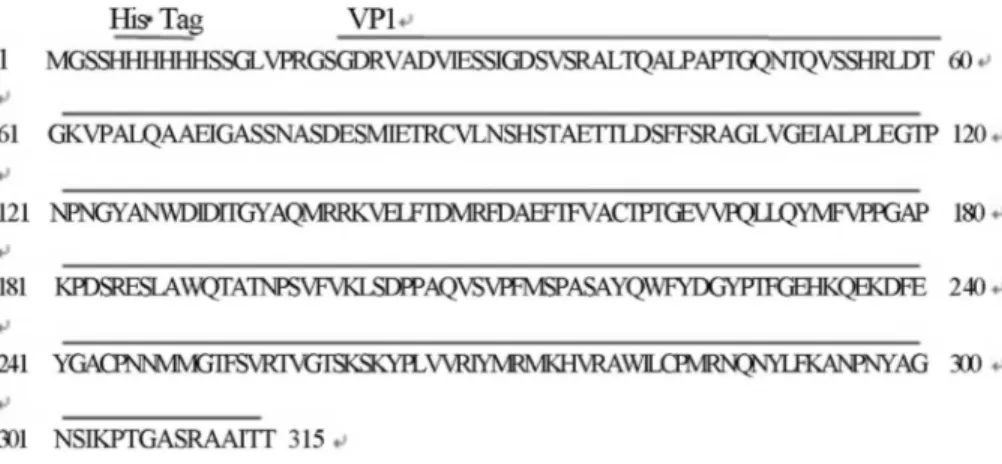
AY465356 (Human enterovirus 71 strain SHZH03). The amino acid sequence of recombinant VPl fusion protein was shown in Figure 1. The NdeI and XhoI restriction endonuclease (Promega, USA) sites in primers were then introduced to allow ligation of the entire VP1 cDNA into pET15b (Novagen, Germany), a prokaryotic expression vector containing an amino-terminal histidine tag.
The expression and purification of VP1 recombinant proteins
The synthesized VP1 gene was verified by DNA se-quencing and restriction endonuclease digestion before ex-pression. CompetentE. coliBL21 (DE3) cells (Novagen, Germany) were transformed with pET15b expression vec-tor harboring VP1 cDNA (pET-VP1). E. coli cells were grown in shaker flasks with LB broth medium containing 50mg/mL ampicillin at 37 °C until optical density (OD) of 0.6 was reached. Then, isopropyl-b -D-thiogalactopyra-noside (IPTG) was added to the LB medium at the final concentration of 1 mM to induce the expression of His-VP1 fusion protein. At 0, 1, 2 and 4 h after induction, the cells were harvested and centrifuged at 3,200 g for 10 min and washed once with PBS. Subsequently, the cells were sub-jected to rapid freeze-thaw treatment at -80 °C twice and then resuspended in PBS buffer. In addition, cell mem-branes were disrupted by sonication at 180 Hz, and then the precipitates and supernatants were collected at 10,310 g for 30 min at 4 °C. The Ni2+ chromatography columns(GE Healthcare, USA) were washed with 25% ethanol and dou-ble-distilled water, and then equilibrated with 30 mL buf-fer. The supernatants were filtered by a 0.22mm-pore-size filter and flow-through chromatography column. 20 mL of buffer containing 500 mmol / L imidazole for elution and collecting 1 mL / tube each. 5mL of samples were taken from each tube and analyzed by 15% SDS-PAGE. The pu-rified protein desalination was completed by HiTrap De-salting column (GE Healthcare, USA) according to the manufacturer’s instructions.
Western blot analysis
The purified VP1 proteins were dissolved in SDS-PAGE sample buffer and then heated at 95 °C for 5 min. The denatured proteins were subjected to SDS-PAGE (15% polyacrylamide) separation and then transferred to the nitrocellulose membrane (Generay, Shanghai) by elec-troblotting for 1 h at 1 Amp. After blocking with diluted Tris-buffered saline containing 0.05% Tween-20 (TBST) and 5% skim milk for 1 h, rabbit anti-His-tag polyclonal an-tibody (Santa Cruz Biotechnology, CA) was added at a di-lution ratio of 1:1,000 in TBST sodi-lution at 37 °C for 1 h. After washing with TBST, goat anti-rabbit IgG (H+L) con-jugated with horseradish peroxidase (Santa Cruz Biotech-nology, CA) as secondary antibody, was added at the dilution ratio of 1:1,000 and incubated at 37 °C for 1 h, and then incubated with 4-chloro-1-naphthol solution for 5-10 min. The cell culture of pET15b/VPl-BL21 without induction was used as the negative control.
The serum cross-reaction of CA16 and other non-EV71 enteroviruses
The purified VP1 protein was examined by Western blot. IgM and IgG positive serum sample of CA16 and 5 se-rum samples collected from patients infected by other enteroviruses (CA4, CA5, CB3, CB5, and echovirus 6) were diluted at a ratio of 1:500. Goat anti-human conju-gated with HRP (Santa Cruz Biotechnology, CA) was used as the secondary antibody. IgM and IgG positive serum samples of EV71 were used as the positive controls.
Detection of EV71 IgG or IgM antibody by ELISA with recombinant VP1 protein
A 96-well microtiter plate was coated with 100mL of recombinant VP1 protein (10mg/mL) in carbonate coating buffer (15 mM Na2CO3, 35 mM NaHCO3, pH 9.6). The plates were incubated at 4 °C overnight, and then washed twice with phosphate-buffered saline containing Tween-20 (PBS-T). The plates were incubated with 1% bovine serum albumin in PBS for 2 h at room temperature to prevent
non-specific binding. Totally 100mL of 1:300 diluted se-rum sample was added to each well and incubated at 37 °C for 1 h. The plate was washed for 4 times with PBS-T, fol-lowed by horseradish peroxidase conjugated goat anti-human IgG or IgM (1: 2,000 dilution, Santa Cruz Biotech-nology, CA). The reaction was developed by 100mL of 3, 3’, 5, 5’-tetramethyl benzidine (TMB) substrate, and then terminated by 100mL of 2 M H2SO4. The OD at 450 nm were determined.
Statistical analysis
The OD values of IgM and IgG antibodies between healthy objects and HFMD patients were compared by un-paired Studentt-tests. A significant difference was consid-ered atPvalue less than 0.05. The sensitivity, specificity, PPV and NPV were evaluated for the diagnostic value of anti-EV71 IgM and IgG antibodies. The diagnostic effi-ciency of EV71 infection between IgM and IgG antibodies was assessed by receiver operating characteristic curve (ROC) analysis.
Results
PCR fluorescence probing assay
Rectal and throat swabs were collected from 176 pa-tients with HFMD .Of these, 91 papa-tients were detected pos-itive by PCR fluorescence probing assay, as well as further verified by virus culture, immunofluorescence test and clinical manifestations.
Construction of recombinant EV71 pET15b/VP1 plasmid for protein expression
In order to construct expression plasmid of pET15b/VP1 according to VP1 gene sequence, the VP1 gene was amplified and sequenced with the expected length of 891-bp VP1 sequence, and then cloned in pET15b vector digested by NdeI and XhoI to obtain recombinant pET15b/VP1 plasmid for protein expression (Figure 2).
Expression of recombinant VP1 protein
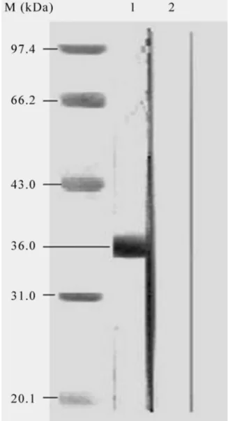
The recombinant pET15b/VP1 plasmid was trans-formed intoE. coliBL-21 (DE3) strain grown on LB me-dium containing ampicillin at 37 °C and the expression of VP1 protein was induced by 1 mM IPTG. The VP1 was produced as a fusion protein with a six-histidine tag at its amino terminus. A band with an approximately 36 kDa from the cell culture induced by IPTG in 15% SDS-PAGE was observed, which is consistent with the expected molec-ular mass of VP1 fusion protein (Figure 3).
Detection of purified VP1 protein
The purified protein was confirmed by Western blot. The result showed that the recombinant VP1 protein re-vealed a specific band with expected molecular mass of 36 kDa, which was specifically recognized by anti-His
poly-clonal antibody. However, in the negative control, empty vector pET-15b could not be induced to express VP1 pro-tein so that it did not have a specific band shown in Western blot (Figure 4).
The serum cross-reaction of EV71 and other enteroviruses
In order to evaluate the cross-interaction, Western blot analyses for recombinant VP1 antigen using serum samples from patients with EV71 and CA16 infections, as well as other 5 enteroviruses including CA4, CA5, CB3, CB5 and echovirus 6 were conducted. The experiments were repeated three times and the results showed that there was no cross-reaction between recombinant VP1 protein and other positive enterovirus serum samples (Figure 5).
Figure 2- The electrophoresis diagram of pET-15b/VP1 and Digestion with NdeI and XhoI. M: Marker, DNA ladder. Lane 1: pET-15b/VP1; Lane 2: pET-15b/VP1 digested by NdeI and XhoI.
Detection of patient sera with HFMD
The serum samples from 91 patients and 64 healthy subjects were determined by ELISA with purified VP1. However, all serum samples tested in this study were nega-tive for rheumatoid factor and antinuclear factor. There were no cross-reaction between recombinant VP1 protein and CA16 positive serum samples as well as 5 samples from patients infected with other enteroviruses. The OD values of anti-EV71 IgM and IgG in patients were signifi-cantly elevated when compared with that of healthy sub-jects (p < 0.001). Compared with PCR fluorescence probing assay, virus culture, immunofluorescence test and clinical manifestation, ROC was determined by OD values of EV71 IgM and IgG antibodies in patients and controls, and their areas under curve (AUC) were 0.958 and 0.929, respectively (Figure 6). If a cutoff value of 0.0955 was
adopted, EV71 IgM antibody had a high specificity (98.4%) and a sensitivity (90.1%), as well as PPV (98.8%) and NPV (88.7%). Among 91 serum samples from patients with EV71 infection in acute phase, 9 serum samples were detected as IgM negative but positive for virus culture. Similarly, among 64 healthy subjects, 1 serum sample was tested positive for IgM antibody (OD = 0.147), however, it was confirmed to be a false positive by PCR fluorescence probing assay, virus culture and indirect immunofluo-rescence tests. Among 80 samples from CA16-infected pa-tients, 5 were detected IgM antibody positive, which showed that the cross reactions existed. In addition, 5 sam-ples from patients infected with other enteroviruses were negative. Therefore, the sensitivity, specificity, PPV and NPV of the VP1 against IgG were 82.4, 89.1, 91.5 and 78.1% at a cutoff value of 0.135 (Table 1).
Discussion
HFMD, a common illness in children, can be caused by many human enteroviruses, such as coxsackievirus A4, A5, A8, A10, A16, B3 and EV71 (Yoke-Fun and Abu-Bakar, 2006; Wu, 2010) Human EV71 and CA16 are two major causative agents of HFMD. EV71 and CA16 infec-tions manifesting as HFMD and herpangina are clinically indistinguishable, but EV71 infection is more frequently associated with serious neurological complications and fa-talities.(Nagataet al., 2004; Liet al., 2005; Xuet al., 2010) Thus, rapid discrimination between EV71 and CA16 is highly desired during HFMD outbreaks. EV71 genome en-codes VPl, VP2, VP3 and VP4 capsid proteins. VPl, VP2 and VP3 are exposed to the virus surface, while VP4 is em-bedded in the virus capsid. In particularly, VPl protein is
Figure 4- Western blot analysis of purified recombinant VP1 protein. M: Protein marker (kDa); Lane 1: purified VP1 protein; Lane 2: pET-15b vec-tor control.
the major neutral epitope of EV71. VPl gene is by far be-coming the major target for molecular epidemiology re-search and vaccine. (Chenet al., 2008; Zhang &Lu,2010) In this study, whole VP1 gene of EV71 was cloned and ex-pressed inE. coli. The VP1 fusion protein with a histidine tag had an estimated molecular mass of 36 kDa, and was expressed inE. coli with IPTG induction. The immuno-genicity of purified VP1 protein was tested by Western blot analysis using human serum samples containing IgG or IgM antibodies of CA16, CA4, CA5, CB3, CB5 and echo-virus 6. Experimental results indicated that, although the serum samples from the patients infected with entero-viruses had a high titer of IgM and IgG, these serum sam-ples did not react with recombinant VP1 fusion protein.
Traditional laboratory diagnosis for EV71 is by cell culture followed by neutralization tests with serotype-spe-cific antisera. (Castroet al., 2005) However, it is
time-consuming and usually needs several weeks. Recently, reverse transcription PCR(RT-PCR) and real-time PCR as-says have been used for EV71 detection. (Chenet al., 2006; Tan et al., 2008; Xiao et al., 2009) Unfortunately, these methods require expensive and special equipments and trained personnel, and can not be applied in primary healthcare agencies. Therefore, the laboratory diagnosis of EV71 infection is not satisfactory. In addition, because EV71 and CA16 infections in patients have similar clinical symptoms and both nucleotide sequences of the genomes have high homology (VP1 homology up to 67%). (Ho, 2000; Li, 2005; Iwai, 2009) it is very important to distin-guish EV71 and coxsackievirus infections to reduce com-plications and mortality in early diagnosis of EV71 infection. (Xuet al., 2010) In the present study, we have de-veloped an efficient, rapid and inexpensive diagnostic kit for EV71 infection through purified VP1 fusion protein to detect IgM or IgG antibody. Among 91 serum samples from patients with EV71 infection in acute phase, 82 serum samples were tested as IgM positive, while 9 serum sam-ples were negative. These 8 infected patients were in the window period so that the IgM serum titer was very low, thus leading to false negative results. On the other hand, among 64 healthy controls, only 1 serum was tested as IgM positive, but this sample was confirmed as false positive re-sult by virus culture and indirect immunofluorescence tests. Nevertheless, this control sample cannot be ruled out as a positive case for EV71. In convalescent phase, 80 serum samples were tested as IgG positive and 11 serum samples were negative among totally 91 tested serum samples. In contrast, among 64 healthy controls, 7 serum samples were tested as IgG positive, which may be due to previous subclinical infection. IgM antibody had a high specificity (98.4%) and sensitivity (90.1%), while the sensitivity and specificity of IgG were 89.1 and 82.4%, respectively. It is confirmed that IgM antibody detection can be used for di-agnosis of acute or primary infection while IgG antibody can only be used for determination of host immune status. Five of 80 samples (6.25%) from CA16-infected patients were detected positive by ELISA with recombinant VP1 protein in which indicated the cross reactions and 0 of 5 samples from patients infected with other enteroviruses in-cluding CA4, CA5, CB3, CB5 and echovirus 6.
Table 1- ELISA analysis of recombinant VP1 protein using serum samples from 91 EV71-infected patients, 80 CA16-infected patients and 64 healthy persons.
Results of ELISA IgM antibodies IgG antibodies
EV71 No. CA16 No. Control No. Total Tested No. Control No. Total
Positive 82 5 1 83 75 7 82
Negative 9 75 63 72 16 57 73
Total 91 80 64 155 91 64 155
Tested: EV71-infected patients. Control: healthy persons. All patients infected with EV71 or CA16 were confirmed by PCR fluorescence probing assay, virus culture, immunofluorescence test, and combined with clinical manifestations.
When rheumatoid factor or antinuclear factor is pres-ent in serum, the detection of specific IgM antibody com-monly leads to false positive results by using ELISA.(Shih et al., 2000) Therefore, IgM detection has a problem in
early infectious diseases because transient production of rheumatoid factor is often observed at the beginning of many infections. We should saturate the sample with aggre-gated IgG or Staphylococcal protein A to pre-treat sera for preventing rheumatoid factor or antinuclear factor bind-ing.(Renaudineauet al., 2005) In this study, these factors weren’t observed in serum samples from patients and healthy subjects.
Taken together, the results of this study suggest that the recombinant VP1 protein is a suitable antigen for the detection of IgM antibody in the early serodiagnosis of EV71 infection.
Acknowledgments
The authors thank Guanghua Luo, Jingting Jiang, Xiaoyi Xu and Zuoya Zheng for their kind help in specimen collection, VP1 cloning, plasmid construction and electro-phoresis analysis.
References
Castro CM, Cruz AC, Silva EEand Gomes Mde L (2005) Molecu-lar and seroepidemiologic studies of Enterovirus 71 infec-tion in the State of Para, Brazil. Rev Inst Med Trop Sao Paulo 47:65-71.
Chan KP, Goh KT, Chong CY, Teo ES, Lau Gand Ling AE (2003) Epidemic hand, foot and mouth disease caused by human enterovirus 71, Singapore. Emerg Infect Dis 9:78-85. Chen HL, Huang JY, Chu TW, Tsai TC, Hung CM, Lin CC, Liu
FC, Wang LC, Chen YJ, Lin MFand Chen CM (2008) Ex-pression of VP1 protein in the milk of transgenic mice: a po-tential oral vaccine protects against enterovirus 71 infection. Vaccine 26:2882-2889.
Chen TC, Chen GW, Hsiung CA, Yang JY, Shih SR, Lai YKand Juang JL (2006) Combining multiplex reverse transcrip-tion-PCR and a diagnostic microarray to detect and differen-tiate enterovirus 71 and coxsackievirus A16. J Clin Micro-biol 44:2212-2219.
Chen Z, Liao Y, Ke X, Zhou J, Chen Y, Gao L, Chen Qand Yu S (2011) Comparison of reverse transcription loop-mediated isothermal amplification, conventional PCR and real-time PCR assays for Japanese encephalitis virus. Mol Biol Rep 38:4063-4070.
Chua BH, Phuektes P, Sanders SA, Nicholls PKand McMinn PC (2008) The molecular basis of mouse adaptation by human enterovirus 71. J Gen Virol 89:1622-1632.
Foo DG, Ang RX, Alonso S, Chow VT, Quak SHand Poh CL (2008) Identification of immunodominant VP1 linear epito-pe of enterovirus 71 (EV71) using synthetic epito-peptides for de-tecting human anti-EV71 IgG antibodies in Western blots. Clin Microbiol Infect 14:286-288.
Herrero LJ, Lee CS, Hurrelbrink RJ, Chua BH, Chua KBand McMinn PC (2003) Molecular epidemiology of enterovirus 71 in peninsular Malaysia, 1997-2000. Arch Virol 148:1369-1385.
Ho M (2000) Enterovirus 71: the virus, its infections and out-breaks. J Microbiol Immunol Infect 33:205-216.
Iwai M, Masaki A, Hasegawa S, Obara M, Horimoto E, Naka-mura K, Tanaka Y, Endo K, Tanaka K, Ueda J, Shiraki K, Kurata Tand Takizawa T (2009) Genetic changes of coxsackievirus A16 and enterovirus 71 isolated from hand, foot, and mouth disease patients in Toyama, Japan between 1981 and 2007. Jpn J Infect Dis 62:254-259.
Lal SK, Kumar P, Yeo WM, Kar-Roy Aand Chow VT (2006) The VP1 protein of human enterovirus 71 self-associates via an interaction domain spanning amino acids 66-297. J Med Virol 78:582-590.
Li CC, Yang MY, Chen RF, Lin TY, Tsao KC, Ning HC, Liu HC, Lin SF, Yeh WT, Chu YTand Yang KD (2002) Clinical manifestations and laboratory assessment in an enterovirus 71 outbreak in southern Taiwan. Scand J Infect Dis 34:104-109.
Li L, He Y, Yang H, Zhu J, Xu X, Dong J, Zhu Yand Jin Q (2005) Genetic characteristics of human enterovirus 71 and coxsa-ckievirus A16 circulating from 1999 to 2004 in Shenzhen, People’s Republic of China. J Clin Microbiol 43:3835-3839. Mao LX, Wu B, Bao WX, Han FA, Xu L, Ge QJ, Yang J, Yuan ZH, Miao CH, Huang XX, Zhang Cand Xu H (2010) Epide-miology of hand, foot, and mouth disease and genotype characterization of Enterovirus 71 in Jiangsu, China. J Clin Virol 49:100-104.
Melnick JL, Tagaya Iand von Magnus H (1974) Enteroviruses 69, 70, and 71. Intervirology 4:369-370.
Nagata N, Iwasaki T, Ami Y, Tano Y, Harashima A, Suzaki Y, Sato Y, Hasegawa H, Sata T, Miyamura Tand Shimizu H (2004) Differential localization of neurons susceptible to enterovirus 71 and poliovirus type 1 in the central nervous system of cynomolgus monkeys after intravenous inocula-tion. J Gen Virol 85:2981-2989.
Ooi MH, Wong SC, Lewthwaite P, Cardosa MJand Solomon T (2010) Clinical features, diagnosis, and management of enterovirus 71. Lancet Neurol 9:1097-1105.
Renaudineau Y, Jamin C, Saraux Aand Youinou P (2005) Rheu-matoid factor on a daily basis. Autoimmunity 38:11-16. Rigonan AS, Mann Land Chonmaitree T (1998) Use of
mono-clonal antibodies to identify serotypes of enterovirus iso-lates. J Clin Microbiol 36:1877-1881.
Schmidt NJ, Lennette EHand Ho HH (1974) An apparently new enterovirus isolated from patients with disease of the central nervous system. J Infect Dis 129:304-309.
Shih SR, Li YS, Chiou CC, Suen PC, Lin TY, Chang LY, Huang YC, Tsao KC, Ning HC, Wu TZand Chan EC (2000) Ex-pression of capsid [correction of caspid] protein VP1 for use as antigen for the diagnosis of enterovirus 71 infection. J Med Virol 61:228-234.
Singh S, Chow VT, Phoon MC, Chan KPand Poh CL (2002) Di-rect detection of enterovirus 71 (EV71) in clinical speci-mens from a hand, foot, and mouth disease outbreak in Sin-gapore by reverse transcription-PCR with universal enterovirus and EV71-specific primers. J Clin Microbiol 40:2823-2827.
Tan EL, Yong LL, Quak SH, Yeo WC, Chow VTand Poh CL (2008) Rapid detection of enterovirus 71 by real-time TaqMan RT-PCR. J Clin Virol 42:203-206.
Wu Y, Yeo A, Phoon MC, Tan EL, Poh CL, Quak SHand Chow VT (2010) The largest outbreak of hand; foot and mouth dis-ease in Singapore in 2008: the role of enterovirus 71 and coxsackievirus A strains. Int J Infect Dis 14:e1076-1081.
Xiao XL, He YQ, Yu YG, Yang H, Chen G, Li HF, Zhang JW, Liu DM, Li XF, Yang XQand Wu H (2009) Simultaneous detec-tion of human enterovirus 71 and coxsackievirus A16 in clinical specimens by multiplex real-time PCR with an inter-nal amplification control. Arch Virol 154:121-125.
Xu F, Yan Q, Wang H, Niu J, Li L, Zhu F, He S, Zhang S, Weng Z, Cheng T, Cai Y, He D, Chen Y, Ge S, Yeo AE, Zhang J, Ng MHand Xia N (2010) Performance of detecting IgM anti-bodies against enterovirus 71 for early diagnosis. PLoS One 5:e11388.
Xu J, Qian Y, Wang S, Serrano JM, Li W, Huang Zand Lu S (2010) EV71: an emerging infectious disease vaccine target in the Far East? Vaccine 28:3516-3521.
Yan JJ, Wang JR, Liu CC, Yang HBand Su IJ (2000) An outbreak of enterovirus 71 infection in Taiwan 1998: a comprehen-sive pathological, virological, and molecular study on a case of fulminant encephalitis. J Clin Virol 17:13-22.
Yoke-Fun C and Abu-Bakar S (2006) Phylogenetic evidence for inter-typic recombination in the emergence of human enterovirus 71 subgenotypes. BMC Microbiol 6:74. Zhang Dand Lu J (2010) Enterovirus 71 vaccine: close but still
far. Int J Infect Dis 14:e739-743.
Zhang Y, Tan XJ, Wang HY, Yan DM, Zhu SL, Wang DY, Ji F, Wang XJ, Gao YJ, Chen L, An HQ, Li DX, Wang SW, Xu AQ, Wang ZJand Xu WB (2009) An outbreak of hand, foot, and mouth disease associated with subgenotype C4 of hu-man enterovirus 71 in Shandong, China. J Clin Virol 44:262-267.
Zhang Y, Zhu Z, Yang W, Ren J, Tan X, Wang Y, Mao N, Xu S, Zhu S, Cui A, Yan D, Li Q, Dong X, Zhang J, Zhao Y, Wan J, Feng Z, Sun J, Wang S, Li Dand Xu W (2010) An emerg-ing recombinant human enterovirus 71 responsible for the 2008 outbreak of hand foot and mouth disease in Fuyang city of China. Virol J 7:94.
Zhu Z, Zhu S, Guo X, Wang J, Wang D, Yan D, Tan X, Tang L, Zhu H, Yang Z, Jiang X, Ji Y, Zhang Yand Xu W (2010) Retrospective seroepidemiology indicated that human enterovirus 71 and coxsackievirus A16 circulated wildly in central and southern China before large-scale outbreaks from 2008. Virol J 7:300.