Joana Catarina Pereira Guedes
Molecular Mechanisms Underlying the
Anticancer Activity of Lactoferrin in Highly
Metastatic Cancer Cell Lines
Joana Cat
arina P
er
eir
a Guedes
Molecular Mechanisms Underl
ying t
he Anticancer Activity
of Lactoferrin in Highl
y Me
tas
Joana Catarina Pereira Guedes
Molecular Mechanisms Underlying the
Anticancer Activity of Lactoferrin in Highly
Metastatic Cancer Cell Lines
Tese de Mestrado
Mestrado em Genética Molecular
Trabalho efetuado sob a orientação de
Professora Doutora Maria Manuela Sansonetty
Gonçalves Côrte-Real
Professora Doutora Lígia Raquel Marona Rodrigues
A poucos momentos de terminar mais uma longa e importante etapa, existem muitas pessoas a quem tenho e devo agradecer por de uma forma ou de outra terem sido essenciais ao longo deste extenso e duro caminho.
Á minha orientadora, professora Manuela Côrte-Real, pela magnífica oportunidade que me deu ao me acolher na sua equipa e neste maravilhoso trabalho. De facto, consigo aprendi e evolui imenso. Obrigada por estar sempre disponível para mim, pelos conselhos e por todas as palavras de motivação. À minha co-orientadora, professora Lígia Rodrigues, pelo apoio e presença constante, pelo abrigo no seu laboratório quando eu não tinha onde trabalhar e por todas as reuniões onde sempre me mostrava que eu era capaz de tudo, bastava querer e lutar por isso. Às duas, tenho tanto a agradecer. Ensinaram-me que na ciência nem tudo corre bem, mas que com esforço e dedicação tudo se consegue. Obrigada por confiarem no meu trabalho e por toda a disponibilidade, fosse semana ou fim-de-semana. Muito, muito obrigada! O mérito é também vosso.
À Cátia, minha mini boss, mentora e amiga, tenho tudo a agradecer. Nunca desististe de mim e deste-me a conhecer tudo aquilo que sabias de uma forma esplêndida, como só tu sabes fazê-lo. Foi, de facto, um prazer trabalhar e aprender contigo. É incrível o amor que pões em tudo aquilo que fazes. Obrigada por me ensinares a ser assim, a ser uma pessoa melhor. Este trabalho é nosso!
Aos melhores companheiros de laboratório, António, Lisandra, Paulinho, João, Anabela, Diana e especialmente à Filipa e ao Nuno, por fazerem da Micro I o espaço ideal para se trabalhar. Por todas as brincadeiras constastes, pelos sorrisos, pela entreajuda e tão bom espírito de equipa que nos carateriza, um obrigada gigante! Fomos de facto uma família super unida, sempre prontos para fazer a festa.
Ao restante pessoal do DB, obrigada pela ajuda constante, pelas conversas nos corredores, os lanches, as trocas de ideias e de materiais. É tao importante a boa relação entre diferentes laboratórios. Obrigada por isso!
À Diana e à Débora, as meninas do grupo LR, que nunca me recusaram ajuda, que estavam sempre a uma chamada de distância e com quem tanto aprendi. As manhas e tardes no LCCT foram tão melhores com vocês lá. Obrigada pelo apoio e pela boa disposição! À Diana, técnica do microscópio, que perdeu horas e horas comigo a descobrir e procurar as mais bonitas imagens das minhas células.
jantares, conversas, pela nossa união. Mas há dúvidas? Somos mesmo a melhor colheita de sempre. Às minhas babes, Clau, Anabela, Cat e Beli por serem o meu maior suporte e nunca me deixarem cair. Estiveram sempre lá para mim, fossem os momentos bons ou menos bons. Possa, que bom que é que é ter-vos comigo, que presente maravilhoso Braga me deu. Um dia vou conseguir agradecer-vos por tudo! Um obrigada sem fim!
Às Evil Queens, Cátia, TT, Popo, Maddie e Olívia. Aos meus meninos Gonçalo, Flores e Abreu. Aos melhores afilhados do mundo, Pipa, Balta e Tiago. Obrigada pela amizade incondicional e indescritível. Um dia disseram-me que os amigos da faculdade seriam para toda a vida. Porra, são mesmo! Nós somos prova viva disso! Estejamos longe ou perto, o que nos une nunca mudará. Obrigada pelo tudo que é tanto.
Aos meus pais e ao Zé, por tudo, tudo. Não tenho sequer palavras para vocês. Tudo aquilo que sou hoje e a pessoa na qual me tornei, a vocês o devo. Se existe alguém que nunca desistiu de mim, que nunca me faltou com uma palavra de apoio e conforto, foram vocês. Sempre me apoiaram em todas as minhas decisões, nunca duvidaram das minhas capacidades e estiveram sempre, sempre presentes em todos os meus momentos. Obrigada também a toda a minha família, porque são a família mais unida de sempre, porque são, sem dúvida, o melhor e maior suporte que tenho. UM OBRIGADA GIGANTE com tantooo amor!
Por fim, ao Centro de Biologia Molecular e Ambiental (CBMA) e ao Centro de Engenharia Biológica (CEB) pela disponibilização de espaço para o desenvolvimento do meu trabalho. Ao programa FEDER através do POFC-COMPETE e à Fundação para a Ciência e Tecnologia (FCT) através dos projetos PEst-OE/BIA/UI4050/2014, UID/BIO/04469/2013 COMPETE 2020 (POCI-01-0145-FEDER-006684) RECI/BBB-EBI/0179/2012 (FCOMP-01-0124-FEDER-027462), FCT-ANR/BEX-BCM/0175/2012 e PTDC/SAU-BMA/121028/2010 pelo financiamento deste trabalho.
ABSTRACT
Cancer is currently one the most lethal disease worldwide and metastases remain the main cause of cancer-associated mortality, which reinforces the importance of developing more targeted and efficient cancer therapies. In this sense, lactoferrin (Lf) has emerged as a safe and effective agent in cancer therapy. Lf is a natural iron-binding protein derived from milk that is present in many tissues and biological fluids. Interestingly, it was found that Lf displays anticancer and anti-metastatic activities against several cancer cell lines. However, Lf cellular targets implicated in its mechanisms of action are poorly elucidated, which limits the usage of Lf in cancer therapy. Hence, unveiling the targets of Lf underlying its anticancer activity is of prime relevance and it will be explored in the current work.
Recently, results of our group showed that bovine Lf (bLf) is preferentially cytotoxic to highly metastatic breast cancer cells through inhibition of the plasmalemmal proton pump V-ATPase, while exhibiting no effect on non-tumorigenic cells. In the present study, we aim to ascertain whether this same mechanism of action could explain the anticancer/anti-metastatic activity of bLf against other types of highly metastatic cancer cells. To this end, three highly metastatic cancer cell lines, reported to display V-ATPase at the plasma membrane, were used: PC-3, MG-63 and MDA-MB-231, prostate, osteosarcoma and breast cancer cell lines, respectively. Results showed that the susceptibility to bLf of PC-3 and MG-63 cancer cell lines was similar to that of the breast cancer MDA-MB-231 regarding both inhibition of cell proliferation and induction of intracellular acidification. Moreover, we found that V-ATPase expression seemingly increased in the three cell lines in comparison to a non-tumorigenic cell line. These data encouraged us to implement several biochemical and analytical approaches, including flow cytometry, western blot, immunofluorescence and confocal microscopy in order to dissect the possible interplay between V-ATPase and bLf in these cell lines. Overall, the results herein obtained may be further explored to develop new cancer therapy strategies against highly metastatic cancers.
RESUMO
Atualmente, o cancro é uma das doenças mais letais no mundo e as metástases constituem a principal causa de mortalidade associada ao cancro, o que reforça a importância do desenvolvimento de terapias mais direcionadas e eficazes. Neste sentido, a lactoferrina (Lf) tem surgido como um agente seguro e eficaz na terapia do cancro. A Lf é uma proteína natural com afinidade para o ferro, derivada do leite, que está presente em muitos tecidos e fluídos biológicos. Curiosamente, foi descoberto que a Lf exibe atividade anticancerígena e anti-metastática contra uma vasta gama de linhas celulares cancerígenas. Contudo, ainda não se conhecem os alvos celulares da Lf que estão implicados no seu mecanismo de ação, o que limita o uso desta proteína na terapia do cancro. Assim, a descoberta dos alvos subjacente à atividade anticancerígena da Lf é de extrema importância e será explorada no presente trabalho.
Recentemente, resultados do nosso grupo demonstraram que a Lf de origem bovina (bLf) é preferencialmente citotóxica para linhas de cancro da mama altamente metastáticas através da inibição da bomba de protões V-ATPase, não tendo qualquer efeito em células não tumorigénicas. No presente estudo, o nosso objetivo é entender se este mecanismo de ação poderá explicar a atividade anticancerígena e anti-metastática da bLf contra outros tipos de células de cancro também altamente metastáticas. Desta forma, para todos os ensaios foram utilizadas três linhas celulares de cancro altamente metastáticas, descritas como tendo V-ATPase na membrana plasmática, nomeadamente: PC-3, MG-63 e MDA-MB-231, linhas celulares de cancro de próstata, osteossarcoma e cancro da mama, respetivamente. Os resultados demonstraram que, à semelhança do que acontece na linha celular de cancro de mama, as linhas celulares PC-3 e MG-63 são também sensíveis à bLf, quer a nível da inibição da proliferação celular quer da indução de acidificação intracelular. Além disso, nas três linhas foi observado um aumento na expressão da V-ATPase em comparação com uma linha não tumorigénica. Estes dados impulsionaram a utilização de várias técnicas bioquímicas e analíticas, incluindo citometria de fluxo, western blot e microscopia de fluorescência e confocal, com o objetivo de entender a relação entre a V-ATPase e a bLf nestas linhas celulares. Em suma, os resultados aqui obtidos poderão ser explorados para o desenvolvimento de novas estratégias de terapia para cancros altamente metastáticos.
Scientific Publications
1. Cátia S. Pereira, Joana P. Guedes, Marília Gonçalves, Luís Loureiro, Lisandra Castro, Hernâni Gerós, Lígia R. Rodrigues, Manuela Côrte-Real (2016) “Lactoferrin selectively triggers apoptosis in highly metastatic breast cancer cells through inhibition of plasmalemmal V-H+-ATPase”. Oncotarget.
DOI 10.18632/oncotarget.11394
2. Joana P. Guedes, Cátia S. Pereira, Lígia R. Rodrigues, Manuela Côrte-Real (2016) "V-ATPase as a target of lactoferrin in highly metastatic cancer cell lines". In prep.
Panel Communications
1. Joana P. Guedes, Cátia S. Pereira, Lígia R. Rodrigues, Manuela Côrte-Real (2016) "Insights on the Molecular Mechanisms Underlying the Anticancer Activity of Lactoferrin in Highly Metastatic Cancer Cell Lines". XIX National Congress of Biochemistry, 8-10/12, Guimarães, Portugal.
2. Cátia S. Pereira, Joana P. Guedes, Marília Gonçalves, Luís Loureiro, Lisandra Castro, Hernâni Gerós, Lígia R. Rodrigues, Manuela Côrte-Real (2016) "Lactoferrin selectively triggers apoptosis in highly metastatic breast cancer cells through inhibition of plasmalemmal V-H+-ATPase". XIX
National Congress of Biochemistry, 8-10/12, Guimarães, Portugal.
3. Cátia S. Pereira, Joana P. Guedes, Marília Gonçalves, Luís Loureiro, Lisandra Castro, Hernâni Gerós, Lígia R. Rodrigues, Manuela Côrte-Real (2016) "Lactoferrin selectively triggers apoptosis in highly metastatic breast cancer cells through inhibition of plasmalemmal V-H+-ATPase". 24th
congress of the European Cell Death Organization - "Cell Death in Health and Disease", 28-30/09, Barcelona, Spain.
Agradecimentos ... iii
Abstract... v
Resumo... vi
Scientific Output ... vii
Table of Contents ... viii
List of Abbreviations and Acronyms ... xi
List of Figures ... xiii
List of Tables ... xiv
Chapter I - Introduction
... 2I.1. CANCER, METASTASIS AND ANTICANCER DRUGS: a brief overview ... 2
I.2. LACTOFERRIN: AN IRON-BINDING GLYCOPROTEIN ... 4
I.2.1. Structure and Synthesis of Lactoferrin ... 5
I.2.2. Biological Functions and Applications of Lactoferrin ... 6
I.2.2.1. Anticancer and Anti-Metastatic Activities of Lactoferrin ... 9
I.3. MECHANISMS UNDERLYING LACTOFERRIN ANTICANCER ACTIVITY ... 9
I.4. V-H+-ATPASE: A MOLECULAR TARGET FOR CANCER THERAPY ... 13
I.4.1. The role of V-H+-ATPase in the tumour microenvironment and metastasis ... 15
I.4.2. Relevance and features of V-H+-ATPase in highly metastatic cancer cell lines ... 15
I.5. LIPID RAFTS: STRUCTURE AND INTERPLAY WITH V-H+-ATPASE ... 17
I.6. Aims ... 19
II.3.1. Preparation of CFSE dye solution in DMSO 2% ... 22
II.3.2. CFSE labelling of cells ... 22
II.4. FITC-Annexin V/Propidium Iodide apoptosis assay ... 23
II.5. Protein Extraction and Quantification ... 23
II.6. Western Blot analysis ... 24
II.7. Intracellular pH measurement ... 25
II.8. Flow cytometric analysis ... 25
II.9. Immunofluorescence and confocal microscopy ... 26
II.10. Evaluation of lipid rafts distribution by filipin staining and fluorescence microscopy ... 26
II.11. Statistical analysis ... 27
Chapter III - Results
... 28III.1. bLf inhibits the cell proliferation of highly metastatic cancer cell lines ... 29
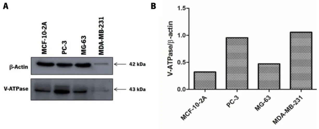
III.2. Highly metastatic cancer cell lines display different V-H+-ATPase expression levels in comparison with a non-tumorigenic breast cell line ... 30
III.3. bLf induces cell death of highly metastatic cancer cell lines ... 31
III.4. bLf induces intracellular acidification in highly metastatic cancer cell lines ... 37
III.5. bLf alters F-actin arrangement of highly metastatic cancer cell lines ... 39
III.6. bLf perturbs lipid rafts distribution of highly metastatic cancer cell lines ... 41
III.7. Methyl- -ciclodextrin protects highly metastatic cancer cells against bLf-induced intracellular acidification and inhibition of cell proliferation ... 43
V.1. FINAL REMARKS ... 54
V.2. FUTURE PERSPECTIVES ... 55
Chapter VI - References
... 58ANOVA ANalysis Of VAriance
ATCC American Type Culture Collection ATP Adenosine TriPhospate
BafA1 Bafilomicin A1
BCECF-AM 2',7'-Bis-(2-CarboxyEthyl)-5-(and-6)-CarboxyFluorescein - AcetoxyMethyl Ester
bLf bovine Lactoferrin BSA Bovine Serum Albumin
Caspase Cystein-dependent aspartate specific protease Cdk Cyclin-dependent kinase
CF Carboxyfluorescein
CFSE CarboxyFluorescein diacetate Succinimidyl Ester ConcA Concanamycin A
DMEM Dulbecco's Modified Eagle's Medium DMSO DiMethyl SulfOxide
DNA DeoxyriboNucleic Acid
DRMs Detergent-Resistant Membranes ECM ExtraCellular Matrix
EFSA-NDA European Food Safety Authority – Panel on Dietetic Products, Nutrition and Allergies
FBS Fetal Bovine Serum
FDA Food and Drug Administration FITC Fluorescein IsoThioCyanate GPI GlycosylPhosphatidylInisotol HBSS Hank’s Balanced Salt Solution hLf human Lactoferrin
Lf Lactoferrin
NPC NasoPharyngeal Cancer PBS Phosphate Buffered Saline PBST Phosphate Buffered Saline Tween PFA ParaFormAldehyde pHcyt cytosolic pH pHe extracellular pH pHi intracellular pH PI Propidium Iodide PS PhosphatidylSerine PVDF PolyVinylidene DiFluoride rLf recombinant Lactoferrin RNA RiboNucleic Acid
ROS Reactive Oxygen Species rpm rotations per minute RT Room Temperature S.D. Standard Derivation
S.E.M. Standard Error of the Mean SDS Sodium Dodecyl Sulfate shRNA Short Hairpin RiboNucleic Acid TME Tumor MicroEnvironment TNT Tunneling NanoTubes
VEGF Vascular Endothelial Growth Factor V-ATPase Vacuolar proton-translocating ATPase
Figure I.1. Structure of lactoferrin. ... 6 Figure I.2. Main biological functions and activities described for Lf. ... 7 Figure I.3. Proposed mechanisms for the anticancer activity of Lf. ... 10 Figure I.4. Model of the structure of the vacuolar V-ATPase expressed in an eukaryotic cell membrane.14
Figure III.1. bLf inhibits the cell proliferation of highly metastatic cancer cell lines. ... 30 Figure III.2. V-ATPase expression levels in highly metastatic cancer cell lines MDA-MB-231, PC-3 and MG-63 in comparison with the non-tumorigenic breast cell line MCF-10-2A. ... 31 Figure III.3. bLf induces cell death of the highly metastatic cancer cell lines MDA-MB-231and PC-3 but not of MG-63. ... 33 Figure III.4. bLf induces an increase in the levels of LC3B-II and Beclin-1 in PC-3 and MG-63 cell lines after 12 h and 24 h of treatment which decreases after 48 h. ... 36 Figure III.5. bLf and ConcA induce intracellular acidification in MDA-MB-231, PC-3 and MG-63 highly metastatic cancer cell lines ... 38 Figure III.6. bLf can alter F-actin rearrangement and/or produce cellular extensions in highly metastatic cancer cell lines ... 40 Figure III.7. bLf perturbs cholesterol-rich lipid rafts distribution in the highly metastatic cancer cell lines………..42 Figure III.8. MβCD protects cells against bLf-induced intracellular acidification and inhibition of cell proliferation in MDA-MB-231, PC-3 and MG-63 cells ... 44
Table I.1. Food and Drug Administration-approved drugs that include natural and natural-derived compounds. ... 3 Table I. 2. Characteristics of transferrin family members ... 4
Table II.1. List of the antibodies and incubation conditions used in the western blot analysis. ... 24
Table VII.1. bLf exhibits similarly cytotoxicity against the three highly metastatic cancer cell lines MDA-MB-231, PC-3 and MG-63 regarding inhibition of cell proliferation... 71
Chapter I
I.1. CANCER, METASTASIS AND ANTICANCER DRUGS: A BRIEF OVERVIEW
Cancer has been defined as a disease that is characterized by a wide range of features, which are summarized in the hallmarks of cancer. These hallmarks include unregulated cell proliferation due to the loss of growth control, escape to signals that control the normal cell behaviour, activation of invasion and metastasis, induction of angiogenesis, among others. Recently, genome instability, deregulation of cellular energetics and escape to immune destruction have been added to those hallmarks (Hanahan and Weinberg, 2011).Currently, cancer is the most lethal disease worldwide, which supports the great need for more accurate and targeted cancer therapies. In 2012, about 14.1 million new cancer cases and 8.2 million deaths due to cancer were reported (World Health Organization, 2014). A recent study performed by Ferlay and colleagues showed that the most frequently diagnosed cancers are lung, breast, colorectal and prostate with incidence rates of 1.8, 1.67, 1.36 and 1.1 million, respectively. Lung, liver and stomach cancers are responsible for the most common cancer-associated deaths (Ferlay et al., 2015). On the other land, metastases are the main cause of cancer-associated mortality. The metastatic process involves two major steps: (i) physical translocation of cancer cells to a distant organ, and (ii) colonization of the translocated cells in this organ (Chaffer and Weinberg, 2011). In the first step, after acquisition of the invasive phenotype, the cancer cells invade the matrix and enter into the bloodstream, transiting to distant organs. Afterwards, when out of circulation, cells invade the foreign tissue, after overcoming resistance by the immune system, and are able to survive in the secondary site as a single cell, promoting the colonization. Therefore, for the development of metastases, cancer cells require not only the integration of multiple events, such as invasion and angiogenesis, but also the adaptation to a foreign microenvironment (Martin et al., 2013).
Combined therapies may be required to overcome cancer. Cancer therapy has been mainly focused on the use of different anticancer drugs, from natural to natural-derived products, attempting to set aside therapeutic measures such as surgery, chemotherapy, radiation and hormones. These conventional treatments are costly, undesirable due to severe side effects and can only extend the patient’s lifespan by a few years. Hence, there has been a continuous search for less expensive and non-toxic natural drugs (Reddy et al., 2003; Prasad and Tyagi, 2014). Indeed, in the recent decades, 78% of the drugs approved by the Food and Drug Administration (FDA) are natural or natural-derived drugs, from
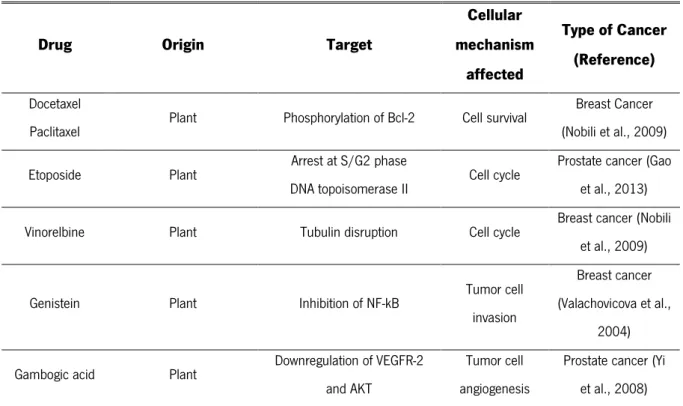
Table I.1. Food and Drug Administration-approved drugs that include natural and natural-derived compounds.
Drug Origin Target
Cellular mechanism affected Type of Cancer (Reference) Docetaxel
Paclitaxel Plant Phosphorylation of Bcl-2 Cell survival
Breast Cancer (Nobili et al., 2009) Etoposide Plant Arrest at S/G2 phase
DNA topoisomerase II Cell cycle
Prostate cancer (Gao et al., 2013) Vinorelbine Plant Tubulin disruption Cell cycle Breast cancer (Nobili
et al., 2009)
Genistein Plant Inhibition of NF-kB Tumor cell invasion
Breast cancer (Valachovicova et al.,
2004) Gambogic acid Plant Downregulation of VEGFR-2
and AKT
Tumor cell angiogenesis
Prostate cancer (Yi et al., 2008)
Several drugs, as specified in Table I.1, derived from natural compounds have been used in the treatment of several types of cancers including prostate, colorectal, lung, breast, stomach, lymphoblastic leukemia and brain tumours and have different targets, such as tubulin, microtubules and DNA topoisomerases(Prasad and Tyagi, 2014). The interaction between the drug and the respective target culminates in one or more of the following consequences: suppression of invasion, inflammation, angiogenesis, proliferation and metastasis of cancer cells (Gupta et al., 2010).
Among the different natural compounds with anticancer activities, some are present in food or are food-derived compounds, commonly referred as nutraceuticals, either of plant or animal origin or produced by microorganisms. Particularly, throughout the years researchers have explored the anticarcinogenic activity of individual milk compounds such as fat components (e.g., linoleic acid, lipids and sphingomyelin, among others) or proteins (e.g., caseins and lactoferrin) (Håkansson et al., 1995; Parodi, 1999; Tsuda et al., 2000; Sah et al., 2015). However, this issue is somewhat controversial. Several authors have reported that it is not clear the beneficial role associated to milk consumption, particularly in women. In 2014, a study demonstrated that high milk administration seems to be associated to the higher mortality incidence in women (Michaëlsson et al., 2014). Moreover, the effect of milk consumption in cancers is also a controversial issue since some evidences indicate its association
(Larsson et al., 2015). On the other hand, other authors reported that milk administration does not affect prostate tumour progression, namely inflammation, fibrosis and invasiveness (Bernichtein et al., 2015). Nevertheless, despite this controversy, some milk compounds and/or milk-derived bioactive peptides have been identified not only as an important part of a normal daily diet, but also as potential agents for cancer prevention and management (Mohanty et al., 2015; Sah et al., 2015). In particular, lactoferrin (Lf), an important protein in the milk whey fraction, was found to have an inhibitory potential against chemically-induced and hereditary carcinogenesis and metastasis (Tsuda et al., 2000; Sah et al., 2015; Pereira et al., 2016). Since this thesis is focused on the study of this natural protein and its anticancer activity, these topics will be further detailed in the following sections.
I.2. LACTOFERRIN: AN IRON-BINDING GLYCOPROTEIN
The transferrin family is a group of single-chain glycoproteins found in both vertebrates and invertebrates with the ability to bind tightly iron and transport it from plasma to cells. Therefore, this family of proteins has a crucial role in the iron metabolism, controlling its levels in biological fluids (Lambert, 2012; Hughes and Friedman, 2014). In vertebrates, transferrins bind and release ferric ions (Fe3+) to
respond to receptor binding and changes in pH (Lambert, 2012). This group includes several proteins with different tissue distribution namely: serum transferrin, ovotransferrin, lactotransferrin, also known as lactoferrin (Lf), and melanotransferrin (Table I.2) (Lambert, 2012; Hughes and Friedman, 2014).
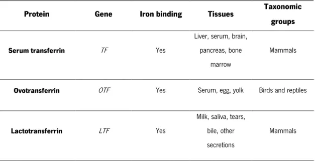
Table I. 2. Characteristics of transferrin family members (adapted from Lambert, 2012).
Protein Gene Iron binding Tissues Taxonomic groups
Serum transferrin TF Yes
Liver, serum, brain, pancreas, bone
marrow
Mammals
Ovotransferrin OTF Yes Serum, egg, yolk Birds and reptiles Lactotransferrin LTF Yes
Milk, saliva, tears, bile, other secretions
Melanotransferrin MFI2 N-lobe only
Melanomas, saliva, sweat, liver,
intestine
Vertebrates
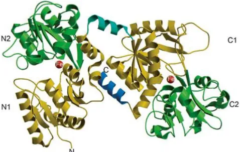
Lf is an 80 kDa protein containing around 700 amino acids. Its three-dimensional structure is composed by a single polypeptide chain folded into two highly homologous iron-binding lobes, namely the C-lobe (carboxyl) and the N-lobe (amino). Each lobe is further divided in two domains known as N1 and N2, C1 and C2 that enclose a deep cleft where the iron-binding site is located. Therefore, each Lf protein is capable of binding two iron ions with the concomitant binding of two carbonate ions (CO32-)
(González-Chávez et al., 2009). The two lobes are connected through a region containing parts of -helix responsible for the flexibility of the molecule (Fig. I.1) (Öztafl and Özgünefl, 2005; Baker and Baker, 2012).
I.2.1. Structure and Synthesis of Lactoferrin
Lf is an 80 kDa protein containing around 700 amino acids. Its three-dimensional structure is composed by a single polypeptide chain folded into two highly homologous iron-binding lobes, namely the C-lobe (carboxyl) and the N-lobe (amino). Each lobe is further divided in two domains known as N1 and N2, C1 and C2 that enclose a deep cleft where the iron-binding site is located. Therefore, each Lf protein is capable of binding two iron ions with the concomitant binding of two carbonate ions (CO32-)
(González-Chávez et al., 2009). The two lobes are connected through a region containing parts of -helix responsible for the flexibility of the molecule (Fig. I.1) (Öztafl and Özgünefl, 2005; Baker and Baker, 2012).
Figure I.1. Structure of lactoferrin. Crystal structure of Lf that illustrates its N1 and C1, N2 and C2 domains in yellow and green, respectively. The -helix that binds the two domains is shown in turquoise, the C-terminal -helix in blue, and the bound iron in red (adapted from Baker and Baker, 2012).
As previously mentioned, Lf has affinity to Fe3+, but this binding is reversible. Accordingly, Lf can
exist free or associated with Fe3+ as apo-Lf and holo-Lf, respectively. Consequently, the Lf molecule can
adopt two different three-dimensional conformations: apo-Lf exhibits an open conformation while holo-Lf presents a closed conformation (Wally and Buchanan, 2007; González-Chávez et al., 2009; García-Montoya et al., 2012). Hence, large-scale conformational changes are associated with the iron binding and release, in which the domains close over the bound ion or open to release it (Baker and Baker, 2012). Lf is synthesized by mucosal epithelial cells and neutrophils during inflammatory processes. As referred before, this protein is present in many tissues and body fluids as well as in many mammalian secretions. Indeed, it can be found in tears, saliva, semen, urine, pancreatic, gastric and vaginal fluids, bronchial and nasal secretions, and at higher concentrations in milk and colostrum, being the second most abundant milk protein after casein (González-Chávez et al., 2009; Alexander et al., 2012; García-Montoya et al., 2012).
I.2.2. Biological Functions and Applications of Lactoferrin
The Lf structure endows it multiple biological activities including: strong antimicrobial activity (Yamauchi et al., 2006; Yen et al., 2011), regulation and maintenance of iron absorption,
anti-inflammatory and anticarcinogenic activities (González-Chávez et al., 2009; García-Montoya et al., 2012). Figure I.2 illustrates some of activities that have been proposed for Lf.
Figure I.2. Main biological functions and activities described for Lf.
Regarding its antimicrobial activity, Lf exhibits a strong toxic effect against a wide spectrum of bacteria (Gram + and Gram -), fungi, yeasts, viruses, protozoa and parasites (Yamauchi et al., 2006; González-Chávez et al., 2009; García-Montoya et al., 2012). There are some mechanisms proposed to explain the Lf antibacterial activity such as iron sequestration from bacterial pathogens, which inhibits their growth (Zarember et al., 2007); direct interaction with bacterial cell surface by an iron-independent mechanism (Farnaud and Evans, 2003; Valenti and Antonini, 2005); and weakening the attachment of certain bacteria to the host cells (Diarra et al., 2003). On the other hand, the anti-viral activity of Lf relies on the inhibition of the internalization and replication of certain viruses in the host cell and the stimulation of the host immune system (Welsh et al., 2011).
Lf is also well known by its immunomodulatory and anti-inflammatory properties. Indeed, Lf is a major component of the secondary granules of neutrophils, being released upon neutrophil activation, which leads to an increase of its levels (Masson et al., 1969). This increase in Lf levels, in turn, promotes the recruitment of leukocytes, cytokines, natural killer (NK) and adaptive cells, and the activation of dendritic cells that allow the control of the excessive inflammation and stimulate the immune response (González-Chávez et al., 2009; García-Montoya et al., 2012). The role of Lf in regulating innate immune responses confirms its crucial function as a first line host defence mechanism against invading pathogens (Puddu et al., 2009).
Anticancer and anti-metastatic activities are two other relevant biological activities of Lf that are the subject of the present study and therefore will be discussed in more detail in the next subtopic.
Based on its multiple biological activities, Lf has been used in several applications. In particular, clinical trials have been conducted to evaluate the effectiveness of Lf administration in the treatment of a large variety of human diseases (González-Chávez et al., 2009; Tomita et al., 2009; García-Montoya et al., 2012). The first clinical application of Lf was in infant formulas to improve the intestinal microbial flora (Roberts et al., 1992), enhance serum ferritin (Chierici et al., 1992) and haematocrit levels and to prevent nosocomial infections (Manzoni et al., 2009). Besides, the activity of Lf and its bioactive peptides have been proved in in vitro an in vivo studies against several pathogens, in the treatment of infections, inflammatory diseases and cancer (González-Chávez et al., 2009; Kozu et al., 2009; García-Montoya et al., 2012). Concerning its administration, the oral intake of Lf has been suggested to be the most promising option as it is easy and safe, and also because more than 60% of the ingested bLf remains intact after the passage through adult human stomach and entrance in the small intestine (Troost et al., 2001; Mayeur et al., 2016).
Lf can also be used as a reliable disease biomarker because its increased levels can be used as a clinical indicator of inflammatory syndromes. In fact, increased levels of fecal Lf have been found in inflammatory bowel disease and allow distinguishing it from irritable bowel disease (Kane et al., 2003; Zhou et al., 2014). Furthermore, it was shown that Lf is downregulated in nasopharyngeal carcinoma (NPC) cells, and its increased expression has therefore been associated with a good prognosis (Zhang et al., 2015b).
The form of Lf that has been mostly used in the health care field, both in prevention and treatment of diseases, is the one from bovine origin. Indeed, bLf has already been approved by the European Food Safety Authority (EFSA) as a safe ingredient for various applications, including for medical purposes (Efsa, 2012; Mayeur et al., 2016). bLf can easily be isolated from cow’s milk by several purification methods and it is produced in large scale by manufacturing companies mainly using a cation-exchange chromatography system (Tomita et al., 2009). However, taking into account the multiple applications of bLf, distinct processes to produce it at a large scale have been exploited. These alternative processes include the production of recombinant Lf (rLf) in bacterial, viral and fungal expression systems, or production using transgenic plants and animals able to express rLf (González-Chávez et al., 2009; García-Montoya et al., 2012).
I.2.2.1. Anticancer and Anti-Metastatic Activities of Lactoferrin
As previously mentioned, anticancer and anti-metastatic activities are among the many different biological activities assigned to Lf. Indeed, the Lf anticancer activity has been observed against a wide range of human cancers in different cell lines, animal models and even in clinical trials (Gibbons et al., 2011; Yin et al., 2013; Zhang et al., 2014a). The Lf anticancer activity was demonstrated for the first time in 1994 in mice. Using this model, this protein was found to strongly decrease the solid tumour growth and inhibit the metastasis occurrence. This study proved the potential of Lf as an anticancer compound, and since then it has been explored for therapeutic purposes (Bezault et al., 1994). Also, in 1995, it was established that the whey fraction of bovine milk could significantly inhibit the development of colon tumours in rats (McIntosh et al., 1995). These pioneer studies and others encouraged scientists to investigate the protein effects on different types of cancer like bladder (Masuda et al., 2000), colon (Fujita et al., 2004), head and neck (Xiao et al., 2004; Wolf et al., 2007), stomach (Xu et al., 2010), lung (Tung et al., 2013) and breast (Duarte et al., 2011; Zhang et al., 2015; Pereira et al., 2016).
Some studies addressed the effect of bLf on colorectal cancer demonstrating that the oral intake of 3 g of bLf per day during 1 year induced a significant delay of the growth of colorectal adenomatous polyps in human patients. These data suggest that bLf inhibits colorectal carcinogenesis and serves as an alternative to the surgical removal of polyps (Kozu et al., 2009; Tsuda et al., 2010). Moreover, due to the in vitro high resistance of non-tumorigenic cell lines to Lf (Gibbons et al., 2015; Pereira et al., 2016) and absence of secondary effects in humans (Gibbons et al., 2011; Yin et al., 2013; Zhang et al., 2014a), bLf exhibits a great potential to be safely used in cancer therapy.
I.3. MECHANISMS UNDERLYING LACTOFERRIN ANTICANCER ACTIVITY
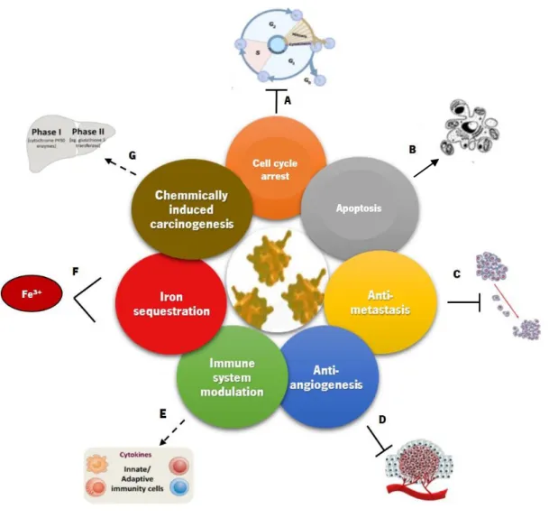
Several researchers along the last years have already provided some mechanistic insights on the anticancer activity of Lf based on its ability to interfere with the cell membrane and its receptors, cell cycle progression and induction of apoptosis, as well as on its anti-metastatic and anti-angiogenesis potential, immunostimulation and iron sequestration capacity (Fig. I.3).Figure I.3. Proposed mechanisms for the anticancer activity of Lf. Legend: Induction Inhibition Modulation Sequestration
Regarding Lf membrane receptors, many studies have demonstrated its presence in cancer cells and each cell type seems to have its specific Lf receptors. The cancer cells that overexpress Lf receptors may be more susceptible to Lf, thus creating an opportunity for the development of cancer-specific drug carriers coated with Lf that may be administered intravenously Moreover, it has been demonstrated an association between Lf and heparan sulfate, in which Lf exhibits conformation-dependent uptake efficiency correlated with efficient binding to heparan sulfate and lipid-induced conformational changes. Accordingly, when heparan sulfate chains are removed, membrane binding and cellular uptake of the peptide are reduced (Duchardt et al., 2009). Furthermore, Lf selective cytotoxicity for cancer cells can be explained by differences in the cellular membrane. The membrane of cancer cells has higher levels of negatively charged molecules when compared with normal cells, which attracts the cationic Lf/Lf-derived peptides. This membrane feature favours a preferentially binding of Lf to cancer cells than to normal cells
(Szachowicz-Petelska and Dobrzynska, 2010). These studies suggest that the membrane can be one of the Lf targets through which it exerts its anticancer activity.
The Lf anticancer activity also relies on its ability to trigger cell cycle arrest (Fig. I.3 A), which occurs predominantly at the G1 phase (Wolf et al., 2007; Wang et al., 2011). Lf can induce this process by decreasing phosphorylated Akt to increase the expression of p21 and p27, which are known inhibitors of the cell cycle, and by stopping the transition of G1 to S of MDA-MB-231 breast cancer cells with decrease in the levels of proteins Cdk2, Cdk4 and cyclin E, and increase in the expression of Cdk inhibitor p21 (Damiens et al., 1999; Xiao et al., 2004). Moreover, this Lf inhibitory effect was reported as cell type-dependent, since in MDA-MB-231 cells the arrest was observed at G2 phase, whereas in MCF-7 cells there was an arrest at G1 phase with low doses of Lf and an arrest at G2 phase with higher doses (Zhang et al., 2014b).
Apoptosis is a highly regulated and conserved process of cell death that involves several events namely cell shrinkage, nuclear fragmentation, chromatin condensation and DNA fragmentation culminating in cell demise and self-destruction without the release of cytosolic contents that trigger an inflammatory response. The regulation of apoptosis is crucial for cell homeostasis and therefore, deregulation of apoptosis can be associated with many pathologies and diseases (Meier and Vousden, 2007; Redza-Dutordoir and Averill-Bates, 2016). Lf-treated cancer cells showed alterations in the proteins involved in apoptosis, such as Bcl-2 family members and p53 (Fig. I.3 B). In human stomach and breast cancer cell lines, the levels of the anti-apoptotic protein Bcl-2 were found to be downregulated by Lf (Xu et al., 2010; Zhang et al., 2014b). Another study showed that, in mice bearing EMT6 breast cancer, apoptosis was induced by decreasing the expression of Bcl-2 and increasing the expression of the pro-apoptotic Bax and the executioner caspase-3 (Wang et al., 2011). Indeed, diet supplemented with bLf decreased carcinogenesis as it increased the expression of fatty acid synthase, 3 and caspase-8, leading to DNA fragmentation in rat colon mucosa (Fujita et al., 2004). Additionally, bLf was found to induce apoptosis and inhibit colony formation of MCF-7 cells, which was associated with mitochondria membrane depolarization, decrease of Bcl-2 levels, cell cycle arrest at G1/G0 phase, and downregulation of CDC25c (Zhang et al., 2015c). Recently, we showed that bLf preferentially inhibits proliferation and induces apoptosis in the highly metastatic breast cancer cell lines MDA-MB-231 and Hs 578T, as well as decreases the extracellular acidification rate and increases intracellular acidification in these two highly metastatic cancer cells (Pereira et al., 2016).
Furthermore, the anticancer activity of Lf also stems from its anti-metastatic potential (Fig. I.3 C), as already mentioned. Several authors demonstrated that Lf is able to reduce or inhibit lung colonization in melanoma and lymphoma cells and colon carcinoma (Bezault et al., 1994; Iigo et al., 1999). According to Iigo and colleagues (Iigo et al., 2004), increased production of IL-18 and activation of NK and T cells to enhance immunity is a consequence of the protective role of Lf against metastasis (Iigo et al., 2004). Recently, Gibbons and colleagues evaluated the proliferation, migration and invasion of two breast cancer cell lines namely MDA-MB-231 and MCF-7 cells when treated with apo-Lf and holo-Lf. Both Lf forms induced a reduction of cell proliferation, as well as the invasion in both cell lines and the inhibition of the expression of survivin, an inhibitor of apoptosis (Gibbons et al., 2015). Therefore, Lf can inhibit tumour metastasis by inhibiting growth and decreasing the number of tumour-induced blood vessels, which indicates that bLf also exhibits anti-angiogenic activity (Fig. I.3 D) (Yeom et al., 2011). Recently, it was shown that bLf inhibits vascular endothelial growth factor (VEGF)-induced angiogenesis since oral administration of bLf in transgenic mice overexpressing the human VEGF-A165 gene suppressed the formation of tumours. This suggests that these effects of Lf might be attributed at least in part to the inhibition of new blood vessel formation. Lf anti-angiogenic properties have important implications in prevention or treatment of angiogenesis-related diseases, such as cancer and chronic inflammatory diseases (Tung et al., 2013; Mayeur et al., 2016).
The ability of Lf to modulate the immune system is another capacity of this protein that may underlie its anticancer activity (Fig. I.3 E). The immunomodulatory activity of Lf is considered a key factor for the in vivo anticancer effects of Lf and involves the modulation of the production of cytokines, such as TNF-α, IL-1b, IL-6 and IL-8, encompassing both innate and adaptive immunity (Yeom et al., 2011; Legrand and Mazurier, 2010). In fact, several studies in animal models established that Lf is capable of modulating the production and activation of cytokines in immune cancer cells (Wolf et al., 2007). On the other hand, when it is released from activated neutrophils, Lf was shown to inhibit lymphocytes proliferation and granulopoiesis, to suppress antibody production and to regulate NK cells. Moreover, at low concentrations, Lf can stimulate NK cells and macrophages against cancer cells by activating a strong Th1 response (Fischer et al., 2006) and promote the recruitment of tumour-infiltrating lymphocytes, such as CD4+ and CD8+, which greatly inhibit the proliferation of cancers (June, 2007).
Another mechanism involved in the anticancer activity of Lf is dependent on Lf iron-binding ability (Fig. I.3 F). Free iron has been found to act as a mutagenic promoter by inducing oxidative damage to nucleic acids and in the gastrointestinal tract (Toyokuni, 2009). Hence, since Lf can tightly bind iron and
hold it in a stable nonreactive form, it can mitigate the local reactive oxygen species (ROS) production thus reducing locally the risk of oxidant-induced carcinogenesis (Rodrigues et al., 2009; Mayeur et al., 2016).
Finally, another mechanism underlying the anticancer activity of Lf was established in models of chemically induced carcinogenesis (Fig. I.3 G). This induction process encompasses two stages, namely initiation and post-initiation. The first stage includes phase I enzymes like cytochrome P450, responsible for the liver detoxification metabolism and activation of carcinogens leading to DNA damage in the specific organs. Liver phase II enzymes, also known as “blocking agents”, suppress this activation and are responsible for detoxication and excretion – post-initiation stage. The compounds that can inhibit this phase by suppressing the proliferation of pre-malignant cells are called “suppressing agents”. Several types of cancers (colon, lung, bladder, among others) in rat and hamster models of chemical carcinogenesis have been inhibited by oral administration of bLf, meaning that bLf acts as a blocking and suppressing agent by inhibiting phase I enzymes and stimulating the phase II enzymes (Tsuda et al., 2002; Mohan et al., 2006).
Although all the mechanisms referred above point to a clear anticancer role of Lf, its cellular targets are still elusive (Rodrigues et al., 2009; Zhang et al., 2014b; Mayeur et al., 2016). More recently, our group identified V-ATPase as a target of Lf. Currently, it is recognized that V-ATPase plays a prominent role in maintaining the tumour microenvironment (TME) acidosis, as well as in the metastatic process. It has therefore been suggested as a target in cancer therapy, as it will be discussed below.
I.4. V-H
+-ATPASE: A MOLECULAR TARGET FOR CANCER THERAPY
The vacuolar-type H+-ATPase (V-H+-ATPase) is an ATP-driven enzyme of the rotary ATPases family
that transforms the energy of ATP hydrolysis in a proton electrochemical potential across different biological membranes, thus mediating a primary active transport of H+ (Beyenbach and Wieczorek, 2006;
Cipriano et al., 2008). A diverse collection of physiological processes depend on V-ATPases, and a number of diseases have been associated with anomalies of these pumps (Bowman and Bowman, 2005).
At the structural and functional level, V-ATPase is a multi-subunit complex comprising two major functional domains known as V1 and V0. The V1 domain (cytoplasmic domain) is soluble and interacts with
consists of a H+-binding ring structure formed by six or more c subunits and at least more three different
subunits identified with small letters (a, d and e) that mediate the transport of H+ across the membrane,
as shown in Fig. I.4 (Forgac, 2007; Marshansky et al., 2014; Rawson et al., 2015).
Figure I.4. Model of the structure of the vacuolar V-ATPase expressed in an eukaryotic cell membrane. Molecular model: cytoplasmic V1 domain (shown in yellow and orange) with eight different subunits identified by capital letters
(A-H) responsible for ATP hydrolysis; and membrane V0 domain (shown in blue and grey) with at least four subunits identified
by small letters (a, c, d and e), responsible for proton translocation across the membrane (Forgac, 2007).
V-ATPase was first discovered in lysosomes and vacuoles of plants and fungi and, later on, in secretory vesicles, endosomes, Golgi-derived, among others (Saroussi and Nelson, 2009). Besides this intracellular localization, V-ATPase is also present at the plasma membrane of cancer cells and specialized cells like osteoclasts, macrophages, etc (Cipriano et al., 2008).
Concerning its physiological role, V-ATPase has been reported to be essential in the acidification of intracellular compartments like endosomes, lysosomes and Golgi apparatus (Sun-Wada et al., 2004; Rawson et al., 2015). Moreover, V-ATPase is essential in cancer cells, namely in metastasizing cells, allowing the invasion process by acidifying the tumour extracellular environment (Sennoune et al., 2004), as it will be discussed below.
I.4.1. The role of V-H+-ATPase in the tumour microenvironment and metastasis
Cancer cells often thrive in a hypoxic microenvironment with an acidic extracellular pH (pHe) when compared with normal cells. Under normal conditions, the low pHe associated with the hypoxic environment is not favourable for cell growth, promoting apoptosis (Gottlieb et al., 1995). To survive in these conditions, cancer cells must display a cytosolic pH (pHcyt) regulatory system, which is crucial to normal cell function providing the optimal pH for many cellular processes like cell growth, while promoting tumorigenesis, metastasis, cell motility (Rofstad et al., 2006) and drug resistance in cancer cells (Martínez-Zaguilán et al., 1999). In fact, pHe in tumours ranges from 6.5 to 6.9, while the intracellular pH (pHi) remains neutral to alkaline, creating an acid-outside pH gradient not observed in normal cells (Wojtkowiak et al., 2011). V-ATPase are normally present in various intracellular organelles, however, in some metastatic cancer cells, they are also functionally expressed at the plasma membrane where they contribute to the acidosis of the TME, playing pivotal roles in tumour invasion and metastasis (Martinez-Zaguilan et al., 1993; Pereira et al., 2016). The link between V-ATPase and tumour metastasis is apparent since the abnormal acidic pH in the TME contributes to the activation, secretion and distribution of proteases like metalloproteinases involved in the digestion of the complex extracellular matrix (ECM) (Fais et al., 2007). In this way, understanding the mechanisms regulating pHi and tumour acidity is of prime importance to develop V-ATPase targeting strategies for cancer therapy.
I.4.2. Relevance and features of V-H+-ATPase in highly metastatic cancer cell lines
Studies in human breast cancer cell lines with distinct metastatic potential have been performed to ascertain the distribution and functional activity of the V-ATPase and relate it with the migration and invasion of these cell lines (Sennoune et al., 2004; Hinton et al., 2009; Capecci and Forgac, 2013). The immunocytochemical results reported by Sennoune and co-workers (2004) demonstrated that highly and lowly metastatic breast cancer cells display a different localization of V-ATPase. Indeed, in the highly metastatic cancer cells, this proton pump was preferentially located at the plasma membrane while in the lowly metastatic cancer cells it had predominantly an intracellular localization. The same authors found that migration and invasion, two hallmarks of cancer, were related with V-ATPase localization/activity in breast cancer cell lines. Moreover, the migration kinetics of highly and lowly
ATPase inhibitor, to evaluate the physiological relevance of this pump at the cell surface. In the absence of this inhibitor, invasion and migration were significantly faster in highly metastatic cancer cells, while in its presence a decrease of these processes was observed. Additionally, no significant effect was found in lowly metastatic cancer cells (Sennoune et al., 2004). Similar results were obtained in human prostate carcinoma cell line PC-3M-2B4 (Xu et al., 2012b). Notably, the a and c subunits seem to be the V-ATPase subunits that more significantly influence the metastasis and proliferation of cancer cells, thus being important factors in the regulation of cancer metastasis (Hinton et al., 2009; Capecci and Forgac, 2013). These studies suggest that the greater migratory and invasive ability of the highly metastatic cells as compared to the lowly ones can be suppressed by V-ATPase inhibitors. Furthermore, at the cell surface this proton pump plays a role in maintaining an alkaline intracellular microenvironment – pHi - favourable for cell growth, conferring them a competitive advantage over normal cells; while maintaining an acidic extracellular microenvironment - pHe - essential for invasion (Xu et al., 2003). In this way, the acquisition of a metastatic and invasive phenotype is associated to the localization of V-ATPase in the plasma membrane.
Recently, in HCT116 and SW480 colon cancer cell lines, Lozupone and colleagues found a new transmembrane protein - TM9SF4 - related with invasive behaviour of metastatic cells and implicated in the V-ATPase activation through the interaction with the ATP6V1H subunit of the V1 domain. TM9SF4
suppression and silencing with small interfering RNAs resulted in a series of cellular alterations related with a reduced V-ATPase activity: (i) decrease of pHi; (ii) alkalization of the intracellular vesicles and (iii) reduction of extracellular acidity through a decrease of proton extrusion. These effects promoted a significant inhibition of the invasive behaviour reported in colon cancer cell lines (Lozupone et al., 2015). Given the contribution of V-ATPase to the acidity of the TME and its recognized importance in breast cancer, inhibitors of this pump have emerged as excellent candidates for breast cancer therapy. The most-well known V-ATPase inhibitors are concanamycin A (ConcA) and BafA1, and treatment with these compounds results in the neutralization of TME. However, several studies have shown that ConcA and BafA1 are extremely toxic in vivo and therefore not suitable for clinical use (Pérez-Sayáns et al., 2009; Spugnini et al., 2015). New V-ATPase inhibitors have then been developed such as indole derivatives (Supino et al., 2008), macrolacton archazolids (Von Schwarzenberg et al., 2013), among others. A recent study from our group with highly metastatic breast cancer cell lines showed that bLf, like ConcA, inhibits V-ATPase in sub-cellular fractions. Thus, bLf was proposed as a V-ATPase inhibitor as it was found to
decrease the extracellular acidification rate and to promote intracellular acidification (Pereira et al., 2016) and selectively induce apoptosis of cancer cells.
Since the preferential expression and activity of V-ATPase at the cell surface has an important role for the development of metastasis and invasiveness of the cancer cells, V-ATPase is considered a potential target in chemotherapy and may be an excellent candidate for anticancer drugs such as bLf.
I.5. LIPID RAFTS: STRUCTURE AND INTERPLAY WITH V-H
+-ATPASE
In 1972, for the first time, a model about the organization and structure of the proteins and lipids in the biological membranes was proposed by Singer and Nicholson, and it was named the fluid mosaic model. According to this model, biological lipid bilayers are two-dimensional fluids favourable to lateral motility allowing free diffusion of proteins in the phospholipid bilayer and distribution along the membrane surface. This fluidity leads to the creation of highly organized structures or membrane domains differing in lipid/protein composition (Singer and Nicolson, 1972). Over time, several experiments indicated that some lipids are essential for creating these levels of order, leading to an “organization of the lipid components of membranes into domains”, supporting the concept of lipid membrane domain (Karnovsky et al., 1982). In 1988, Simons and van Meer explained the different lipid composition providing the basis for the “lipid rafts hypothesis” (Simons and van Meer, 1988). After a decade, intracellular trafficking and signal transduction mechanisms were hypothetically associated with lipid rafts (Simons and Ikonen, 1997). Later on, stabilization of the lipid rafts was associated with the presence of cholesterol, its key lipid component, within a liquid-ordered phase (Simons and Sampaio, 2011). Therefore, lipid rafts were initially described as ordered domains created by lateral separation of sphingolipids with different molecular composition and properties relative to the surrounding membrane (Sonnino and Prinetti, 2012). This notion has then emerged in several studies in cell biology being involved in a great variety of cellular and biological functions. Such ordered lipid microdomains are enriched in glycosylphosphatidylinisotol (GPI)-anchored proteins as well as in lipids, especially cholesterol and sphingolipids (sphingomyelin and gangliosides), in which the cholesterol tightly interlaces the saturated fatty acyl chains of the sphingolipids forming these organized microdomains. These structures are small, dynamic and heterogeneous and have affinity to several proteins such as Src family kinases (SFKs) and transmembrane proteins such as CD44 (Murai, 2012; Mollinedo and Gajate, 2015). Lipid rafts are
membranes (DRMs) (Staubach and Hanisch, 2011; Sonnino and Prinetti, 2012). However, some experimental evidences appear to contradict the principles underlying the lipid rafts notion, leading to some controversy around this subject. For example, a recent study in live cell plasma membrane demonstrated that GPI-anchored proteins do not reside in ordered domains, since it is not observed the formation of a connective phase with altered membrane fluidity (Sevcsik et al., 2015). For these reasons, the concept of lipid rafts, mainly its true nature and role, still generates some debate among scientists and it is currently being redefined. Nevertheless, lipid rafts have been implicated in various cellular processes, namely associated to cancer and V-ATPase.
At the functional level, these lipid structures represent authentic signalling platforms responsible for signal transduction and protein trafficking. Therefore, they are implied in many signalling pathways namely related with cancer progression like cell migration and adhesion. Indeed, the deregulation of raft-dependent signalling was shown to favour tumour progression (Murai, 2012; Wang et al., 2013; Mollinedo and Gajate, 2015). In particular, a study with breast cancer cells showed that lipid rafts may regulate several processes involved in cancer progression, namely at the initial stages of tumour growth and progression to a migratory and metastatic phenotype (Babina et al., 2011). Murai and colleagues demonstrated that lipid rafts play a critical role in the localization and functionality of CD44, a major cell adhesion molecule expressed in cancer cells. When cells were treated with a lipid-raft-disrupting agent methyl- -cyclodextrin (M CD) or filipin, CD44 shedding increased suggesting that low lipid rafts levels disturbs the CD44 membrane localization, which is essential for enhanced cancer cell adhesion and migration (Murai, 2012). Moreover, a recent study performed in human melanoma cells confirmed these evidences through the evaluation of the actin cytoskeleton and cell adhesion. In this case, lipid rafts not only regulate the dynamics of actin cytoskeleton, but also the disruption of its integrity with M CD, promoted alterations on cell morphology such as actin fiber formation and inhibition of the adhesion disassembly (Wang et al., 2013). In our laboratory, we have also studied breast cancer cell lines treated with bLf and M CD in which cholesterol-rich lipid rafts are re-distributed (Pereira C, 2014 master thesis). Importantly, V-ATPase has been identified as a component of the lipid rafts in highly metastatic melanoma cells (Baruthio et al., 2008), osteoclasts (Ryu et al., 2010) and coronary arterial epithelial cells (Xu et al., 2012a). Focusing on melanoma cells, Baruthio and colleagues demonstrated that V-ATPase is present in the lipid rafts fraction of highly metastatic cells, while appearing to be inconspicuous in non-metastatic cells, thus suggesting that there is an interplay between this proton pump and the lipid rafts (Baruthio et al., 2008). This interplay is still unknown but there are some studies that explain the
V-ATPase transport into lipid rafts. In coronary arterial epithelial cells, V-V-ATPase was found to provide an acidic microenvironment around the lipid rafts when it is assembled and transported into these microdomains, promoting the formation of ceramide-enriched signalling platforms and amplification of the lipid raft-associated signals. In the presence of BafA1, V-ATPase was inhibited impeding the lipid rafts clustering. Given this evidence, it was concluded that when V-ATPase co-localizes with lipid rafts it has a crucial role in the structural and functional stability of these domains (Xu et al., 2012a). However, additional evidence support that this interaction is reciprocal since V-ATPase activity is regulated by the interaction with specific lipidic environments (Lafourcade et al., 2008).
In this work, the interplay between V-ATPase and lipid rafts will be explored in different highly metastatic cancer cell lines after treatment with bLf.
I.6. AIMS
Our previous results showed that the selective anticancer activity of bLf against highly metastatic breast cancer cells, in comparison with lowly metastatic and non-tumorigenic cell lines, occurs through inhibition of the plasmalemmal V-ATPase (Pereira et al., 2016). Herein, we aimed to ascertain whether this same mechanism of action of bLf could underlie its activity against other types of highly metastatic cancer cells. To accomplish our goal, we selected three highly metastatic cell lines: a prostate cancer cell line (PC-3), an osteosarcoma cell line (MG-63), and the previously characterised breast cancer cell line (MDA-MB-231), the latter used as a positive control, and defined the following specific aims to:
- compare the susceptibility of these three cell lines to bLf in terms of inhibition of cell proliferation and uncover the mechanism underlying bLf-induced cell death;
- evaluate a possible relation between cell susceptibility and the levels of V-ATPase of the three cell lines and compare them with the non-tumorigenic breast cell line MCF-10-2A;
- assess whether the susceptibility of PC-3 and MG-63 cell lines to bLf is associated with alteration in the intracellular pH;
- study the effect of bLf on F-actin arrangement;
unravel the effect of the pretreatment of cells with the lipid rafts disruption agent methyl -cyclodextrin (MβCD) in the response to bLf treatment;
Chapter II
II.1. Chemicals and Solutions
Bovine lactoferrin (bLf) was obtained from DMV (Veghel, The Netherlands). The protein was dissolved in phosphate buffered saline (PBS) (1.37 M NaCl, 2.7 mM KCl, 10 mM Na2HPO4, 1.8 mM KH2PO4, pH 7.4) to obtain the different concentrations used throughout this study. According to the manufacturer, the protein purity is about 80% with 3.5% moisture and 21% iron-saturated.
Concanamycin A (ConcA), methyl- -ciclodextrin (M CD), paraformaldehyde (PFA), cisplatin, etoposide, filipin and LC3B, β-actin and caspase-3 antibodies were purchased from Sigma-Aldrich. BCECF-AM (2’, 7’-Bis-(2-Carboxyethyl)-5-(and-6)-Carboxyfluorescein, Acetoxymethyl Ester) and Alexa Fluor 488-Phalloidin were obtained from Molecular Probes. CFSE probe (Carboxyfluorescein diacetate succinimidyl ester), and FITC Annexin V apoptosis detection kit were acquired from BD Bioscience. Secondary antibodies - goat anti-rabbit IgG or goat anti-mouse IgG were obtained from Jackson ImmunoResearch. Beclin-1 antibody was acquired from Cell Signalling and V-ATPase C1 antibodies were purchased from Santa Cruz Biotechnology. Vectashield mounting medium was acquired from Biosystems.
II.2. Cell lines and culture conditions
Human prostate cancer cell line PC-3 (CRL-1435; ATCC), human osteosarcoma cell line MG-63 (CRL-1427; ATCC) and human breast cancer cell line MDA-MB-231 (HTB-26; ATCC) were grown in Dulbecco’s modified Eagle’s medium (DMEM), purchased from Biochrom - Merck Millipore, supplemented with 10% fetal bovine serum (FBS), acquired from the same company, and 1% zell shield (Minerva Biolabs). MCF-10-2A (CRL-10781), a normal breast cell line, was grown in DMEM-F12 medium supplemented with 5% horse serum, 1% zell shield, 500 ng/mL hydrocortisone, 100 ng/mL cholera toxin, 20 ng/mL epidermal growth factor and 0.01 mg/mL insulin.
Cells were kept in cryovials with a freezing mixture containing 5% dimethyl sulfoxide (DMSO), 10% fetal bovine serum (FBS) and DMEM without supplements, and stored in liquid nitrogen. In order to perform the experiments bellow described, frozen cells were thawed and maintained in culture in a 37 ºC incubator with a humidified atmosphere containing 5% CO2.
For each experiment, cells were collected from the culture flask, washed with PBS 1× and incubated with trypsin 0.05% (v/v). Trypsin was inactivated by fresh medium and the cell suspension was
experiment, cells were seeded in 6-well plates at an appropriate concentration: 1 × 10⁵ at 24 h and 7.5
× 104 cell/ml at 48 h for PC-3 cell line, 9 × 10⁴ at 24 h and 5 × 10⁴ cell/ml at 48 h for MG-63 and MDA-MB-231 cell lines, and 7.5 × 10⁴ cell/ml at 48 h for MCF-10-2A cell line. All the compounds under study
were added to the wells only when cells reached at least 80% confluence.
II.3. Assessment of cell proliferation by carboxyfluorescein diacetate
succinimidyl ester (CFSE)
The CFSE probe was used to assess cell proliferation. This non-fluorescent compound is metabolized by cellular esterases and the resulting fluorescent carboxyfluorescein (CF) binds covalently to long-lived intracellular molecules emitting green fluorescence when excited at 488 nm. The cellular fluorescence is diluted in each cell division. The daughter cells exhibit half the fluorescence intensity of their mother cells (Lyons et al., 2013).
II.3.1. Preparation of CFSE dye solution in DMSO 2%
To prepare a 10 mM stock solution of CFSE dye, 90 µL of DMSO 2% were added to a CFSE vial. Afterwards, the CFSE working solution (1.25 µM) was distributed into single-use aliquots that were frozen at -20 ºC protected from light.
II.3.2. CFSE labelling of cells
CFSE labelling was performed before cells’ seeding. Briefly, cells were collected from the culture flask and washed with PBS 1× to remove all the serum proteins to avoid probe inactivation in the presence of those proteins (Quah et al., 2007). Cell concentration was estimated using a Neubauer chamber. Next, 1 × 10⁵ cells/ml were resuspended in PBS 1× and were further incubated with the CFSE dye (final
concentration: 20 µM) for 15 min in a 37 ºC water bath. Afterwards, cells were rinsed with PBS 1×, the correct amount of complete culture media to obtain the same cell concentration was added and cells were plated in 6-well plates. After 24 h of adherence, cells were treated solely with medium (negative control), 50 µM cisplatin (MG-63 and MDA-MB-231 cell lines) or 60 µM etoposide (PC-3 cell line) as positive controls, and 175 µM bLf. Cisplatin and etoposide were the selected controls for these cell lines because they induce apoptosis and are anti-tumour agents currently used in clinical use for the treatment
of this type of cancers (Carrle and Bielack, 2006; Gao et al., 2013, respectively). Cells were harvested and the CF median fluorescence intensity was analysed by flow cytometry using the FL1 channel at time points 0, 24, 48 and 72 h after treatment. At the moment of seeding, a sample from the labelled cell suspension was also collected and analysed to ensure the correct staining of the cells. In the assays with MβCD, cells were pre-treated with 0.5 mM MβCD 2 h before incubation with bLf.
The FlowJo 7.6 software was used to analyse the data, namely to obtain the medians and the histograms. All data (medians) were normalized to the time point 0 h, which corresponds to the maximum fluorescence intensity. GraphPad Prism version 6.0 was used to elaborate the graphics.
II.4. FITC-Annexin V/Propidium Iodide apoptosis assay
The Annexin V/Propidium iodide (AV/PI) apoptosis assay was performed to characterize bLf-induced cell death. Cells were seeded in 6-well plates, two wells per condition – negative control (cells without treatment), positive control (treated with 60 µM etoposide for PC-3 cell line, and 50 µM cisplatin for MG-63 and MDA-MB-231 cell lines), 175 µM bLf and 10 nM ConcA (used as positive control for V-ATPase inhibition). After an adherence period of 24 h, cells were incubated in the absence or presence of the different compounds. After 24 and 48 h of treatment, cells were harvested, rinsed with PBS 1× and centrifuged at 2500 rpm for 5 min. Pellets were resuspended in PBS 1× for cell counting to obtain a final concentration of 2 × 10⁵ cells per condition. The corresponding amount of cellular suspension was
transferred to microtubes correctly identified – autofluorescence, AV or PI mono-stainings, and AV/PI double staining – and centrifuged at 2500 rpm for 7 min. The pellets were resuspended in Binding Buffer 1× (BD PharmigenTM), transferred to cytometer tubes and incubated with 1 µL of AV-FITC and 1 µL of PI
for 15 min in the dark. Binding buffer 1× was added to the cells in order to obtain a final volume of 500 µL. Acquisition was performed in a flow cytometer using the FL1 and FL4 channels. Data were analysed using the FlowJo 7.6 software.
II.5. Protein Extraction and Quantification
Cells seeded in 6-well plates were harvested by trypsinization, centrifuged at 2000 rpm for 5 min, and resuspended in 40-60 µL ice-cold RIPPA buffer [50 mM Tris HCl pH=7.4, 150 mM NaCl, 2 mM
orthovanadate (Na3VO4), 1 mM phenylmethylsulfonyl fluoride (PMSF) and 4% protease cocktail inhibitor],
and kept on ice for 20 min. Then, cell extracts were centrifuged at 14000 rpm for 15 min and the supernatant containing the total proteins was transferred to new microtubes, previously identified. The soluble protein concentration was determined using the Bio-Rad DC protein assay (Bio-Rad Laboratories). The protein concentration of each sample was determined using a calibration curve obtained with solutions of bovine serum albumin (BSA) with increasing concentrations – from 5 mg/mL to 0.25 mg/mL.
II.6. Western Blot analysis
For western blot analysis, 35 µg or 50 µg (depending on the assay) of protein were separated by a 12.5% sodium dodecyl sulfate (SDS)-polyacrylamide gel electrophoresis. Afterwards, protein bands were transferred onto PVDF (polyvinylidene difluoride) membranes. Next, to avoid non-specific interactions, membranes were blocked in 5% non-fat milk in PBS-Tween 0.1% solution (PBST 1×) for 1-2 h with agitation at RT. Membranes were then incubated overnight at 4 °C with the primary antibodies, namely monoclonal anti- -actin, anti-active caspase 3, anti-LC3B, anti-Beclin-1 and anti-V-ATPase (Table II.1).
Table II.1: List of the antibodies and incubation conditions used in the western blot analysis. Antibody
(Host)
Size of the
target (kDa) Dilution
Temperature (ºC)
Incubation
time Manufacturer Anti-β-Actin
(Mouse) 42 1:1000 4 Overnight Sigma Aldrich Active
anti-caspase 3 (Rabbit)
17-19 1:2000 4 Overnight Sigma Aldrich
Anti LC3B
(Rabbit) 16-18 1:1000 4 Overnight Sigma Aldrich Beclin-1
(Rabbit) 60 1:1000 4 Overnight Cell Signalling V-ATPase C1
(Mouse) 43 1:25 4 Overnight Santa Cruz Biotechnology Anti-Rabbit
(Goat) - 1:2000 RT 1 hour Jackson ImmunoResearch Anti-Mouse
Next, membranes were incubated with secondary antibodies Peroxidase AffiniPure goat anti-rabbit IgG or goat anti-mouse IgG (1:2000; Jackson ImmunoResearch). Chemiluminescence detection was performed using the ECL detection system (Millipore-Merck) in a ChemiDOCTM XRS system (BioRad).
II.7. Intracellular pH measurement
Measurements of pHi were performed with the pH-sensitive probe BCECF-AM. This non-fluorescent ester is a substrate of intracellular esterases and the fluorescence of BCECF varies according to the pH. Changes in the cytosolic pH were monitored by changes in the ratio of green/red fluorescence intensities (FL1/FL4), since this ratio value is independent of the probe concentration and exclusively dependent of pHi(Ozkan and Mutharasan, 2002).
Cells were seeded in 6-well plates and treated with only medium, 175 µM bLf and 10 nM ConcA, for 24 h and/or 48 h. After this time, cells were tripsinized, washed with HBSS – Hank’s Balanced Salt Solution (10 × concentrated solution: 1379 mM NaCl, 53.33 mM KCl, 3.36 mM Na2HPO4-7H2O, 55.55
mM D-glucose and 4.41 mM KH2PO4) and centrifuged. The pellets were resuspended in HBSS 1× and
incubated with 50 µM BCECF-AM from a stock solution of 161.3 µM for 30 min at 37 ºC, and protected from light. After incubation, samples were analysed in a flow cytometer. For the assays with MβCD, cells were pre-treated with 0.5 mM MβCD during two hours before the additional treatments. The % of cells exhibiting intracellular acidification were estimated from the percentage of cells displaying a FL1/FL4 ratio lower than control cells. Data were analysed using the FlowJo 7.6 software.
II.8. Flow cytometric analysis
Flow cytometry analysis was performed in an Epic® XLTM (BeckmanCoulter) flow cytometer
equipped with an argon-iron laser with emission of a 488 nm beam at 15 mW. Red fluorescence was collected through a 560 nm short-pass dichroic, a 640 nm long-pass and another 670 nm long-pass filter. Green fluorescence was collected through a 488 nm blocking filter, 525 nm band-pass filter and a 550 nm long-pass dichroic. For each experiment, 20.000 events were evaluated for each sample and data were analysed using the FlowJo 7.6 software.