Article
J. Braz. Chem. Soc., Vol. 24, No. 4, 663-668, 2013. Printed in Brazil - ©2013 Sociedade Brasileira de Química 0103 - 5053 $6.00+0.00
A
*e-mail: [email protected]
Ficusonic Acid: a New Cytotoxic Triterpene Isolated from
Maytenus royleanus
(Wall. ex M. A. Lawson) Cufodontis
Ala Ud Din,*,a Ghias Uddin,aNusrat Hussainb and Mohammad Iqbal Choudaryb
aCenter for Phytomedicine and Medicinal Organic Chemistry, Institute of Chemical Sciences,
University of Peshawar, Peshawar-25120, Pakistan
bInternational Center for Chemical and Biological Sciences, HEJ Research Institute of Chemistry,
University of Karachi, Karachi-75270, Pakistan
A investigação fitoquímica das raízes de Maytenus royleanus resultou no isolamento de um novo triterpeno citotóxico, denominado ácido ficusônico, ou ácido 3β,21β -diidroxiolean-12-en-29-óico, juntamente com três compostos conhecidos, ácido 3α,22β-diidroxiolean-12-en-29-óico, ácido salaspérmico e ácido ortosfênico, relatados pela primeira vez nesta espécie. Suas estruturas foram estabelecidas com base em técnicas espectroscópicas. A atividade citotóxica do composto ácido 3β,21β-diidroxiolean-12-en-29-óico foi avaliada contra duas linhagens de células de câncer: PC-3 (próstata) e HeLa (cervical). O ácido 3β,21β-diidroxiolean-12-en-29-óico apresentou fraca atividade contra PC-3 (IC50 = 35,42 µmol L
-1), todavia contra HeLa (IC
50 = 20,47 µmol L -1) sua atividade foi moderada.
Phytochemical investigation of the roots of Maytenus royleanus resulted into the isolation of a new cytotoxic triterpene ficusonic acid, 3β,21β-dihydroxyolean-12-en-29-oic acid, together with three known compounds, 3α,22β-dihydroxyolean-12-en-29-oic acid, salaspermic acidand orthosphenic acid,reported for the first time from this source. Their structures were established on the basis of extensive spectroscopic techniques. The cytotoxic activity of compound 3β,21β-dihydroxyolean-12-en-29-oic acidwas evaluated against two cancer cell lines, PC-3 prostate and HeLa cervical cancer lines. 3β,21β-dihydroxyolean-12-en-29-oic acidshowed weak activity against PC-3 (IC50 = 35.42 µmol L
-1) however against HeLa (IC
50 = 20.47 µmol L -1), its activity was moderate.
Key words: Celastraceae, Maytenus royleanus, roots, cytotoxicity, ficusonic acid
Introduction
The family Celastraceae (also called Chingithamnceae, Canotinceae, Goupiaceae and Siphonodentaceae) is a large family of 90-100 genera and about 1300 species which are widely distributed in the world having a wide range of uses in folk medicine.1 In China and
South America, species of Celastraceae have been used for the treatment of stomach disorder, fever, cancer, arthritis and as insecticidal.2,3 Particularly, the genus
Maytenus is used as insecticide,4 anticancer,5 for cure of
skin problems and rheumatism,6,7 and in Canary Island,
it has been applied by shepherds for extreme fatigue.8
The species of the genus Maytenus (Celastrales order, Celastraceae family) have proven to be a rich source of the
structurally diverse cytotoxic compounds: maytensinoids,9
quinoid triterpenes,10-12 sesquiterpene polyesters13 and
sesquiterpene pyridine alkaloids.14Maytenus royleana is
a widely distributed thorny shrub of Pakistan Northern region and commonly known as “sur azghee”. Literature survey reports no previous phytochemical study of this plant. In this work, it is reported the isolation of a new triterpene 3β,21β-dihydroxyolean-12-en-29-oic acid (1), which was named ficusonic acid, together with three known compounds, 3α,22β-dihydroxyolean-12-en-29-oic acid(2),15 salaspermic acid(3)16 and orthosphenic acid(4).17
screening for antiproliferative activity, the dichloromethane fraction (FB) was found cytotoxic which become prelude for our future study. Large scale extraction of Maytenus royleana (13 kg) resulted into the isolation of one new (1, C30H48O4, m/z 472) and three known compounds
[(2, C30H48O4, m/z 472), (3, C30H48O4, m/z 472) and
(4, C30H48O5, m/z 488)] (Figure 1).
Results and Discussion
The known data was dereplicated by comparing the UV and MS (mass spectra) data with reported compounds in Dictionary of Natural Product (DNP) database.18 A
number of hits was observed in DNP for four major peaks (m/z 472, 472, 472 and 488 having different UV values and retention time) in the LC-MS (liquid chromatography-mass spectrometry) profile of the FB fraction. However due to excellent activity of crude fractions, the peaks were selected for purification and characterization.
The structure of ficusonic acid (1) commenced by establishing of the molecular formula C30H48O4
through HREIMS (high resolution electron impact mass spectroscopy) m/z 472.3602 (calcd. 472.3553 for C30H48O4). The IR spectrum showed absorption bands at
3358 (hydroxyl), 3045 (C−H olefinic), 1760 (carboxylic carbonyl) and 1381 (gem-dimethyl group) cm-1. 13C Nuclear
magnetic resonance (NMR) and distortionless enhancement by polarization transfer (DEPT) spectra indicated a total of 30 carbons including seven tertiary methyls, nine methylenes, six methines (three sp3 hybridized, two
oxymethines at d 76.0 and 79.7 and one olefinic carbon at
d124.3) and eight quaternary carbons (six sp3 hybridized,
one olefinic d 144.7 and one carboxylic carbon at d 182.3) (Table 1). In the 1H NMR spectrum, one proton triplet
at d 5.25 (J 3.5 Hz, H-12) displayed the presence of a trisubstituted double bond and two oxygenated methines resonated at d 3.14 (dd, J 11.5, 4.5 Hz, H-3) and 3.51 (dd,
J 12.5, 4.5 Hz, H-21) (Table 1). The 1H NMR spectrum
showed seven tertiary methyl signals as expected for olean-12-en skeleton with a secondary hydroxyl substituent.18 The
characteristic pentacyclic triterpene fragmentation pattern was observed in the EIMS spectrum of 1 (Figure 2a). These spectral data suggested that compound 1 was based on dihydroxyolean-12-enoic acid skeleton.18-20 The 7 degree of
unsaturations evident in the molecular formula was satisfied by one carbonyl group, one olefinic bond and five rings.
The difficult task was to determine the positions of two hydroxyl groups. A proton resonated at d 3.14 (dd, J 11.5, 4.5 Hz, H-3) connected with carbon at d79.7 (C-3) in the HMQC correlation spectrum which showed the attachment of one hydroxyl group at this carbon. This attachment was further confirmed by HMBC (heteronuclear multiple bond correlation) correlations of Me-23 (dH 0.97) and Me-24 (dH 0.96) to C-3 (dC 79.7), C-4 (dC 38.0) and C-5 (dC 48.0), and of CH2-1 (Ha, m, 1.90, Hb, m, 1.62) to C-3
(dC 79.7), C-5 (dC 48.0) and C-25 (dC 16.1). Another supporting evidence for the hydroxyl group attachment at C-3 was provided by 1H-1H COSY correlations of H-2
(m, 1.05) with H-3 (dd, 3.14, J 11.5, 4.5 Hz) and H-1 (m, 1.62). The second hydroxyl group of compound 1
was assigned to C-21 on the basis of HMBC and COSY correlation spectra instead to C-22 as in compound 2. An oxymethine proton resonated at d 3.51 (dd, J 12.5, 4.5 Hz, H-21) showed long range HMBC correlations with carbon atoms appeared at d 182.3 (C-29), 25.2 (C-30), 40.0 (C-19) and 41.5 (C-22). The COSY correlation spectrum indicated correlations of proton at d 3.51 (dd,
J 12.5, 4.5 Hz, H-21) with d 1.23 (s, H-30), 2.24 (dd,
J 14.0, 12.5 Hz, H-22a) and 2.03 (dd, J 14.0, 4.5 Hz,
H-22b). A detailed analysis of
1H-1H COSY and HMBC
data (Figures 2a-2b), when coupled with information from the 1H, 13C NMR and mass fragmentation pattern
(Figure 2c), led to the conclusion that the structure of the compound 1 was 3,21-dihydroxyolean-12-en-29-oic acid.
The relative configuration of two hydroxyl groups at C-3 and C-21 were determined by splitting pattern,
coupling constant and NOESY (nuclear Overhauser effect spectroscopy) correlation spectrum. Both the carbinol protons in compound 1 appearing as doublet of doublet (dd, 3.14, J 11.5, 4.5 Hz, H-3 and dd, 3.51, J 12.5, 4.5 Hz, H-21) revealed the equatorial positions of two hydroxyl groups at C-3 and C-21 instead of triplet for axial position. The splitting pattern and large coupling constant of the signal at
d 3.14 (dd, J 11.5, 4.5 Hz, H-3) suggested a β-configuration for hydroxyl group at C-3, otherwise it would appeared as
a broad singlet.19 The β-configuration of the two hydroxyl
groups was deduced from NOESY correlation spectrum; the proton at d 3.14 (H-3) correlated with protons at d 0.96 (H-24) and d 1.18 (H-27); the proton at d0.85 (H-28) with protons at d 2.24 (H-22β) and d1.23 (H-30); the proton at
d 3.51 (H-21α) with protons at d0.85 (H-18) and d 2.03 (H-22α).
A detailed analysis of the NMR spectral data and comparison with related compounds in the literature18-20
Table 1. 1H and 13C NMR data and HMBC correlations of compound 1
in CD3ODa
Carbon No. dC dH / ppm (multi, integral, J / Hz) HMBC
1 38.2 t 1.90 (m, 1Ha) 3, 5
1.62 (m, 1Hb) 4, 10 2 26.6 t 1.73 (d, 1Ha, 9.0)
1.05 (m, 1Hb)
3 79.7 d 3.14 (dd, 1H, 11.5 and 4.5) 4 38.0 s 2.03 (dd, 1H, 13.5 and 4.0) 5 48.0 d 2.03 (dd, 1H, 13.5 and 4.0)
6 19.5 t 1.56 (m, 1Ha) 10
1.43 (m, 1Hb)
7 33.7 t 1.58 (m, 1Ha) 5, 14
1.35 (m, 1Hb)
8 39.6 s
9 49.6 d 1.63 (m, 1H)
10 39.9 s
11 20.2 t 1.24 (m, 1Ha) 13
1.20 (m, 1Hb)
12 124.3 d 5.25 (t, 1H, 3.5) 9, 14
13 144.7 s
14 43.2 s
15 24.7 t 1.88 (m, 2H) 13, 17
16 27.9 t 1.62 (m, 1Ha) 14, 18
1.57 (m, 1Hb)
17 41.2 s
18 56.6 d 0.85 (s, 1H) 14, 16
19 40.0 t 1.64 (m, 1Ha) 1.01 (m, 1Hb)
20 43.9 s
21 76.0 d 3.51 (dd, 1H, 12.5 and 4.5) 17, 19, 29 22 41.5 t 2.24 (dd, Ha, 14.0 and 12.5) 18, 20
2.03 (dd, 1Hb, 14.0 and 4.5)
23 28.7 q 0.97 (s, 3H) 3, 5
24 17.4 q 0.96 (s, 3H) 3, 5
25 16.1 q 0.78 (s, 3H) 1, 9
26 16.3 q 1.00 (s, 3H) 9, 14
27 26.7 q 1.18 (s, 3H) 7, 15
28 21.0 q 0.98 (s, 3H) 16, 22
29 182.3 s
30 25.2 q 1.23 (s, 3H) 29
aSpectra were recorded at 500 MHz for 1H NMR and 125 MHz for 13C NMR.
suggested that the structure of 1 as 3β,21β -dihydroxyolean-12-en-29-oic acid (ficusonic acid).
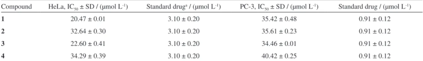
Ficusonic acid(1) was tested against two cancer cell lines, HeLa (cervical cancer cells) and PC-3 (prostate cancer cells) for its antiproliferative activity along with three known compounds 2-4. The activity of the four compounds 1-4 against PC-3 was weak with IC50 values
35.42, 35.61, 34.46 and 40.42 µmol L-1, respectively (Table
2); however in case of HeLa compounds 1 and 3 showed moderate or significant activity with IC50 values 20.47 and
22.60 µmol L-1, respectively.
Conclusions
The chemical investigation of the roots of Maytenus royleanus resulted in the isolation of four compounds: 3β,21β-dihydroxyolean-12-en-29-oic acid (1), 3α,22β-dihydroxyolean-12-en-29-oic acid(2), salaspermic acid(3) and orthosphenic acid(4). These compounds were reported for the first time from this plant, including one new compound (1). All four compounds were evaluated for their cytotoxic activity against two cancer cell lines.
Experimental
General experimental procedures
Melting point was determined on Buchi 535 apparatus and the IR data (KBr) were taken on Bruker Vector 22 spectrophotometer, νmax in cm
-1. Optical
rotation analysis was recorded on Jasco-P2000 digital polarimeter in MeOH at room temperature. UV data was taken from 996 photodiode array detector connected to LC-MS instrument. 1H NMR (500 MHz, CD
3OD) and 13C NMR (125 MHz, CD
3OD) spectra were recorded
on Bruker 500, while the chemical shift values were presented in ppm (d) and coupling constant (J) in Hz. For dereplication analysis, the LC-MS profile of FB was developed by HPLC-ELSD-UV-MS system (column C18;
solvent system: acetonitrile and water (0.1% formic acid in both); gradient system: 10-100% for 30 min). The
LC-MS data for confirming the purity of the compounds (MeOH/H2O with 0.1% FA) was recorded by using a
Phenomenex Luna column C18 RP (5 µm, 150 × 4.6 mm)
on Sedex 55 ELSD (evaporative light-scattering detector), 996 photodiode array detector in combination with ESI-TOF-MS (+) (time-of-flight electrospray ionization mass spectrometry). During purification on HPLC, the column used was Phenomenex 5µm Luna C18 RP column
(250 × 10 mm). ESI (+ve) MS and HRESIMS (high resolution electrospray ionization mass spectrometry) spectra were recorded on mariner ESI-TOF-MS (+). For open gravity column and thin layer chromatography, silica gel 60 column, mesh size 70-230 (E. Merck, 0.063-0.200 mm) and silica gel 60 PF254 (E. Merck) were used, respectively.
Plant material
The roots of Maytenus royleanus were collected from the Buner district, Khyber Pukhtoonkhwa, Pakistan during the month of June 2008. The plant was identified by taxonomist Mr. Ambara Khan (Degree College Daggar, Buner). The voucher specimen (Bot. 10068) was deposited in the Herbarium of Department of Botany, University of Peshawar, Khyber Pukhtoonkhwa (KPK), Pakistan.
Extraction and isolation
The air dried roots (13 kg) of Maytenus royleanus were repeatedly extracted (X3) with 80% MeOH/H2O at room
temperature after every 24 h. The combined extract was concentrated under vacuum at 40 °C, to obtain brownish thick syrup that constituted the crude aqueous methanolic extract (100 g). This was first partitioned according to our standard laboratory procedure into five fractions: FA, FB, FC, FD and FE. The crude extract suspended in distilled water and defatted (X3) with petroleum ether afforded fraction FA (35 g). The polarity of aqueous phase was changed to 10% MeOH/H2O by addition of MeOH
(200 mL) followed by extraction with dichloromethane (DCM) and concentrated under vacuum to obtain DCM
Table 2. Antiproliferative activity (IC50 in µmol L-1 ) of four compounds against Hela and PC-3 cell lines
Compound HeLa, IC50 ± SD / (µmol L-1) Standard druga / (µmol L-1) PC-3, IC
50 ± SD / (µmol L-1) Standard drug / (µmol L-1)
1 20.47 ± 0.01 3.10 ± 0.20 35.42 ± 0.48 0.91 ± 0.12
2 32.64 ± 0.30 3.10 ± 0.20 35.61 ± 0.23 0.91 ± 0.12
3 22.60 ± 0.41 3.10 ± 0.20 34.46 ± 0.01 0.91 ± 0.12
4 34.29 ± 0.39 3.10 ± 0.20 40.42 ± 0.25 0.91 ± 0.12
soluble fraction FB (11.5 g). The polarity of aqueous layer was changed to 50% MeOH/H2O by addition of MeOH
(1.8 L) and extracted with DCM to obtain fraction FC (7 g). By addition of MeOH in the aqueous layer, it was changed to approximately 70% MeOH/H2O which was
further extracted with DCM to get DCM soluble fraction FD (10 g), while the remaining DCM insoluble phase was named aqueous methanolic fraction (FE).
FB (11.5 g) was subjected to column chromatography (CC) on silica gel 60 (70-230 mesh, Merck) and eluted with solvents n-hexane and ethyl acetate increasing in order of polarity. As a result, seven sub-fractions (FD1F1 to FD1F7) were obtained after combination of the fractions on the basis of TLC profile. Sub-fraction FD1F6 (125 mg) was subjected to further CC on silica gel 60 (70-230 mesh, Merck), n-hexane, n-hexane-EtOAc, EtOAc solvent systems and elution with 23% n-hexane/EtOAc furnished 4 (22 mg). Fraction FD1F3 was a mixture of three compounds based on LC-MS profile which was subjected to HPLC (Phenomenex Luna column C18 RP
(5 µm, 150 × 4.6 mm; 0.1% acidic (formic acid) gradient solvent system (10-100 MeCN/H2O in 30 min) furnished
three compounds; 1 (8 mg), 2 (5 mg) and 3 (20 mg) (see Figure S1b in the Supplementary Information (SI) section).
Ficusonic acid(1)
White amorphous powder with molecular formula C30H48O4 (8 mg); mp 185-189 °C; [α]D
29.7−115 (c 0.1,
MeOH); IRfilm KBr cm-1 3358, 2945, 1760; EIMS m/z
(%) 55 (100), 95 (90), 120 (80), 246 (80), 264 (45), 217 (45), 185 (35), 454 (5); HRESIMS m/z 472.3602 (calcd. 472.3553 for C30H48O4); for
1H and 13C NMR see
Table 1.
Antiproliferative assays
Antiproliferative results of the compounds 1-4, were determined by the MTT [3-(4,5-dimethyl-2-thiazolyl)-2,5-diphenyltetrazolium bromide] assay on two different cancer lines. HeLa (cervical cancer lines) and PC-3 (Prostate cancer lines) are shown in Table 1. Cells were grown in DMEM (Dulbecco’s modified eagle medium) for PC-3 and MEM (minimal essential medium) for HeLa, containing 10% FBS (Fetal Bovine Serum) and 2% antibiotic (penicillin and streptomycin) and maintained at 37 oC with 5% CO
2 level
for 24 h in flask. Cells (1 × 10⁵ cells mL-1) were placed in
a 96 well flat bottom plates for 24 h incubation to allow for cell attachment. Various concentrations of sample varying from 100-1 µmol L-1 were added into the well and incubated
for 48 h. The IC50 values were calculated and at least three
independent experiments were carried out for each sample.
Doxorubicin was used as positive control in this assay for both PC-3 and HeLa.21
Supplementary Information
All the NMR (1H and 13C NMR, COSY, NOESY,
HMBC and HMQC) and MS data for compound 1, bioactivity results and extraction schemes (Figures S1-S15) are available free of charge at http://jbcs.sbq.org.br as PDF file.
Acknowledgement
This work was financially supported by Higher Education Commission (HEC) of Pakistan. We would like to thank Mr. Ambara Khan for plant taxonomic identification and ICCBS, University of Karachi, Pakistan for their help in providing NMR facilities.
References
1. Muñoz, O.; Peñaloza, A.; González, A. G.; Ravelo, A. G.; Crespo, A.; Bazzocchi, I. L.; Alvarenga, N. L. In Celastraceae, Bioactive Metabolites; Atta-Ur-Rah-man, ed.; Series Studies in Natural Products, Elsevier Science: Amsterdam, Holland, 1996, p. 739.
2. Schaneberg, B. T.; Green, D. K.; Sneden, A. T.; J. Nat. Prod.
2001, 64, 624.
3. Perestelo, N. R.; Jimenez, I. A.; Tokuda, H.; Hayashi, H.; Bazzocchi, I. L.; J. Nat. Prod. 2010, 73, 127.
4. Gonzalez, A. G.; Jimenez, I. A.; Ravelo, A. G.; Belles, X.; Piulachs, M. D.; Biochem. Syst. Ecol.1992, 20, 311.
5. Reider, P. J.; Roland, D. M.; The Alkaloids; Academic Press: New York, USA, 1984.
6. Flores, F. A.; Advances in Economic Botany; The New York Botanical Garden: New York, USA, 1984.
7. Gonzalez, J.G.; Delle Monache, G.; Delle Monache, F.; Marini-Bettolo, G. B.; J. Ethnopharmacol.1982, 5, 73. 8. Gonzalez, A. G.; Jimenez, I. A.; Ravelo, A. G.; Bazzochi, I. L.;
Tetrahedron1993, 49, 6637.
9. Kupchan, S. M.; Komoda, Y.; Court, W. A.; Thomas, G. J.; Smith, R. M.; Karim, A.; Gilmore, C. J.; Haltiwanger, R. C.; Bryan, R. F.; J. Am. Chem. Soc. 1972, 94, 1354.
10. Nakanishi, K.; Gullo, V. P.; Miura, I.; Govindachari, T. R.; Viswanathan, N.; J. Am. Chem. Soc. 1973, 95, 6473.
11. Itokawa, H.; Shirota, O.; Ikuta, H.; Morita, H.; Takeya, K.; Iitaka, Y.; Phytochemistry 1991, 30, 3713.
12. Shirota, O.; Morita, H.; Takeya, K.; Itokawa, H.; Iitaka, Y.; J. Nat. Prod. 1994, 57, 1675.
13. Itokawa, H.; Shirota, O.; Morita, H.; Takeya, K.; J. Nat. Prod.
14. Monache, D. F.; Marini-Bettolo, B. G.; Bernays, A. E.; Z. Angew. Entomol.1984, 97, 406.
15. Kutney, J. P.; Hewitt, G. M.; Lee, G.; Piotrowska, K.; Roberts, M.; Rettig, S.; Can. J. Chem.1992, 70, 1455.
16. Wiswanathan, N. I.; J. Chem. Soc., Perkin Trans 1 1979, 2, 349. 17. Zhang, W. J.; Pan, D, J.; Zhang, L. X.; Shao, Y. D.; Acta
Pharmacol. Sin. 1986, 21, 592.
18. Bruning, R.; Wagner, H.; Phytochemistry 1978, 17, 1821.
19. Nakagawa, H.; Takaishi, Y.; Fujimoto, Y.; Duque, C.; Garzon, C.; Sato, M.; Okamoto, M.; Oshikawa, T.; Ahmad, U. S.; J. Nat. Prod. 2004, 67, 1919.
20. Nakano, K.; Oose, Y.; Takaishi, Y.; Phytochemistry 1997, 46, 1179.
21. Forguson, L. R.; Mutat. Res.1994, 307, 395.