Evaluation of mandibular length in
subjects with Class I and Class II
skeletal patterns using the cervical
vertebrae maturation
Abstract: The aim of this study was to compare the mandibular size in boys and girls with Class I and Class II skeletal patterns, taking into consideration the bone maturation stage, as deined by the cervical ver-tebrae maturation. One hundred and sixty cephalometric radiographs were obtained from subjects (aged between 7 and 12 years) with Class I or Class II skeletal patterns, according to the ANB angle and WITS ap-praisal. The Class I sample consisted of 80 subjects (40 boys, 40 girls). The Class II sample also consisted of 80 subjects (40 boys, 40 girls). On a cross-sectional basis, mandibular length (Co-Gn) was compared be-tween groups and genders. The bebe-tween-stages changes were also evalu-ated, with the cervical vertebrae analysis used for establishing the bone maturation stages at CS2, CS3, CS4 and CS5. The results were statisti-cally analyzed by the Kruskal-Wallis test. The mandibular length dif-fered between skeletal patterns only at the earlier stages of development. In the Class I pattern, the mandibular lengths of boys were greater than those of girls at stages CS2, CS4 and CS5, whereas in the Class II pat-tern, the mandibular lengths of boys were greater than those of girls at stages CS2, CS3 and CS4. The present results indicate a sexual dimor-phism in the mandibular length at almost all stages of bone maturation, in exception of the CS5 stage in Class II.
Descriptors: Mandible; Cervical vertebrae; Growth and development. Rodrigo Generoso(a)
Elaine Cristina Sadoco(b) Mônica Costa Armond(c) Gustavo Hauber Gameiro(d)
(a) Professor; (b)MSc in Orthodontics
– Department of Orthodontics, School of Dentistry, Vale do Rio Verde University, Três Corações, MG, Brazil.
(c) Professor, Department of Oral Diagnosis,
School of Dentistry, Vale do Rio Verde University, Três Corações, MG, Brazil.
(d)Professor, Department of Physiology,
Federal University of Rio Grande do Sul, Porto Alegre, RS, Brazil.
Corresponding author:
Gustavo Hauber Gameiro
Universidade Federal do Rio Grande do Sul Rua Sarmento Leite, 500
Porto Alegre - RS - Brazil CEP: 90050-170
E-mail: [email protected]
Received for publication on Sep 30, 2008 Accepted for publication on Jan 19, 2009
Introduction
Moreover, almost all studies found statistically sig-niicant correlations between hand-wrist and skel-etal maturation of the cervical vertebrae,4,9-12 but racial variations in these relationships were also re-ported.10,11
Class II malocclusion is a commonly observed clinical problem, occurring in about a quarter of the young Brazilian population13 and in up to a third of the United States population.14 Despite the sub-stantial prevalence of Class II malocclusion as an orthodontic problem, review of the related litera-ture showed no consensus with regard to the growth changes of the mandible in untreated subjects with Class II malocclusion, when compared with subjects with normal occlusion. Some studies reported that subjects with Class II malocclusion had shorter man-dibles at both infantile and adolescent ages.15-18 In contrast, others19,20 found no differences in mandib-ular growth in Class II subjects from the deciduous dentition through the permanent dentition. Howev-er, the results in most of these studies were based on longitudinal growth changes related to the subjects’ chronologic ages or the dentition stages, which ac-cording to many authors, are not reliable predictors of a patient’s stage of skeletal development.7,9,21-23
To our knowledge, only one study compared the craniofacial dimensions in subjects with normal oc-clusion and Class II malococ-clusion, considering the cervical vertebrae method as a biological indica-tor of individual skeletal maturity to analyze the results.24 These authors showed that subjects with Class II malocclusion had smaller increases in man-dibular length at the growth spurt, and this dento-skeletal disharmony did not tend to self-correct with growth. However, the possible differences in the mandibular size according to gender were not inves-tigated in this study.
On this background, the aim of the present study was to compare mandibular size and cervical ver-tebrae maturation in subjects of both genders with Class I and Class II malocclusions.
Materials and Methods
The total sample (160 cephalometric radio-graphs) was obtained at the Orthodontic Depart-ment, School of Dentistry, Vale do Rio Verde
Uni-versity (UNINCOR). This study was designed as a cross-sectional research project. All the participants were aged between 7 and 12 years with no orth-odontic treatment at the time of study. They were divided in Class I or Class II skeletal patterns, ac-cording to the ANB angle and WITS appraisal. Class II subjects had the ANB angle greater than 4° and the linear distance between AO and BO (WITS appraisal) greater than 1 mm. They also presented an overjet greater than 2.5 mm. Class I subjects had the ANB angle between 0 and 3°, with normal dis-tances between AO and BO (–1 to 1 mm). The Class I sample consisted of 80 subjects (40 boys, 40 girls). The Class II sample also consisted of 80 subjects (40 boys, 40 girls). This study was approved by the re-search ethics committee of UNINCOR.
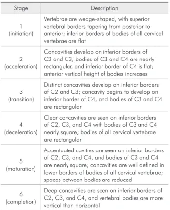
Cervical vertebrae stages were determined by the Hassel and Farman4 (1995) modiication of the cri-teria of Lamparski25 (1972), which established the 6 maturational stages by the observation of the bodies of the second, third, and fourth cervical vertebrae (Table 1).
Table 1 - Six stages in the evaluation of cervical vertebrae maturation according to the method of Hassel and Farman.
Stage Description
1 (initiation)
Vertebrae are wedge-shaped, with superior vertebral borders tapering from posterior to anterior; inferior borders of bodies of all cervical vertebrae are flat
2 (acceleration)
Concavities develop on inferior borders of C2 and C3; bodies of C3 and C4 are nearly rectangular, and inferior border of C4 is flat; anterior vertical height of bodies increases
3 (transition)
Distinct concavities develop on inferior borders of C2 and C3; concavity begins to develop on inferior border of C4, and bodies of C3 and C4 are rectangular
4 (deceleration)
Clear concavities are seen on inferior borders of C2, C3, and C4 with bodies of C3 and C4 nearly square; bodies of all cervical vertebrae are rectangular
5 (maturation)
Accentuated cavities are seen on inferior borders of C2, C3, and C4, and bodies of C3 and C4 are nearly square; concavities are well defined in lower borders of bodies of all cervical vertebrae; spaces between bodies are reduced
6 (completion)
The stages CS2, CS3, CS4 and CS5, in which signiicant skeletal growth occurs, were used in the present study to divide the sample according to their skeletal stage (Table 2).
Total mandibular length (Co-Gn) was measured on cephalograms traced by 1 investigator and veri-ied for landmark location and anatomical contours by another. Any disagreements were resolved by retracing the landmark or structure to the satis-faction of both observers. The measurements were performed manually in a blinded manner, and Dahl-berg’s formula was used to calculate the intra-op-erator error, by re-counting 10 randomly selected radiographs ifteen days later. The formula revealed values below 1.0, indicating suficient accuracy of the measurements.
All assessments were performed in a darkened room with a radiographic illuminator to ensure con-trast enhancement of the bone images.
Descriptive statistics were obtained for total mandibular length in both Class I and Class II sam-ples at the different maturation stages. Because of the small subsample sizes, the Kruskal-Wallis test was used to compare the mandibular lengths in the following situations: (1) Class II vs. Class I samples at CS2, CS3, CS4 and CS5 (2) male vs. female in each sample and (3) between-stage changes (CS2-CS3, CS3-CS4, CS4-CS5) in Class I and Class II samples. The signiicance level was set at p < 0.05.
Results
The total mandibular lengths (Co-Gn) in Class I and Class II subjects at the CS2, CS3, CS4 and CS5 stages are shown in Table 3. Class I boys presented greater mandibular lengths than Class II boys at stages CS2 and CS3. The differences at stages CS4 and CS5 were not statistically signiicant in this group. In the female group, Class I girls presented a greater mandibular length than Class II girls only at stage CS3. At the other stages, the differences were not statistically signiicant (Table 3).
The comparisons between genders revealed dif-ferent results depending on the skeletal pattern eval-uated. In the Class I pattern, the mandibular lengths of boys were greater than those of girls at stages CS2, CS4 and CS5. In the Class II pattern, the man-dibular lengths of boys were greater than those of girls at stages CS2, CS3 and CS4 (Table 3).
The comparisons of between-stage changes (CS2-CS3, CS3-CS4, CS4-CS5) in Class I and Class II samples are shown separately for boys (Table 4), Table 2 - Sample distribution in relation to bone maturation
stage in subjects with Class I and Class II skeletal patterns.
Bone maturation stage
Groups according to skeletal pattern
Class I Class II
CS2 Male (n = 10) Female (n = 10)
Male (n = 10) Female (n = 10)
CS3 Male (n = 10) Female (n = 10)
Male (n = 10) Female (n = 10)
CS4 Male (n = 10) Female (n = 10)
Male (n = 10) Female (n = 10)
CS5 Male (n = 10) Female (n = 10)
Male (n = 10) Female (n = 10)
Skeletal pattern
Class I Class II
(maturation stage) (Gender) (mandibular length)
CS2 Male 112.1 ± 5.0*
# 108.6 ± 4.3#
Female 103.2 ± 3.0 103.0 ± 3.6
CS3 Male 115.1 ± 5.9* 110.0 ± 3.9
#
Female 110.6 ± 2.9* 106.1 ± 3.3
CS4 Male 122.6 ± 5.1
# 121.0 ± 5.7#
Female 116.2 ± 4.0 113.4 ± 3.4
CS5 Male 127.4 ± 6.4
# 127.4 ± 2.7
Female 118.4 ± 3.9 122.2 ± 6.9
* P < 0.05 Cl I vs. Cl II; #P < 0.05 Male vs. Female.
and girls (Table 5). The between-stage differences were similar in boys with Class I and Class II pat-terns. The mandibular length was greater at stage CS4 than at CS2 and CS3; also, this measure was greater at stage CS5 than at CS4 (Table 4). In girls with Class I pattern, the mandibular lengths at CS4 and CS5 were greater than at CS3 and CS2. The dif-ferences between CS4 and CS5 were not signiicant in this group. On the other hand, the mandibular lengths at stages CS2 and CS3 in Class II girls were similar, while this measure was greater at stage CS5 than at CS4, and at CS4 it was also greater than at CS3 and CS2 (Table 5).
Discussion
In orthodontics and dentofacial orthopedics, each patient’s skeletal maturation period is an important factor to be considered in order to better take ad-vantage of his/her growth potential. In recent years, many authors have supported the eficacy of the cer-vical vertebrae analysis to assess skeletal age, which would represent a valid instrument to calculate the speed of growth and skeletal maturation.1-12 Our aim in this study, was to analyze mandibular length in
subjects with Class I and Class II skeletal patterns, considering their skeletal maturation stage. Sexual dimorphism in this measure was also evaluated.
The present study showed that the mandibular lengths in subjects with skeletal Class II pattern can differ from those with skeletal Class I pattern. These differences were found only at the initial stages of bone maturation, as observed in the male group at stages CS2 and CS3, and in the female group at stage CS3. These results are consistent with those of previous studies, and they show that dentoskel-etal characteristics of Class II malocclusion are es-tablished early in development.15,16,18,26 The peak in mandibular growth seems to occur between CS3 and CS4, as previously reported.27,28
In relation to the absence of statistical differ-ence between Class I and Class II patterns at the later stages of development (CS4 and CS5), our results are in accordance with those of Bishara et al.19 (1997) and Bishara20 (1998),in which Class II subjects had shorter mandibles when compared with normal subjects only in the earlier stages of development. But the differences were not signii-cant when the permanent dentition had completely
Skeletal pattern
Class I Class II
(maturation stage) (Gender) (mandibular length)
CS2 Male 112.1 ± 5.0 108.6 ± 4.3
CS3 Male 115.1 ± 5.9 110.0 ± 3.9
CS4 Male 122.6 ± 5.1 121.0 ± 5.7
CS5 Male 127.4 ± 6.4 127.4 ± 2.7
Statistical comparisons
between-stages (p < 0.05) (CS2=CS3) < CS4 < CS5 (CS2=CS3) < CS4 < CS5 Table 4 - Mean ± standard
deviation (mm) of mandibular length (Co-Gn) in boys with Class I and Class II skeletal patterns regarding the between-stages differences.
Skeletal pattern
Class I Class II
(maturation stage) (Gender) (mandibular length)
CS2 Female 103.2 ± 3.0 103.0 ± 3.6
CS3 Female 110.6 ± 2.9 106.1 ± 3.3
CS4 Female 116.2 ± 4.0 113.4 ± 3.4
CS5 Female 118.4 ± 3.9 122.2 ± 6.9
Statistical comparisons
between-stages (p < 0.05) (CS2=CS3) < (CS4=CS5) (CS2=CS3) < CS4 < CS5 Table 5 - Mean ± standard
erupted. These indings might suggest the possibil-ity of a late “catch up” growth period occurring in Class II subjects. However, these studies were based on longitudinal growth changes related to the sub-jects’ dentition stages, which seems not to be a re-liable indicator of skeletal maturation.21-23 On the other hand, Stahl et al.24 (2008) compared the lon-gitudinal craniofacial growth changes in untreated subjects with Class II malocclusion with those in subjects with normal occlusion from the prepuber-tal through the postpuberprepuber-tal stages of development, as deined by the cervical vertebrae maturation method. These authors found that the deiciency in mandibular growth in Class II subjects is signii-cant not only at the growth spurt, but that it is also maintained at a postpubertal observation. Consid-ering that our indings are based on cross-sectional data, the possibility that Class II dentoskeletal dis-harmony does not have a tendency to self-correct with growth should not be neglected.
Even though the present data are cross-sectional, they represent the irst attempt to investigate possi-ble gender differences of mandibular sizes in Class I and Class II subjects, within the same stage of bone maturation. Our results indicate that boys with the Class I pattern had greater mandibles than girls at stages CS2, CS4 and CS5, whereas in the Class II pattern the boys had greater mandibles than girls at stages CS2, CS3 and CS4. The similar mandibular lengths observed in Class II boys and girls at stage CS5 indicates the possibility of a “catch up” period in mandibular growth in Class II girls at this later stage (CS5) of bone maturation. This inding can also be observed when the between-stages differ-ences in boys and girls are compared (Tables 4 and 5). Although the between-stages differences in Class I and Class II boys are very similar, a signiicant in-crease in mandibular length from stage CS4 to CS5 was observed only in the Class II girls, but not in the Class I girls. These results suggest that Class II girls can present a delay in the mandibular growth, and this information has important orthodontic clinical implications. A substantial number of treat-ment protocols in dentofacial orthopedics beneits from the inclusion of the period of accelerated
man-dibular growth. For example, functional appliances have been shown to be more effective when used in the peak of mandibular growth rather than earlier.29 However, some authors have stated that because of the high variability in mandibular growth, ortho-dontists should take care to make this prediction for the individual patient.2 Therefore, the variables in-volved in the skeletal maturation should be known to improve the effectiveness of this biological index as an orthodontic tool in clinical practice.
Racial variations in the relationships between skeletal maturity established by different methods of evaluation were previously reported.10,11,25 These variations could be due to the predominant ethnic origin, climate, nutrition, socioeconomic level and urbanization.30 The outcomes of the present study suggest that the gender and the type of skeletal mal-occlusion are also important factors to be consid-ered in the skeletal maturity evaluation of a patient. Although most of the studies report that the peak in mandibular growth occurs between the CS3 and CS4 stages,27,28 this does not seem to be the case in Class II girls, in whom signiicant “delayed mandib-ular growth” could occur at stage CS5. This possible “catch up” growth of the mandible in Class II girls does not mean that the Class II will change to Class I, but this inding is extremely relevant in order to avoid equivocal prediction of the peak in mandibu-lar growth and, consequently, underestimate growth potential in these patients. Nevertheless, some limi-tation must be considered in this study, such as the lack of evaluation of regional growth and remodel-ing of the mandible. Moreover, the sample for our study did not include all types of Class II subjects who would be encountered in clinical practice, and the conclusions therefore cannot be extended to pa-tients with different problems.
Conclusions
Subjects with skeletal Class I and Class II pat-terns had different mandibular lengths at the earlier stages of bone maturation.
A sexual dimorphism in this measure was ob-served at almost all stages, in exception of the CS5 stage in Class II.
•
References
1. Baccetti T, Franchi L, McNamara JA Jr. An improved ver-sion of the cervical vertebral maturation (CVM) method for the assessment of mandibular growth. Angle Orthod. 2002;72(4):316-23.
2. Flores-Mir C, Burgess CA, Champney M, Jensen RJ, Pitcher MR, Major PW. Correlation of skeletal maturation stages determined by cervical vertebrae and hand-wrist evaluations. Angle Orthod. 2006;76(1):1-5.
3. Santos EC, Bertoz FA, Arantes F de M, Reis PM, de Bertoz AP. Skeletal maturation analysis by morphological evaluation of the cervical vertebrae. J Clin Pediatr Dent. 2006;30(3):265-70.
4. Hassel B, Farman AG. Skeletal maturation evaluation us-ing cervical vertebrae. Am J Orthod Dentofacial Orthop. 1995;107(1):58-6.
5. Caldas M de P, Ambrosano GM, Haiter Neto F. New formula to objectively evaluate skeletal maturation using lateral cepha-lometric radiographs. Braz Oral Res. 2007;21(4):330-5. 6. Mito T, Sato K, Mitani H. Predicting mandibular growth
potential with cervical vertebral bone age. Am J Orthod Den-tofacial Orthop. 2003;124(2):173-7.
7. Hellsing E. Cervical vertebral dimension in 8-11 and 15 year old children. Acta Odontol Scand. 1991;49(4):207-13. 8. Franchi L, Bacetti T, McNamara JA Jr. Mandibular growth
as related to cervical vertebral maturation and body height. Am J Orthod Dentofacial Orthop. 2000;118(3):335-40. 9. O’Reilly M, Yanniello G. Mandibular growth changes and
mat-uration of cervical vertebrae. Angle Orthod. 1988;58(2):179-84.
10. Garcia-Fernandez P, Torre H, Flores M, Rea J. The cervi-cal vertebrae as maturational indicators. J Clin Orthod. 1998;32(4):221-5.
11. Roman PS, Palma JC, Oteo D, Nevado E. Skeletal maturation determined by cervical vertebrae development. Eur J Orthod. 2002;24(3):303-11.
12. Uysal T, Ramoglu SI, Basciftci FA, Sari Z. Chronologic age and skeletal maturation of the cervical vertebrae and hand-wrist: is there a relationship? Am J Orthod Dentofacial Or-thop. 2006;130(5):622-8.
13. Grando G, Young AA, Vedovello Filho M, Vedovello SA, Ramirez-Yañez GO. Prevalence of malocclusions in a young Brazilian population. Int J Orthod Milwaukee. 2008;19(2):13-6.
14. McLain JB, Proffit WR. Oral health status in the United States: prevalence of malocclusion. J Dent Educ. 1985;49(6):386-96.
15. Kerr WJ, Hirst D. Craniofacial characteristics of subjects with normal and postnormal occlusions – a longitudinal study. Am J Orthod Dentofacial Orthop. 1987;92(3):207-12.
16. Baccetti T, Franchi L, McNamara JA Jr, Tollaro I. Early den-tofacial features of Class II malocclusion: a longitudinal study from the deciduous through the mixed dentition. Am J Orthod Dentofacial Orthop. 1997;111(5):502-9.
17. Ngan PW, Byczek E, Scheick J. Longitudinal evaluation of growth changes in Class II division 1 subjects. Semin Orthod. 1997;3(4):222-31.
18. Varrela J. Early developmental traits in Class II malocclusion. Acta Odontol Scand. 1998;56(6):375-7.
19. Bishara SE, Jakobsen JR, Vorhies B, Bayati P. Changes in den-tofacial structures in untreated Class II division 1 and normal subjects: a longitudinal study. Angle Orthod. 1997;67(1):55-66.
20. Bishara SE. Mandibular changes in persons with untreated and treated Class II division 1 malocclusion. Am J Orthod Dentofacial Orthop. 1998;113(6):661-73.
21. Björk A, Helm S. Prediction of the age of maximum pubertal growth in body height. Angle Orthod. 1967;37(2):134-43. 22. Hägg U, Taranger J. Maturation indicators and the pubertal
growth spurt. Am J Orthod. 1982;82(4):299-309.
23. So LLY. Skeletal maturation of the hand and wrist and its cor-relation with dental development. Aust Orthod J. 1997;15(1):1-9.
24. Stahl F, Baccetti T, Franchi L, McNamara JA Jr. Longitudi-nal growth changes in untreated subjects with Class II Di-vision 1 malocclusion. Am J Orthod Dentofacial Orthop. 2008;134(1):125-37.
25. Lamparski D. Skeletal age assessment utilizing cervical ver-tebrae [thesis]. Pittsburgh: University of Pittsburgh; 1972. 26. Anderson DL, Popovich F. Lower cranial height vs craniofacial
dimensions in Angle Class II malocclusion. Angle Orthod. 1983;53(3):253-60.
27. Franchi L, Baccetti T, McNamara JA Jr. Mandibular growth as related to cervical vertebral maturation and body height. Am J Orthod Dentofacial Orthop. 2000;118(3):335-40. 28. Gu Y, McNamara JA. Mandibular growth changes and
cer-vical vertebral maturation. A cephalometric implant study. Angle Orthod. 2007;77(6):947-53.
29. Baccetti T, Franchi L, Toth LR, McNamara JA Jr. Treatment timing for Twin-block therapy. Am J Orthod Dentofacial Or-thop. 2000;118(2):159-70.