Influence of initial occlusal severity on time and efficiency of Class I
malocclusion treatment carried out with and without premolar extractions
Ruben Leon-Salazar1, Guilherme Janson2, José Fernando Castanha Henriques2, Vladimir Leon-Salazar3
Introduction: The aim of this retrospective study was to compare the occlusal outcomes, duration and efficiency of Class I malocclusion treatment carried out with and without premolar extractions in patients with different degrees of initial malocclusion severity. Methods: Complete records of 111 patients were obtained and divided into two groups: Group 1 consisted of 65 patients at an initial mean age of 13.82 years old treated with four premolar extractions; whereas Group 2 consisted of 46 patients at an initial mean age of 14.01 years old treated without extractions. Two subgroups were obtained from each group (1A, 1B, 2A and 2B) with different degrees of malocclusion severity according to the initial values of PAR index. Compatibility was assessed using chi-square and t-tests. The subgroups were compared by means of Analysis of Variance (ANOVA).The variables that might be related to treatment duration and efficiency were assessed using the multiple linear regression analysis. Results: Initial malocclusion severity was positively related to the amount of occlusal correction and consequently to a higher efficiency index. Moreover, extraction protocol showed a positive relationship with treatment duration and a negative relationship with treatment efficiency. Conclusion: Extraction and non-extraction protocols for correction of Class I malocclusion provide similar satisfactory results; however, the extrac-tion protocol increases the overall treatment duraextrac-tion. Orthodontic treatment is more efficient in cases with high initial malocclusion severity treated with a non-extraction protocol.
Keywords:Class I malocclusion. Efficiency. Time. Tooth extraction.
How to cite this article: Salazar R, Janson G, Henriques JFC, Leon-Salazar V. Influence of initial occlusal severity on time and efficiency of Class I malocclusion treatment carried out with and without premolar extrac-tions. Dental Press J Orthod. 2014 July-Aug;19(4):38-49. DOI: http://dx.doi. org/10.1590/2176-9451.19.4.038-049.oar
Submitted: March 22, 2009 - Revised and accepted: June 29, 2009
Contact address: Ruben Leon-Salazar
Al. Dr. Octávio Pinheiro Brizola, 9-75 ‒ CEP: 17043-101 ‒ Bauru/SP — Brazil 1 Masters student in Orthodontics, School of Dentistry — USP/ Bauru.
2 Full professor, School of Dentistry — USP/ Bauru.
3 PhD resident in TMD and Orofacial Pain, School of Dentistry — University of Minnesota.
» The authors report no commercial, proprietary or financial interest in the products or companies described in this article.
DOI: http://dx.doi.org/10.1590/2176-9451.19.4.038-049.oar
Introdução: o objetivo desse estudo retrospectivo foi comparar os resultados oclusais, o tempo e o grau de eficiência do tratamento da má oclusão de Classe I realizado com e sem extrações em pacientes que apresentavam diferentes tipos de severidade oclusal inicial. Métodos: a amostra foi composta pelas documentações de 111 pacientes, divididas em dois grupos: Grupo 1 (n = 65), com idade inicial média de 13,82 anos, tratados com extrações; Grupo 2 (n = 46), com idade inicial média de 14,01 anos, tratados sem extrações. De cada grupo, foram obtidos dois subgrupos (1A, 1B, 2A e 2B) com severidades oclusais diferentes (alta e baixa), de acordo aos valores iniciais do índice PAR. A avaliação da compatibilidade foi realizada por meio do teste qui-quadrado e do teste t. Os subgrupos foram comparados por meio da análise de variância (ANOVA) e foi realizada a análise de regressão linear múltipla para avaliação das variáveis que poderiam estar relacionadas com o tempo e com a eficiência do tratamento. Resultados: a severidade oclusal inicial esteve diretamente relacionada à quantidade de sua correção e, consequentemente, à obtenção de um maior índice de eficiência; por outro lado, a utili-zação do protocolo de extrações de pré-molares mostrou uma relação direta com o tempo de tratamento e inversa com a eficiência do tratamento. Conclusão: no tratamento da má oclusão de Classe I, podem ser obtidos resultados oclusais satisfatórios com uma maior quantidade de correção das alterações oclusais nos casos com maior severidade inicial, e um maior tempo de tratamento quando o tratamento envolve extrações dentárias.
INTRODUCTION
Assessing treatment outcomes by means of oc-clusal indexes allows us to understand the effects dif-ferent types of appliances, techniques and treatment
protocols produce on dental occlusion,1,4,8,13,20,28,33
treatment time2,3,10,21,34 and efficiency. In this context,
efficiency is described as the achievement of the best
results within a shorter period of time.19,31
Some authors have observed the inluence of dental extractions on correction of initial malocclusion se-verity, showing better occlusal results when a
non-ex-traction protocol was used.6 However, they observed
that in Class II malocclusion cases, the protocol that included the extraction of two maxillary premolars yielded better occlusal outcomes than the
non-extrac-tion and the four-premolar extracnon-extrac-tion protocols.19,20
Regarding treatment time, the literature gener-ally highlights dental extractions as one of the main
factors for increased treatment time.6,10,34 Contrary to
those findings, Beckwith et al3 stated that the
differ-ence in treatment time between extraction and non-extraction protocols is not significant. Other authors also assessed the influence of malocclusion severity on treatment time and found no relation between treatment duration and initial malocclusion
sever-ity.16. Nevertheless, other studies have shown that
there is a direct correlation between initial
malocclu-sion severity and the treatment duration.6,10
Unfortunately, these previous studies used mixed samples that included different types of malocclusions and treatment protocols. Therefore, the applicability of their findings is limited and cannot be extrapolated to Class I malocclusion.
The objective of this study was to compare the oc-clusal outcomes, treatment duration and eiciency of two diferent protocols for Class I malocclusion: non-extraction and four-premolar non-extractions, in order to elucidate the efects of dental extractions on orthodon-tic treatment performed for this speciic malocclusion.
MATERIAL AND METHODS
Material
The sample of this retrospective study comprised patients with Class I malocclusion and similar pre-treatment characteristics who were treated with four premolars extractions or without extractions. Pa-tients were selected from the Master’s and Postgraduate
Orthodontic programs at the School of Dentistry of University of São Paulo, Bauru, Brazil. In selecting the sample, the following inclusion criteria were applied:
» Class I malocclusion treated without extrac-tions or with extraction of four premolars, two maxillary and two mandibular.
» Presence of all permanent teeth up to the first molar.
» Presence of crowding not greater than 8 mm. » Absence of supernumerary teeth.
» Absence of impacted teeth.
» Absence of abnormalities in tooth size and / or shape.
» Treatment with full fixed Edgewise appliances. » No history of orthognathic surgery.
» Full orthodontic records available for review. The sample comprised the initial and final orth-odontic records of 111 patients who were divided into two groups according to the extraction protocol used as part of the orthodontic treatment.
Group 1 consisted of 65 patients, 24 males (36.92%) and 41 females (63.08%), with initial mean age of 13.82 years old (ranging from 10.69 to 22.04 years), who had Class I malocclusion and were treat-ed with extraction of four premolars, two maxillary and two mandibular (Tables 4 and 5).
Group 2 consisted of 46 patients treated with-out extractions, 16 males (34.78%) and 30 females (65.22%) with initial mean age of 14.01 years old (ranging from 11.04 to 21.54 years) (Tables 4 and 5). Both groups were treated with full fixed appliances using the simplified Edgewise technique.
Since previous studies have shown that severity of malocclusion could influence the treatment
dura-tion,6,10 we further divided each group, based on their
initial occlusal index, into two subgroups with differ-ent malocclusion severity (high and low). Thus, the four subgroups, two in each group, with high and low initial malocclusion severity had the following characteristics (Table 6).
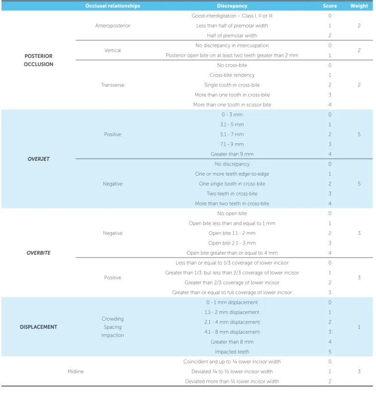
Table 1 - Criteria applied to score each component of PAR index9.
Table 2 - Description of variables used.
Occlusal relationships Discrepancy Score Weight
POSTERIOR
OCCLUSION
Anteroposterior
Good interdigitation – Class I, II or III 0
2 Less than half of premolar width 1
Half of premolar width 2
Vertical No discrepancy in intercuspation 0 2 Posterior open bite on at least two teeth greater than 2 mm 1
Transverse
No cross-bite 0
2 Cross-bite tendency 1
Single tooth in cross-bite 2 More than one tooth in cross-bite 3 More than one tooth in scissor bite 4
OVERJET
Positive
0 - 3 mm 0
5
3.1 - 5 mm 1
5.1 - 7 mm 2
7.1 - 9 mm 3
Greater than 9 mm 4
Negative
No discrepancy 0
5 One or more teeth edge-to-edge 1
One single tooth in cross-bite 2 Two teeth in cross-bite 3 More than two teeth in cross-bite 4
OVERBITE
Negative
No open bite 0
3 Open bite less than and equal to 1 mm 1
Open bite 1.1 - 2 mm 2 Open bite 2.1 - 3 mm 3 Open bite greater than or equal to 4 mm 4
Positive
Less than or equal to 1/3 coverage of lower incisor 0
3 Greater than 1/3. but less than 2/3 coverage of lower incisor 1
Greater than 2/3 coverage of lower incisor 2 Greater than or equal to full coverage of lower incisor 3
DISPLACEMENT
Crowding Spacing Impaction
0 - 1 mm displacement 0
1 1.1 - 2 mm displacement 1
2.1 - 4 mm displacement 2 4.1 - 8 mm displacement 3
Greater than 8 mm 4
Impacted teeth 5
Midline
Coincident and up to ¼ lower incisor width 0
3 Deviated ¼ to ½ lower incisor width 1
Deviated more than ½ lower incisor width 2
ABBREVIATIONS DESCRIPTION
PARi Initial PAR index
APINH Initial amount of mandibular crowding
AGE Age at the beginning of treatment
PARf Final PAR index
PARi-PARf Improvement of occlusal discrepancy PC-PAR Improvement of occlusal discrepancy (percentage)
Time Treatment duration in months
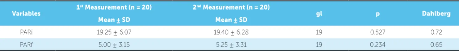
Table 3 - Results of systematic and random errors assessed using depended t-test and Dahlberg’s formula.
Table 4 - Compatibility of groups.
Table 5 - Comparison of the initial characteristics using t-test.
Variables 1
st Measurement (n = 20) 2nd Measurement (n = 20)
gl p Dahlberg
Mean ± SD Mean ± SD
PARi 19.25 ± 6.07 19.40 ± 6.28 19 0.527 0.72
PARf 5.00 ± 3.15 5.25 ± 3.31 19 0.234 0.65
Sex GROUP 1 (Extraction) n = 65 GROUP 2 (Non-extraction) n = 46 Total
Females 41 30 71
Males 24 16 40
Total 65 46 111
X2 = 0.535 GL = 1 p = 0.817
VARIABLES GROUP 1 (Extraction) n = 65 GROUP 2 (Non-extraction) n = 46 DF p
Mean ± SD Mean ± SD
PARi 19.92 ± 8.08 17.89 ± 6.96 109 0.170
CROWDING 4.94 ± 1.59 4.32 ± 1.87 109 0.065
AGE 13.82 ± 2.11 14.01 ± 1.78 109 0.620
females with a initial mean age of 13.88 ± 1.06 years (minimum 11.90, maximum 15.82). Subgroup 2B (Low severity; n =15) comprised 4 males and 11 females with a initial mean age of 14.09 ± 1.45 years (minimum 11.40, maximum 15.99).
Methods
Clinical records
Patients’ orthodontic records were used to obtain the demographic and clinical information included in the analysis: sex, date of birth, age at treatment onset, Table 6 - Results of ANOVA and Tukey’s test regarding the initial characteristics of subgroups 1A, 1B, 2A e 2B. Subgroups were classified according to their initial malocclusion severity.
* Statistically significant: P < 0.05.
Variables
GROUP 1 (Extraction) GROUP 2 (Non-extraction)
ANOVA Subgroup 1A High
Severity n = 22
Subgroup 1B Low
Severity n =22
Subgroup 2A High
Severity n =15
Subgroup 2B Low
Severity n = 15
Mean ± SD Mean ± SD Mean ± SD Mean ± SD F p
PARi 29.09a ± 5.32 11.68b ± 2.40 25.60a ± 5.15 10.87b ± 2.97 97.28 0.000* CROWDING 5.17 ± 1.67 4.64 ± 1.62 4.08 ± 2.39 4.19 ± 1.72 1.35 0.265
proposed treatment protocol, including extraction and non-extraction of premolars, and length of active orthodontic treatment.
To estimate total treatment time, the starting date was defined as the date when placement of first mo-lar bands or first direct bonding occurred, whereas final date was defined as the date when orthodontic retainers were delivered.
Dental cast analysis
Assessment of mandibular crowding
The amount of mandibular crowding was calcu-lated based on the difference between the arch perim-eter (circumference measured from the mesial of one permanent first molar to its antimere) and the sum of the mesio-distal width of all mandibular permanent
teeth except molars.26
Calculation of occlusal index
The occlusal index was calculated according to the weighted Peer Assessment Rating (PAR index)
advo-cated by DeGuzman et al9 which includes the
assess-ment of ive occlusal features (posterior occlusion, over-jet, overbite, midline and maxillary tooth displacements) with well-deined measurement criteria (Table 1).
The scores for PAR index calculation30 are
record-ed according to the following:
1. Posterior occlusion.
Posterior occlusion, also described as “buccal seg-ment relationship” in the original PAR index, com-prises the zone from the distal anatomical contact point of canine to the mesial anatomical contact point of irst permanent molar. Posterior dental relationship is assessed in three planes of space and scores are given to anteroposterior, vertical and transverse discrepan-cies according to Table 1. These scores are added and the inal value is multiplied by two. Each posterior segment, right and let, is recorded separately.
2. Overjet.
Positive or negative overjet is recorded using the most prominent surface of any central or lateral incisors as reference. During this measurement, the ruler is held parallel to the occlusal plane and radial to the line of the arch. The magnitude of the overjet is transformed into a score according to Table 1 and then multiplied by 5.
3. Overbite.
Overbite is recorded as the proportion of the lower incisor crown that is covered by upper incisors or the amount of open bite, in millimeters, taking as reference the tooth with greater overlap. The score obtained according to Table 1 is then multiplied by 3.
4. Midline.
Discrepancy of maxillary midline is assessed in relation to lower central incisors using the score in Table 1 which is then multiplied by 3.
5. Maxillary tooth displacement.
Displacements such as crowding, spacing and im-pacted teeth are recorded in the maxillary anterior re-gion, only. These occlusal features are recorded con-sidering the shortest distance between contact points of adjacent teeth parallel to the occlusal plane. These measurements are transformed into scores and added according to the criteria defined in Table 1. A tooth is considered impacted when the space available for this tooth is less than 4 mm.
We calculated the PAR index for each of the pre-treatment and post pre-treatment dental casts (n = 222) us-ing the criteria described above and usus-ing the scores speciied in Table 1. PAR index was termed initial PAR (PARi) when obtained from the pretreatment models, and inal PAR (PARf) when calculated in post-treat-ment casts. The higher the numerical value obtained in these indexes, the more severe the malocclusion, be-cause PAR index is obtained by applying scores to the intra-arch (e.g. crowding) and inter-arch (e.g. overbite, overjet, crossbite, midline) dental relationships as well as by using an ordinal scale starting at 0 for a normal value. All measurements in the initial and inal casts were ob-tained using a digital caliper (Mitutoyo, Kawasaki, Ja-pan) with accuracy closed to 0.1 mm.
Assessing changes in occlusal discrepancy
Changes in occlusal discrepancy produced by each treatment protocol were calculated by subtracting PARf from PARi values (PARi - PARF). The numerical re-duction in the index accounted for occlusal changes
di-rectly related to treatment protocol.29,30 In addition, the
percentage of PAR reduction (PcPAR) during treatment was calculated to verify the amount of improvement
For this calculation, we applied the following mathematical formula:
PcPAR =
PARi-PARf
x 100
PARi
Treatment efficiency index (TE)
Treatment efficiency was defined as the greatest occlusal index change produced within the shortest treatment time. It was calculated using the following formula, in which the denominator is the total
treat-ment time expressed in months:19,31
T
E=
PcPAR
TIME
Statistical analysis
Errors of the method were assessed by repeating the measurements on 20 initial and 20 inal dental casts randomly selected from the sample. Repeated measure-ments were taken approximately one month ater the irst occlusal index calculation (Table 3). The formula
proposed by Dahlberg7 (S2 = Σd2/2n) was applied to
es-timate random errors, while paired t-test was used to
analyze systematic errors.18
Initial compatibility regarding gender distribution between the two study groups was assessed using the non-parametric chi-square test (Table 4). T-test was also used to assess other baseline characteristics, such as age, malocclusion severity, and amount of mandibu-lar crowding (Table 5). Subgroups 1A, 1B, 2A and 2B were compared using Analysis of Variance (ANOVA). Tukey’s test was used to investigate the hypothesis that severity of PARi inluences the treatment duration (Ta-bles 6 and 7). Multiple linear regression was used to as-sess the inluence of initial malocclusion severity, man-dibular crowding and the extraction/non-extraction protocols over treatment eiciency (Tables 8 and 9). All statistical analyses were performed using Statistica sot-ware. P value ≤ 0.05 was considered signiicant.
RESULTS
No systematic errors18 were found for repeated
mea-surements one month ater the initial assessment.
Ran-dom errors7 were considered negligible (Table 3).
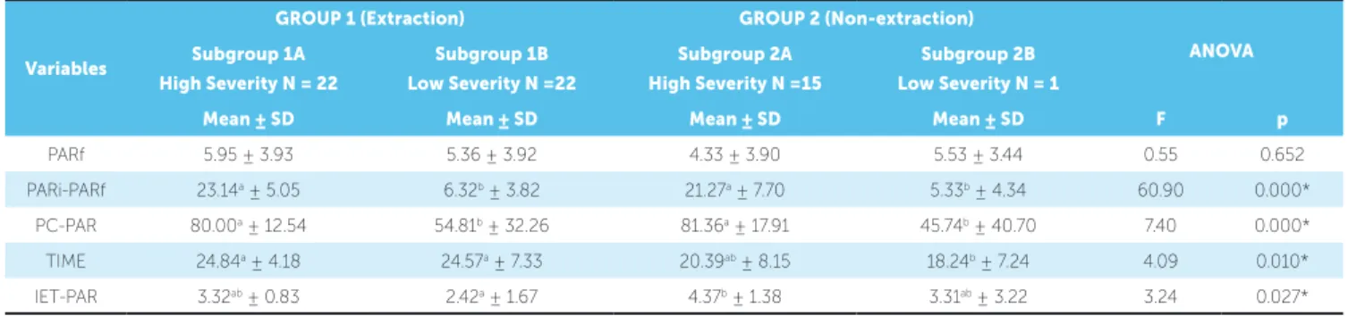
Groups were compatible regarding age, sex, man-dibular crowding and PARi (Tables 4 and 5). As shown in Table 6, malocclusion severity was signiicantly dif-ferent between subgroups with high PARi (1A, 2A) and low PARi (1B, 2B) severity. The diference be-tween PARi was of approximately 16 points. Sub-groups were compatible in all other variables.
Final occlusal outcome, assessed by means of PARf, was similar in all subgroups (Table 7). How-ever, numerical and proportional reduction in the occlusal index was significantly greater in subgroups with high PARi (1A, 2A) than in subgroups with low PARi (2A, 2B). The treatment duration for the non-extraction subgroups was about four (2A) to six (2B) months less than the extraction subgroups; however, treatment time was only significantly reduced in sub-group 2B that started with a low PARi. Treatment was also more efficient in the group with high maloc-clusion severity treated without extractions (Table 7). Multiple linear regression analysis showed that of the three variables evaluated (PARi, CROWDING and PROTOCOL), only the treatment protocol with extractions showed significant positive correlation with treatment duration (Table 8). Regarding treat-ment efficiency, initial malocclusion severity showed a positive influence on the efficiency index, while treatment protocol influenced it negatively (Table 9).
DISCUSSION
Sample and compatibility
The overall objective of this study was to compare two different treatment protocols for Class I maloc-clusion. For this reason, the sample only included patients with Class I malocclusion who were treated with or without extraction of four premolars. We fo-cused on this specific type of malocclusion because the compatibility of groups regarding initial maloc-clusion severity decreased the risk of bias. As showed in previous studies, treatment length and efficiency varies according to the amount of initial
anteropos-terior discrepancy.6,19,31,34 Distribution of sex, age,
PARi, and mandibular crowding were also compat-ible between groups, which reduced the risk of con-founding and selection bias.
extractions showed greater amount of crowding. There-fore, we only included patients with initial mandibular crowding not greater than 8 mm in order to eliminate
the inluence of this variable on the results of our study.32
Cases that had their initial treatment plan changed during the course of treatment (e.g. non-extraction cases that ended up with extractions) were excluded from the study to avoid the inluence of this factor on
treatment duration.21
The aforementioned inclusion and exclusion cri-teria were applied to 4000 clinical charts belong-ing to the Department of Orthodontics’ archives of the School of Dentistry — University of São Paulo/ Bauru. A sample of 111 subjects was obtained. Con-sidering that the incidence of Angle Class I
maloc-clusion is of approximately 55 %,15 we expected to
come up with a larger study sample. However, the meticulous application of these criteria resulted in
Table 7 - Comparison of occlusal changes, treatment duration and treatment efficiency between subgroups 1A, 1B, 2A e 2B.
* * Statistically significant: P < 0.05.
Variables
GROUP 1 (Extraction) GROUP 2 (Non-extraction)
ANOVA Subgroup 1A
High Severity N = 22
Subgroup 1B Low Severity N =22
Subgroup 2A High Severity N =15
Subgroup 2B Low Severity N = 1
Mean ± SD Mean ± SD Mean ± SD Mean ± SD F p
PARf 5.95 ± 3.93 5.36 ± 3.92 4.33 ± 3.90 5.53 ± 3.44 0.55 0.652 PARi-PARf 23.14a ± 5.05 6.32b ± 3.82 21.27a ± 7.70 5.33b ± 4.34 60.90 0.000*
PC-PAR 80.00a ± 12.54 54.81b ± 32.26 81.36a ± 17.91 45.74b ± 40.70 7.40 0.000* TIME 24.84a ± 4.18 24.57a ± 7.33 20.39ab ± 8.15 18.24b ± 7.24 4.09 0.010* IET-PAR 3.32ab ± 0.83 2.42a ± 1.67 4.37b ± 1.38 3.31ab ± 3.22 3.24 0.027*
Table 8 - Multiple regression analysis using treatment duration as a dependent variable.
Table 9 - Multiple regression analysis using treatment efficiency as a dependent variable. SE: standard error
R2= 0.1297. Length of treatment=22.75 + 0.33(Protocol) + 0.080(Crowding) + 0.031 (PARi). *Protocol: 0 – Non-extraction; 1 – Extraction.
R2=0,1149. Treatment efficiency = 2.075 - 0.261(Protocol) - 0.057(Crowding) + 0.238 (PARi). *Protocol: 0 – Non-extraction; 1 – Extraction.
Variables Coeicient SE t p
(Constant) 22.748 2.657 8.562 0.0000
PARi 0.031 0.087 0.344 0.7316
CROWDING 0.080 0.389 0.870 0.3864
PROTOCOL 0.332 1.369 -3.593 0.0005*
Variables Coeicient SE t p
(Constant) 2.075 0.639 3.247 0.0016
PARi 0.238 0.021 2.589 0.0110*
CROWDING -0.057 0.094 -0.615 0.5397
the elimination of a large number of potential par-ticipants with Class I malocclusion. The study sam-ple reduced even further because some of the clinical charts did not have the orthodontic documentation that met the specific needs of this study.
METHODS
We used the PAR occlusal index to quantify both, pre-treatment discrepancy and post-treatment oc-clusal outcomes, since its accuracy and reliability has been previously validated. Moreover, the PAR index is an objective and user friendly analysis method that
has been extensively used in similar studies.1,4,8,9,13,19,30
Besides allowing us to compare our findings with previous studies, the use of PAR index allowed us to investigate the compatibility of both groups
regard-ing the severity of initial malocclusion,4,9,19,29,30 as
well as the numerical and the percentage of improve-ment obtained in each group at the end of treatimprove-ment. We associated the percentage of occlusal improve-ment with treatimprove-ment duration in order to obtain an index capable of objectively expressing the degree of
treatment efficiency (TE), as previously described by
other studies.19,31
Lack of signiicant systematic errors and reduced value of random errors found in this study relect stan-dardization and accuracy of measurements (Table 3) and are related to the calibration of the examiner prior to data collection. The simple and objective assessment of dental casts by means of PAR index also allowed us to obtain a high degree of precision and reproducibility.
COMPARISON BETWEEN GROUPS AND VARIABLES
Post-treatment occlusal outcomes
The comparison of PARf values between sub-groups showed that inal occlusal relationships in all subgroups were similarly satisfactory (Table 7). PARf index of non-extraction and extraction groups ranged from 4.33 to 5.95, thus showing a good occlu-sal outcome in all patients regardless of severity of their initial occlusal discrepancy. These values were similar to those found in prior studies conducted with Class
I patients. For instance, Birkeland et al4 found PARf
values of 5.9 and 6.2 for cases treated with and without extractions, respectively. Likewise, other authors, such
as Willems et al,38 Freitas et al,8,13 found pooled PARf
values of 5.1, 6.32, and 5.65, respectively. However, these studies used diferent criteria for sample
selec-tion. Birkeland et al4 included cases treated with ixed
appliances in both maxillary and mandibular arches and cases treated with ixed appliances in a single arch.
Willems et al38 included patients treated with
remov-able orthodontic or functional orthopedic appliances and cases treated with ixed appliances in one or two
dental arches. Freitas et al8,13 only included patients
treated with extraction of four premolars and ixed ap-pliances in both dental arches.
Several other studies have assessed the amount of correction of initial occlusal discrepancy using PAR index. Nevertheless, these studies included different types of malocclusion in their samples be-cause their main objectives were to audit the qual-ity of orthodontic treatment provided and the factors that may influence treatment duration and efficiency provided in private practice, university-based and hospital-based clinics. Some of these studies
report-ed similar PARf values,6,12,17,24 while others showed
higher PARf11,12,24,33 and lower PARf values than our
study.25,28,31,39 The difference in samples and
method-ology prevented us to make further comparison with these studies.
The high PARf values found in some previous studies were related to cases treated with removable
functional orthopedic appliances,11,12,24,33 or cases
re-ceiving treatment for only one of the dental arches, or the use of historical samples that do not show
re-sults as good as recently treated cases.11 Additionally,
the experience of the treatment provider was also found to be directly associated with the quality of
occlusal outcomes.11,12,28,31,33 Moreover, the common
characteristic among studies that showed low PARf values was that orthodontic treatment was provided by a certified specialist, regardless of being a private or public setting.
Treatment-related occlusal improvement
the amount of occlusal improvement has been
previ-ously reported.4,6,12,13,24,27,33 The higher the initial
oc-clusal discrepancy, the greater reduction in PARf and the greater percentage of occlusal improvement.
For instance, Robb et al31 observed percentages of
PAR index reduction of 84.5% in adults and 88.1% in
adolescents while Woods, Lee and Crawford,39 found
82.2% and 87.2% of occlusal improvement in patients treated with and without extractions, respectively. These high levels of occlusal improvement could be attributed to the inclusion of patients with high PARi (24.9 to 26.6)
treated by specialist in private practices.11,12,28,33
Treatment duration
To analyze treatment duration, we took into ac-count initial malocclusion severity (high and low) and differences between subgroups regarding age, sex, mandibular crowding, and dentition. Our results showed that patients treated without extractions had overall shorter treatment than those treated with ex-traction of four premolars. However, difference was significant only in the non-extraction subgroup with low severity (Table 7). As shown in the multiple re-gression analysis and in agreement with previous
studies,6,10,34 extractions had a direct incremental
ef-fect on the length of treatment (Table 8). Our results corroborate the conclusions by Turbill, Richmond
and Wrigh,34 indicating that treatment with
extrac-tions in Class I malocclusion has an additional phase (closure of extraction spaces) as compared to treat-ment without extractions, thus resulting in increased total treatment time.
Previous studies have shown that there is a direct influence of initial malocclusion severity over treat-ment duration, meaning that severe malocclusions
require longer treatment time,6,10. Our findings,
how-ever, are similar to Grewe and Hermanson’s16 results:
we did not find a significant correlation between ini-tial malocclusion severity and treatment duration.
Total treatment time found in our study was simi-lar to several other studies investigating Class I
mal-occlusion. For instance, Wes Fleming et al37 reported
a treatment time of 20.6 ± 6 months for
non-extrac-tion cases, whereas Freitas et al8,13 and Nakamura23
reported treatment times of 24.96, 25.08 and 28.95 months, respectively, for patients treated
with extrac-tions. Kocadereli22 found greater treatment time for
non-extraction and extraction cases, 26.35 ±13.25 months and 31.53 ± 14.10 months, respectively.
In-terestingly, Germec and Taner14 found that
border-line Class I cases treated with extraction lasted 24.8 ±6.9 months while those treated with stripping lasted 17 ± 4 6 months. On the other hand, Skidmore et
al32 reported that treatment duration of their Class I
sample was 21.9 months, despite the fact that they used different treatment protocols. Minor variations in treatment time between studies are probably re-lated to differences in methodology and sample.
Treatment efficiency
Treatment efficiency is defined as the satisfactory occlusal relationship obtained within the shortest treatment time, assuming that the outcomes meet clinician’s and patient’s expectations. Treatment ef-ficiency index allowed us to objectively assess and compare the degree of efficiency of the two protocols used in this study.
Initial malocclusion severity
Similarly to previous studies,4,6,12,13,24,27,33 our
re-sults revealed a direct relationship between initial malocclusion severity and its correction, as analyzed numerically and in percentage (Table 7). Thus, knowing that the percentage of correction (PcPAR) has a direct relationship with efficiency, it would be expected that initial severity also influence it, as shown by multiple regression analysis (Table 9). Therefore, treatment efficiency was positively in-fluenced by high initial occlusal discrepancy (sub-groups 1A and 2A) and negatively influenced by low initial severity (subgroups 1B and 2B).
Treatment duration
While the occlusal changes resulting from treat-ment have a proportional relationship with effi-ciency ratio, treatment duration showed an inversely
proportional relationship.19,31 Thus, the lower
val-ues in the length of treatment in subgroups treated without extractions resulted in higher values for the efficiency ratio.
Treatment protocol
The multiple regression analysis showed a di-rect relationship between the extraction protocol and longer treatment time (Table 8), and an in-verse relationship with the efficiency ratio (Ta-ble 9). This result was expected, as several stud-ies have also shown a direct relationship between the number of extractions and a longer treatment
time,6,10,20,34 which suggests that extractions
nega-tively influence treatment efficiency.
Therefore, the significant greater efficiency found in the subgroup with high PARi values treated with-out extractions (subgroup 2A) was mainly due to the positive influence of a high value of initial severity
and treatment protocol. An opposite effect was ob-served in the subgroup with low PARi treated with extractions (subgroup 1B), which showed a low ef-ficiency index.
Clinical considerations
Extraction of permanent teeth for orthodontic
purposes has been used for a long time.5,36 However,
the controversy surrounding its use is far from be-ing resolved. The popularity of extraction and non-extraction protocols have alternated in orthodontic
history,5,36 showing a “pendulum” efect, i.e., favoring
one protocol for a period of time and then the other in the next period. New appliances and techniques have also inluenced the use of tooth extraction as part of the orthodontic treatment (e.g. cephalometry,
expand-ers, distalization, brackets, archwire alloys).36
Current-ly, the search for better esthetic, functional and stable results has decreased this discussion, and extractions are more accepted as means and not as objectives of orthodontic treatment. Its use has also decreased and it is only considered ater careful evaluation of all factors
involved in each particular case.19,20,22,35
In this study, we found that initial malocclusion severity did not significantly influence the duration of orthodontic treatment. However, initial severity was directly related to the amount of its correction and, as a consequence, to a higher degree of effi-ciency, which corroborates the results reported in previous studies.4,6,12,13,24,27,33
Extraction of premolars as part of Class I treat-ment showed a direct relationship with treattreat-ment duration and an inverse relationship with treatment efficiency. This positive relationship between the extraction of premolars and treatment duration had
already been observed in other studies;6,10,34
sample, types of malocclusion, and appliances used limited the application of their results to specific sit-uations, such as treatment of Class I malocclusion.
Moreover, Beckwith et al3 showed that there was no
relationship between extractions and an increased treatment time, making it difficult to generalize these conflicting results.
The treatment objectives regarding the occlu-sal outcomes in all subgroups were the same (tooth alignment, ideal overjet and overbite, and mainte-nance of Class I molar relationship). Therefore, the main difference between groups was whether or not their treatment included extraction of four premo-lars. The greater treatment time in the extraction group could be explained by the need for an addi-tional phase that involved closure of the extraction space by retraction of maxillary and mandibular an-terior teeth. The size of the remaining extraction
space depended on the amount of initial crowding.34
This study confirms the positive influence of ini-tial malocclusion severity on treatment efficiency and the negative influence of dental extractions on
orthodontic treatment duration. Clinicians can ex-pect satisfactory occlusal outcomes with a greater amount of correction in cases with severe occlusal discrepancy and a longer treatment time when it in-volves dental extractions. Our findings can be used to inform patients and parents about the expected treat-ment time for correction of Class I malocclusion. Ad-ditionally, it can be used to calculate professional fees.
CONCLUSIONS
The methodology and results of this study led us to the following conclusions:
1. Occlusal outcomes were satisfactory and similar in the four subgroups evaluated, regardless of the pro-tocol (extraction or non-extraction) used.
2. Initial malocclusion severity showed a signifi-cant direct relationship with the amount of occlusal improvement and with the efficiency ratio; but no in-fluence on orthodontic treatment duration.
1. Andrews LF. The six keys to normal occlusion. Am J Orthod. 1972;62(3):296-309.
2. Angle EH. Classiication of malocclusion. Dent Cosmos. 1899;41:248-64; 350-7.
3. Capelozza Filho L, Silva Filho O, Ozawa T, Cavassan A. Individualização de braquetes na técnica de straight-wire: revisão de conceitos e sugestão de indicações para uso. Rev Dental Press Ortod Ortop Facial. 1999;4(4):87-106. 4. Ceylan I, Baydas B, Bolukbasi B. Longitudinal cephalometric changes in
incisor position, overjet, and overbite between 10 and 14 years of age. Angle Orthod. 2002;72(3):246-50.
5. Cotton WN, Takano WS, Wong WM. The Downs analysis applied to three other ethnic groups. Angle Orthod. 1951;21(4):213-20.
6. Downs WB. Variations in facial relationship: their signiicance in treatment and prognosis. Am J Orthod. 1948;34:812-40.
7. Engel G, Spolter BM. Cephalometric and visual norms for a Japanese population. Am J Orthod. 1981;90(1):48-60.
8. Fêo OS, Interlandi S, Martins DR, Almeida RR. Avaliação cefalométrica da inclinação dos lábios e relações com a estrutura dento-esquelética. Estomat Cult. 1971;5(2):166-77.
9. Fernandes TMF. Estudo comparativo do padrão cefalométrico de jovens mestiços nipo-brasileiros - Grandezas tegumentares e esqueléticas [dissertação]. Bauru (SP): Universidade de São Paulo; 2009.
10. Hayasaki SM, Henriques JFC, Janson G, Freitas MR. Inluence of extraction and nonextraction orthodontic treatment in Japanese-Brazilians with class I and class II division 1 malocclusions. Am J Orthod Dentofacial Orthop. 2005;127(1):30-6.
11. Houston WJ. The analysis of errors in orthodontic measurements. Am J Orthod. 1983;83(5):382-90.
12. Interlandi S. O cefalograma padrão do curso de pós-graduação de Ortodontia da Faculdade de Odontologia da USP. Rev Fac Odontol S Paulo. 1968;6(1):63-74.
13. Ioi H, Nakata S, Nakasima A, Counts AL. Anteroposterior lip positions of the most-favored Japanese facial proiles. Am J Orthod Dentofacial Orthop. 2005;128(2):206-11.
14. Iwasawa T, Moro T, Nakamura K. Tweed triangle and soft-tissue consideration of Japanese with normal occlusion and good facial proile. Am J Orthod. 1977;72(2):119-27.
15. Ludwig M. A cephalometric analysis of the relationship between facial pattern, interincisal angulation and anterior overbite changes. Angle Orthod. 1967;37(3):194-204.
REFERENCES
16. Margolis HI. The axial inclination of the mandibular teeth. Am J Orthod Oral Surg. 1943;29(10):571-94.
17. Merriield LL. The proile line as an aid in critically evaluating facial esthetics. Am J Orthod. 1966;52(11):804-22.
18. Miura F, Inoue N, Suzuki K. Cephalometric standards for japanese according to the steiner analysis. Am J Orthod. 1965;51(4):288-95.
19. Miyajima K, McNamara Jr JA, Kimura T, Murata S, Iizuka T. Craniofacial structure of Japanese and European-American adults with normal occlusions and well-balanced faces. Am J Orthod Dentofacial Orthop. 1996;110(4):431-8.
20. Parker CD, Nanda RS, Currier GF. Skeletal and dental changes associated with the treatment of deep bite malocclusion. Am J Orthod Dentofacial Orthop. 1995;107(4):382-93.
21. Pepicelli A, Woods M, Briggs C. The mandibular muscles and their importance in orthodontics: a contemporary review. Am J Orthod Dentofacial Orthop. 2005;128(6):774-80.
22. Pinzan A. Estudo cefalométrico longitudinal das medidas SNA, Nperp-A, SNB, SND, Nperp-P, ANB, SN.GoGn, SN.Gn, PoOr.GoMe e BaN.PtGn, em jovens leucodermas brasileiros de ambos os sexos, com oclusão normal dos 5 aos 11 anos [tese]. Bauru (SP): Universidade de São Paulo; 1994. 23. Raddi I. Determinação da linha “I” em xantodemas nipo-brasileiros, dos 12
aos 18 anos e 6 meses, com “oclusão normal” [dissertação]. Bauru (SP): Universidade de São Paulo; 1988.
24. Steiner CC. Cephalometrics for you and me. Am J Orthod. 1953;39(10):729-55.
25. Takahashi R. Padrão cefalométrico FOB-USP para jovens nipo-brasileiros com oclusão normal [dissertação]. Bauru (SP): Universidade de São Paulo; 1998.
26. Taylor WH, Hitchcock HP. The Alabama analysis. Am J Orthod. 1966;52(4):245-65.
27. Tweed CH. Frankfort Mandibular Incisor Angle (FMIA) in diagnosis treatment planning and prognosis. Angle Orthod. 1954;24(3):121-69.
28. Uesato G, Kinoshita Z, Kawamoto T, Koyama I, Nakanishi Y. Steiner cephalometric norms for Japanese and Japanese-Americans. Am J Orthod. 1978;73(3):321-7.
29. Williams R. The diagnostic line. Am J Orthod. 1969;55(5):458-76. 30. Williamson EH, Caves SA, Edenield RJ, Morse PK. Cephalometric analysis: