From conventional to self-ligating bracket systems: Is it possible to
aggregate the experience with the former to the use of the latter?
Anderson Capistrano1, Aldir Cordeiro2, Danilo Furquim Siqueira3, Leopoldino Capelozza Filho3, Mauricio de Almeida Cardoso3, Renata Rodrigues de Almeida-Pedrin3
How to cite this article: Capistrano A, Cordeiro A, Siqueira DF, Capelozza Filho L, Cardoso MA, Almeida-Pedrin RR. From conventional to self-ligating bracket systems: Is it possible to aggregate the experience with the former to the use of the latter? Dental Press J Orthod. 2014 May-June;19(3):139-57. DOI: http://dx.doi.org/10.1590/2176-9451.19.3.139-157.sar
» Patients displayed in this article previously approved the use of their facial and intraoral photographs.
Contact address: Anderson Capistrano
Av. Engenheiro Domingos Ferreira, 3647, apto. 3101 - Boa Viagem CEP: 51.020-035 – Recife/PE – Brazil — E-mail: [email protected] » The authors report no commercial, proprietary or financial interest in the
prod-ucts or companies described in this article.
Submitted: March 20, 2014 - Revised and accepted: April 10, 2014
1 Professor of Occlusion and Orthodontics, School of Dentistry of Recife
(FOR—PE).
2 Masters student in Orthodontics, Sacred Heart University (USC). 3 Professor, Department of Orthodontics, Undergraduate and Postgraduate
Program, USC.
Introduction: Orthodontics, just as any other science, has undergone advances in technology that aim at improving treatment efficacy with a view to reducing treatment time, providing patients with comfort, and achieving the expected, yet hardly attained long-term stability. The current advances in orthodontic technology seem to represent a period of transition between conven-tional brackets (with elastic ligatures) and self-ligating brackets systems. Scientific evidence does not always confirm the clear clinical advantages of the self-ligating system, particularly with regard to reduced time required for alignment and leveling (a relatively simple protocol), greater comfort for patients, and higher chances of performing treatment without extractions — even though the number of extractions is more closely related to patient’s facial morphological pattern, regardless of the technique of choice. Orthodontics has recently and brilliantly used bracket individualization in compensatory treatment with a view to improving treatment efficacy with lower biological costs and reduced treatment time. Objective: This paper aims at presenting a well-defined protocol employed to produce a better treatment performance during this period of technological transition. It explores the advantages of each system, particularly with regards to reduced treatment time and increased compensatory tooth movement in adult patients. It particularly addresses compensable Class III malocclusions, comparing the system of self-ligating brackets, with which greater expansive and protrusive tooth movement (maxillary arch) is expected, with conventional brackets Capelozza Prescription III, with which maintaining the original form of the arch (mandibular arch) with as little changes as pos-sible is key to yield the desired results.
Keywords: Orthodontic brackets. Angle Class III malocclusion. Facial pattern. DOI: http://dx.doi.org/10.1590/2176-9451.19.3.139-157.sar
Introdução: a Ortodontia passa, como toda ciência, por constantes evoluções tecnológicas que buscam aumentar a efetividade da abordagem terapêutica, visando a diminuição do tempo de tratamento, o aumento do conforto para os pacientes, bem como a obtenção da tão almejada, e pouco alcançada, estabilidade em longo prazo. O estágio atual de desenvolvimento tecnológico da Ortodontia representa, ao que tudo indica, uma fase de transição entre os sistemas convencionais de ligação (com módulos elásticos) e os chamados autoligáveis. As evidências científicas nem sempre consubstanciam a clara percepção clínica das vanta-gens desse sistema, no que diz respeito a um menor tempo de alinhamento e nivelamento, uma relativa simplificação técnica, maior conforto para os pacientes, além do aumento da capacidade de tratamento sem extrações — embora essa indicação esteja mais ligada à avaliação do padrão morfológico facial, e menos a qualquer escolha técnica. Desde um passado recente e não menos brilhante, a Ortodontia vem utilizando a individualização de braquetes para tratamentos compensatórios, buscando aumentar a efetividade da abordagem terapêutica, com menores custos biológicos e menor tempo de tratamento. Objetivo: o presente artigo tem como objetivo apresentar um protocolo bem definido de melhor aproveitamento dessa fase de transição tecnológica, buscando explorar o que cada sistema tem de melhor, principalmente sob a óptica da redução do tempo de tratamento e aumento da capacidade de movimentação dentária compensatória em pacientes adultos. Especificamente, serão abordadas as más oclusões de Classe III compensáveis, usando o sistema de braquetes autoligáveis onde se deseja maior capacidade de movimento expansivo e protrusivo (arcada superior) e braquetes convencionais Prescrição III Capelozza® onde a manutenção da forma com mínima mudança (arcada inferior) é imprescindível para a obtenção dos resultados almejados.
INTRODUCTION
Patients with Class III facial pattern and severe Angle Class III malocclusion pose difficulties for the clinical management of sagittal relationship between maxilla and mandible. Should surgery not be an op-tion, clinical management is mainly concerned about guiding the mechanics, since its onset, in order to produce effects that meet the compensatory charac-teristics of the pattern. In the mandibular arch: re-stricted buccal tipping of incisors; maintenance of reduced mesial angulation of anterior lower teeth (except for canines that are usually distally angulated and, now, will be uprighted); and, in the transversal plane, respect to mandatory dentoalveolar compensa-tory mechanism — a sine qua non condition for trans-verse adjustment between the arches. As for the ther-apeutic management of the maxillary arch, it highly welcomes transverse gains, increased mesial angula-tion of canines and controlled protrusion.
Analysis of Class III dental arches reveals that com-pensatory changes must be proportional to the degree of malocclusion. As these patients nearly always un-dergo a functional routine, at least temporarily, the exception will be if these compensatory changes do not occur.1
The essence of compensatory treatment performed with these patients is to adapt the concept of normality for the occlusal relationship which is strongly inluenced by the degree of sagittal discrepancy between the arches. In these cases, the therapeutic goals are completely in-dividualized and treatment protocol must respect the adapted concept of normality for the occlusal relation-ship. At compensatory treatment completion, maxillary incisors will be more protruded and buccally tipped in accordance with esthetic limitations; the maxillary arch will be more expanded or with a decreased lingual in-clination of posterior teeth; and all upper teeth will be more mesially angulated. All these goals are set for the maxillary arch with a view to increasing its circumfer-ence and length. Conversely, opposite goals are set for the mandibular arch: mandibular incisors as well as pos-terior teeth more lingually tipped, with decreased me-sial angulation for all other teeth.2
Orthodontics has continuously sought to improve the eiciency of treatment in the attempt to reduce its duration and chair time. Although average treatment lasts between 1 and 2 years, there is an ongoing attempt
to reduce it. To this end, several techniques and appli-ances — including surgical procedures, vibratory stim-ulation, greater use of individualized archwires and brackets, as well as less frequent indications for tooth extraction — will still be recommended. This article explores three important aspects of such continuous progression: bracket individualization, self-ligating systems and mechanical customization used to achieve greater therapeutic eicacy.3
WHEN TO TREAT?
Despite not being the primary objective of this arti-cle, it is worth noting that the compensatory approach of skeletal Class III patients must safely begin, at least theoretically, in patients whose mandibular growth has ceased. Patient must present signs of skeletal ma-turity — for girls, 24 months ater menarche; whereas for boys, there must be signs of full pubescence, such as voice alterations and facial hair. Such signs may be conirmed by carpal radiograph which reveals that the patient has achieved Haag &Taranger’s4 stage IJ — an
indication that compensatory orthodontic treatment may begin or that there is a need for corrected treat-ment by means of orthognathic surgery. Unlike com-pensatory treatment of skeletal Class II malocclusions, should orthodontic treatment be performed before the patient achieves the stage of skeletal maturity, treat-ment stability is not guaranteed even if satisfactory oc-clusal correction is achieved.5
CHOOSING BRACKETS AND LIGATION SYSTEMS IN EACH ONE OF THE DENTAL ARCHES
In order to facilitate one’s understanding of the treatment protocol presented in this article, it is im-portant to divide the choice of brackets and ligation system in accordance with each dental arch.
Maxillary arch
The influence of therapeutic goals over the me-chanical management of self-ligating systems is strengthened by a convenient method that includes the use of stops. They are little extensions of telescopic tubes or U-shape 2 to 3-mm open hooks normally positioned in the midline with the primary objective of avoiding distal sliding of wire, which would invari-ably injure the patient. In the context of the treatment protocol presented in this paper, it is recommended that the stops be placed in the mesial surface of max-illary first molars with a view to favoring incisors pro-trusion and canine mesialization.12 The possibility of
fully exploring this capacity of producing expansion and protrusion within a shorter period of time and in a more effective and, perhaps, more biological man-ner is what explains our choice of using a self-ligating system to treat the maxillary arch.
Mandibular arch
Individualized brackets were reintroduced and spread in Brazilian literature by Capelozza Filho et al.13 This type of bracket created an irrevocable
culture of customization in Orthodontics which aims at fully respecting the morphology of patient’s origi-nal malocclusion and, as a result, setting individual therapeutic goals. Capelozza® Prescription III
brack-ets require considerably limited angulation (which certainly is the most important factor for customiza-tion), with zero degree for canines and incisors and increased lingual incisor torque (-6°). For this reason, they are an excellent treatment option to maintain or increase (in a controlled manner) the compensatory features naturally present at the mandibular arch in Class III. This set of brackets aims at minimizing pro-trusion and eliminating retroclination, which is key to achieve success of compensatory treatment con-ducted with this type of patient. Nevertheless, cus-tomization is clearly not restricted to the choice of brackets. It includes careful bonding, proper selection of more restricted diagrams for the mandibular arch, properly fitted wires and Class III elastic mechanics, all of which decisively participate in preserving what deserves to be kept and highlighting what should be increased.2,5,13 Particularly with regards to diagram, it
seems important to consider that it is determined in an objective manner, that is, respecting the essence of the arch which, in Class III patients, tends to present Increase in treatment efficacy is defined as the
achievement of results which are as good as or better than those obtained by conventional treatment, espe-cially within a shorter period of time. Additionally, increased productivity brings along major benefits for both clinician and patient. In orthodontic treatment, these benefits include a reduced number of visits, re-duced chair time, more comfortable treatment, clini-cal procedures that can be easily performed by the orthodontist, a decreased need for extractions, less invasive treatment procedures and minimized feel-ings of pain and anxiety for the patient. Additionally, other factors associated with treatment conclusion could also be included, namely: less decalcification or root resorption or even better occlusal outcomes. The major gains of self-ligating systems, which lay the groundwork for approaches that opt for this type of treatment, are as follows: safe and complete posi-tioning of the arch into the slot of the self-ligating bracket, which allows greater control of tooth move-ment; less resistance to sliding between the bracket and the arch, which increases the expansive capacity of the system; quicker arch removal and placement with a consequent reduction in chair time.8
Transverse expansion produced by self-ligating systems is explained by low friction between the bracket and the leveling arch. This fact was dem-onstrated by a study conducted with 20 patients in which the authors used non-conventional low fric-tion elastometers. Their results revealed significant transverse expansion during alignment and leveling without further protrusion.9
On the other hand, another research assessed pa-tients treated with passive, active and conventional brackets and found no significant differences for the distance between canines, premolars and molars. It is worth noting that no statistically significant difference was observed in the three groups assessed. Further-more, in the group treated with passive self-ligating brackets, the distance between canines as well as first and second premolars had slightly higher values in comparison to the other groups.10
an increase in the distance between canines and a de-crease in the posterior width of the mandibular arch.16
According to the literature, mandibular canines of Class III patients present an average difference in angulation of approximately 5 degrees in a distal di-rection, in comparison to Class I patients. For this reason, Class III patients tend to promote a natural compensation of mandibular incisors. Conversely, their maxillary canines present a smaller difference in mesial angulation of 2 degrees. In short, Class III patients have less tipped mandibular canines (-1.75o)
in comparison to Class I patients (3.5o). These
val-ues are very close to those suggested for compen-sation brackets (Prescription III®): zero degree for
mandibular canines.14
There is a tendency towards lingual inclination of mandibular incisors in cases of naturally compensated Class III malocclusion, since incisors inclination tends to promote a movement of opposite direction and which is compensatory to the maladjustment that results from a maxillomandibular skeletal imbalance. In other words, Pattern III, Class III patients have maxillary incisors more buccally tipped and mandib-ular incisors with increased lingual inclination.14
It is difficult to preserve the natural compensatory characteristics of the mandibular arch with the use of self-ligating brackets because, if we compare the de-gree of expansion achieved by self-ligating and con-ventional systems, it is clear that there is a stronger tendency for the former to increase the width of the arch.15 Therefore, since this effect does not agree with
treatment primary objective — which consists of pre-serving the transverse dimension of the mandibular arch — the treatment protocol reported herein chose to use the system of conventional brackets.
CASE REPORT 1
A 36-year and 9 month-old female, Caucasian patient sought orthodontic treatment with a chief complaint of anterior crossbite and mandibular prog-nathism. Her clinical examinations revealed a great difference between maximal intercuspation (MI) and centric relation (CR) in the anteroposterior and vertical direction, with a major impact on face and occlusion (Fig 1). With a view to performing a safe morphological analysis, an acrylic interocclusal device was manufactured (Fig 2) with the mandible in CR,
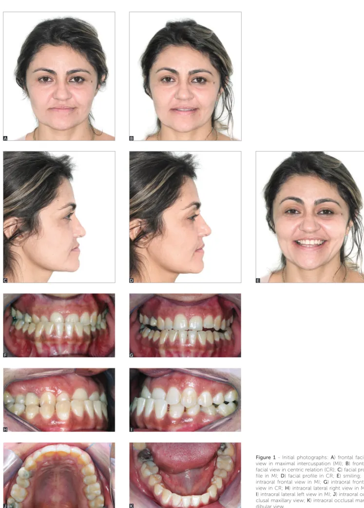
given that this position favored qualitative analysis and, as a result, improved prognosis for a compen-satory treatment. For this reason, two analyses were carried out in order to obtain patient’s facial morpho-logical diagnosis: one in MI, and another in CR. Her frontal facial analysis in MI revealed little asymmetry, chin deviated to the right, severe anterior proclina-tion, good zygomatic projecproclina-tion, compressive labial seal and decreased lower third. Nevertheless, in CR, analysis revealed that vertical shortening and facial asymmetry were minimized, and a more balanced face without signs of chin deviation (Figs 1A, 1B). Profile analysis confirmed the aforementioned char-acteristics, both in MI and CR, as well as an in-creased chin-neck line in MI. Nasolabial angle was closed partially due to the compensation of maxillary incisors, but, especially in MI, due to forced labial seal and consequent decreased ALFH (Figs 1C, 1D). Smile analysis revealed good incisors exposure with normal inclination and slight deviation of the occlusal plane, which was later justified by unilateral crossbite on the right side (Fig 1E).
Occlusal assessment in MI revealed a sagittal rela-tionship between maxilla and mandible of ¾ of Class III on the right side and ¼ on the left side, with ante-rior and posteante-rior crossbite on the right side without involving second molars. Mandibular incisors were retroclined at a clearly compensatory position as a re-sult of a decreased maxillomandibular step. Mid lines coincided with the facial midline (Fig 1F, 1K).
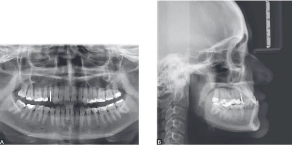
A panamoramic radiograph confirmed the pres-ence of all permanent teeth, with third molars in oc-clusion and a periodontal condition that was consis-tent with patient’s age. Tooth #14 had a provisional crown as well as an intracanal post and presented fa-vorable conditions for orthodontic treatment onset (Fig 3A).
Figure 1 -Initial photographs: A) frontal facial view in maximal intercuspation (MI); B) frontal facial view in centric relation (CR); C) facial pro-file in MI; D) facial profile in CR; E) smiling; F) intraoral frontal view in MI; G) intraoral frontal view in CR; H) intraoral lateral right view in MI. I) intraoral lateral left view in MI; J) intraoral oc-clusal maxillary view; K) intraoral occlusal man-dibular view.
A
C
F
H
J
B
D E
G
I
Figure 2 -Acrylic resin device used for occlusal fixation in CR.
Figure 3 -Initial photographs: A) Panoramic radiograph; B) Profile radiograph.
A B
Ater collecting all necessary occlusal, functional, cephalometric and face-morphology examinations, and evincing a deviation from CR to MI, we came up with the following diagnosis: adult patient, mild skeletal Class III, brachyfacial, borderline for Short Face and with an acceptable facial pattern. Class III relationship on the right side and ¼ on the let side, with anterior and posterior crossbite on the right side. Well-positioned maxillary incisors and retroclined mandibular incisors in relation to the bone. Patient’s morphological analysis of the face in CR (Figs 1B, 1D) reinforced the need for compensatory treatment that aimed at increasing volume in the maxillary arch and restricting the mandibular arch. The absence of crowding in the mandibular arch favored such treat-ment goal, although it hindered an increase in circum-ference in the maxillary arch.
Treatment plan included the use of Damon MX®
standard self-ligating brackets (Ormco), with torque of +12o applied to central incisors, +8o to lateral
in-cisors and 0o to canines, respecting the need for
in-creasing volume in the maxillary arch within esthetic limits — which could be exceeded with the use of high torque brackets (+17°, +12° and +6°, respective-ly) or by producing, by means of low torque brack-ets (+7° and +3°, for central and lateral incisors), a weaker protrusion, insufficient to correct crossbite. The mandibular arch received Capelozza®
Prescrip-tion III brackets (Abzil, 3M™). At first, mandibular incisors were not included in order to avoid protru-sion (given that anterior lower crowding was quite discrete) which could have been produced by initial and random leveling of lower teeth (Fig 4).
Capelozza® Prescription III brackets (Abzil, 3M™)
have a very positive characteristic that favors the ther-apeutic goal recommended for this patient: maxi-mum preservation of the mandibular arch or, in small proportions, a modest increase in the natural compensatory characteristics. Torque of -6o would be
applied to mandibular incisors to this end. In other words, mandibular incisors would be severely lin-gually tipped by the mechanics to which brackets would contribute. Although there was no intention of further using rectangular wires in the mandibular arch, this procedure does not break with the concept of maintaining the original form of the arch. Without a doubt, the key factor to achieve treatment success in this compensatory game is the absolute economy of angulations provided by brackets with no angulation bonded from canine to canine, which results in little protrusion and requires less space during leveling.
All aforementioned alterations are favorable in com-pensatory cases of skeletal and dental Class III, as they favor good overbite as well as functional anterior guidance.
With a view to enhancing the position of mandib-ular orthopedic stability and deconstructing maximal intercuspation, ixed stops made of composite resin were bonded to the lingual surface and incisal third of mandibular incisors with balanced and uniform occlusal contact with antagonist teeth (Fig 4E). This measure favors buccal movement of teeth involved in crossbite, stimulates extrusion of posterior teeth with-in the posterior with-interocclusal space created to produce gain in vertical dimension of occlusion (VDO), and, at the same time, improves treatment mechanical eicacy
by producing an efect of occlusal unlocking.
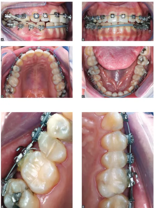
Also, with a view to directing movement towards the areas of interest, which were carefully inves-tigated, stops were bonded to the mesial surface of first molars and the arch was adjusted with a space of 2 mm between the wire and the bracket. In other words: The wire was mesially fitted on first molars and passed 2 mm away from the maxillary incisors, thereby stimulating protrusion of these teeth. Ad-ditionally, elastomers were placed on premolars and canines so as to concentrate the expansion in the an-terior region, thereby meeting the primary treatment objective (Fig 5). After correcting anterior crossbite, mechanics was directed towards teeth #14 and 15.
With a view to maintaining the compensatory
Figure 4 -Intraoral photographs at the beginning
of complete levelling in the maxillary arch and par-tial levelling in the mandibular arch.
Figure 5 -Photographs depicting right and left
maxillary quadrants, highlighting stops place-ment and the use of elastic ligatures.
C A
A
E D
B
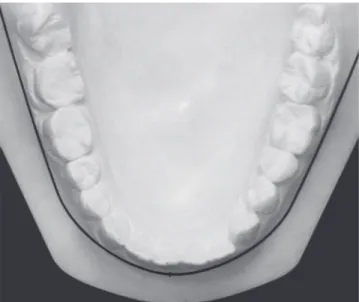
characteristics, which are also related to the form of the arch, C6A7 diagram was chosen (Fig 6) for favoring slight retroclination of mandibular incisors and protrusion of maxillary incisors. Additionally, in the posterior region, it respected the mandato-ry dentoalveolar compensatomandato-ry mechanism of the mandibular arch.16
Two months ater the onset of leveling in the maxil-lary arch, a mandibular appliance was installed with im-mediate use of Class III 5/16-in rubber bands supported by hooks placed on maxillary irst molars and man-dibular canines. This measure immediately prevented mandibular protrusion and, at the same time, produced space gain necessary for future leveling of mandibular incisors without stimulating buccal inclination.
Without a doubt, this treatment phase was the most diicult in terms of mechanics, given that any careless procedure could worsen anteroposterior relationship between the maxilla and the mandible and, as a result,
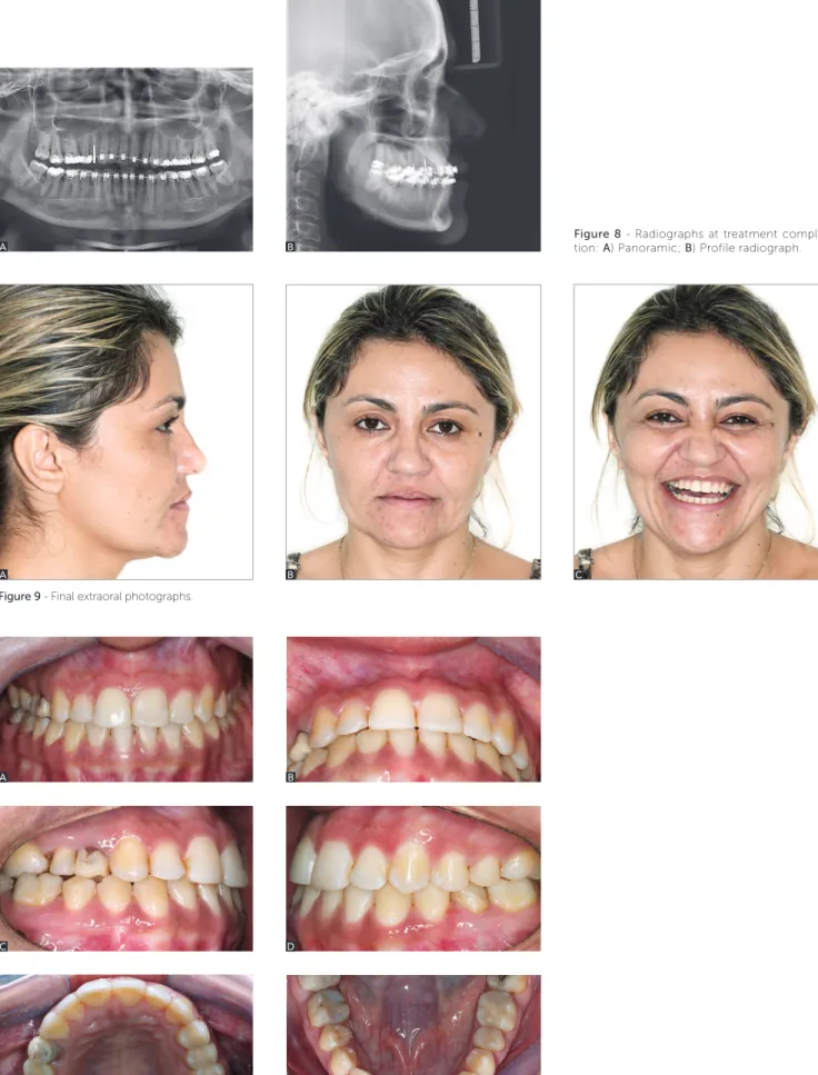
create greater demand for treatment of potential side ef-fects. From this initial phase on, maxillary leveling was conducted with a sequence of archwires evolving to 0.019 x 0.015-in steel wire. As for the mandibular arch, the sequence of archwires stopped at 0.018 steel wires because rectangular wires were not necessary for addi-tional angulation or inclination reading. During the for-matting phase of the mandibular arch, morphology was consistent with the initial treatment goals. Moreover, even if individualized brackets were used, they were not completely customized and, for this reason, their maxi-mum expression may not suit this type of patient (Fig 7). In the inal treatment phase, panoramic and lateral radiographs were taken with a view to assessing tooth positioning and potential biological costs inherent to orthodontic treatment (Fig 8).
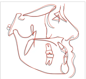
Figure 9 shows slight, yet major improvements in lip contact. It also depicts decreased asymmetry initially shown at maximal intercuspation in fron-tal view. Figure 10 shows good occlusal relationship achieved after removing the appliance.
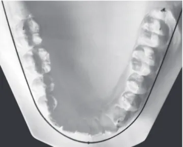
Figure 11 shows initial and final cephalometric tracings superimposition at treatment completion, which allowed an accurate analysis of the mechanisms that enabled occlusal adjustment. Improvements were achieved due to a set of several small adjustments, namely: correction of discrepancy between CR and MI, retroclination of mandibular incisors and protru-sion of maxillary incisors. All these factors added up to magnify the positive impacts on patient’s occlusion and face as well as to allow transverse expansion of the maxillary arch and crossbite correction.
Treatment lasted for 15 months, with a total num-ber of 10 visits since the appliance was firstly installed in the maxillary arch until it was removed.
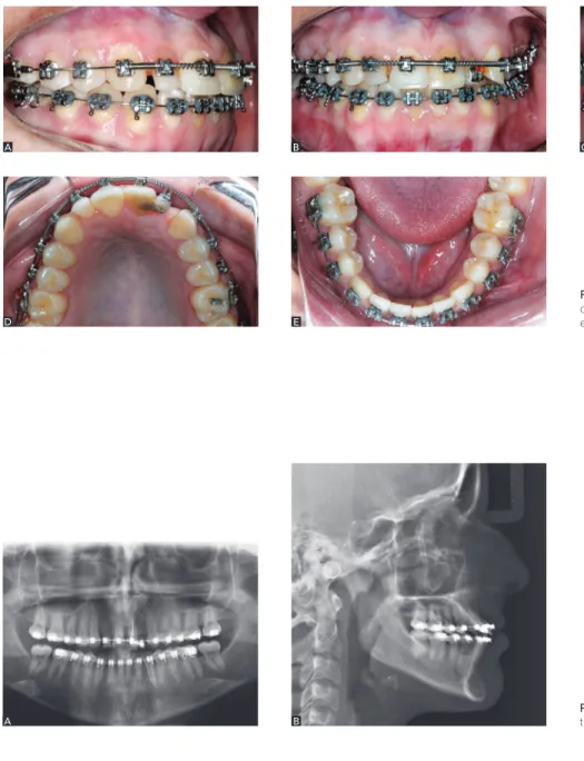
Figure 6 - C6A7 objective anatomic individual diagram.
Figure 7 -Intraoral photographs at the end of leveling in the maxillary arch (with 0.019 x 0.025-in wire) and in the mandibular arch (with 0.018” steel wire).
C
Figure 8 -Radiographs at treatment comple-tion: A) Panoramic; B) Profile radiograph.
Figure 10 -Final intraoral photographs.
Figure 9 -Final extraoral photographs.
A A
B B
C
A B
E F
Figure 11 - Initial (black) and final (red) cephalometric tracings superimposition.
CASE REPORT 2
A 26-year and 2-month-old female, Caucasian patient sought orthodontic treatment with chief complaint of lack of space for implant placement at tooth #22 site and small-sized tooth #12. Her profile analysis revealed maxillary deficiency and unsatisfactory lip contact with her lower lip ahead her upper lip and open nasolabial angle (Fig 24A). Her frontal facial analysis revealed a balanced face with good acceptability (Fig 12 B). Her smile was characterized by lack of space, disproportional max-illary lateral teeth and tooth #21 darkened by end-odontic treatment (Fig 12C).
Her occlusal analysis revealed Class III subdivi-sion malocclusubdivi-sion of ¼ on the right side, crossbite on tooth #12 as well as decreased overbite and overjet. Her mandibular arch showed evident compensation, with retroclined incisors and mandibullar canines with no mesial angulation. Mid lines coincided with the facial midline (Fig 12D, 12H).
Panamoramic radiograph confirmed maxillary and mandibular third molars as well as tooth #22 agenesis corrected by an adhesive prosthesis bonded to teeth #21 and 23. She presented general periodontal con-dition that favored orthodontic treatment (Fig 13A). From a skeletal standpoint, the cephalogram re-vealed a negative maxillomandibular step with mild
maxillary deficiency and differences between palatal and mandibular planes. Maxillary incisors were buc-cally tipped as expected. However, mandibular inci-sors counteracted occlusal analysis as they were well positioned in the symphysis (Fig 13B).
Diagnosis was as follows: adult patient, mild skeletal Class III, dolichofacial with acceptable fa-cial pattern, espefa-cially from frontal view. Class III relationship on the right side, with anterior cross-bite on tooth #12, decreased overcross-bite and overjet, agenesis of tooth #22 and increased buccal tipping of maxillary incisors.
Patient’s self-perception of facial normality in frontal view reinforced the need for compensatory treatment while eliminating the need for absolute cor-rective treatment by means of orthognathic surgery for maxillary advancement. In this context, treatment plan was directed towards the protocol presented herein: the use of Damon MX® (Ormco) self-ligating
brackets. Unlike case 1, high torque prescription was chosen for the maxillary arch (CI +17 o, IL +10o,
C +7o) as it required greater protrusion and
expan-sion, both of which were justified by more expres-sive buccal torque applied to the maxillary arch and Capelozza® Prescription III brackets (Abzil, 3M™) used in the mandibular arch.
In case 2, the greatest challenge was to increase overbite and overjet while opening spaces for appropri-ate rehabilitation of maxillary lappropri-ateral incisors without producing the efect of reversing the incisal curvature at smiling — which is quite common in cases requir-ing major compesantion of maxillary incisors. In order to control such efect, Prescription III brackets were used in the mandibular arch with brackets more cervi-cally bonded on maxillary lateral and central incisors.
Figure 12 -Initial extra and intraoral photographs.
A
D
G
B C
E F
H
Figure 13 -Initial radiographs: A) Panoramic
ra-diograph; B) Profile radiograph.
Figure 14 -Initial treatment approach with level-ing of maxillary arch performed with stops placed on the mesial surface of molars, elastic ligatures on anchorage teeth and the use of 0.014-in heat-activated with anterior slack.
A
D
B
E
C
in the maxillary arch — 4 months after treatment onset. In other words, it was installed after the max-illary arch form was established, which used to be limited, and was now more expanded and defined. In order to favor greater anterior overbite, maxillary second molars were not included in leveling. This set of actions is definitively in accordance with the therapeutic goals previously established for patient’s compensatory treatment (Fig 14).
C4A7 diagram was chosen (Fig 15) for providing greater freedom to improve the form of the mandibu-lar arch, which was allowed by the great demand for space in the maxillary arch.
The sequence of wires used in the maxillary arch was as follows: 0.014” heat-activated; 0.014 x 0.025-in heat-activated; 0.017 x 0.025-in TMA; 0.019 x 0.025-in TMA and 0.019 x 0.025-in steel wire. As for the mandibular arch, 0.014 NiTi super-elastic and 0.016 NiTi supersuper-elastic wires were me-sially fitted, followed by 0.018” steel wire installed with omega loops.
Figure 16 shows the effect produced with the use of open and closed springs to equalize the space nec-essary for proper rehabilitation of teeth #12 and #22. Final panoramic and profile radiographs not only certify safe and trustful results, but also confirm in-traosseous space gain for future implant placement on tooth #22 site (Fig 17).
Treatment produced considerable improvements and discreet, yet extremely positive benefits for the face. Thus, it proves the protocol adopted herein
to be efficient with regard to the therapeutic goals previously established (Fig 18).
Cephalometric tracings superimposition helps us understand that right choices were made with a view to achieving functional and esthetic balance of a malocclusion that presents compensatory char-acteristics inherent to both skeletal Class III and sagittal relationship between maxilla and mandible aggravated by agenesis in the anterior region of the maxillary arch (Fig 19).
Treatment lasted for 18 months, with a total num-ber of 11 visits.
Figure 16 -Final leveling phase with open and closed springs installed to increase in circumfer-ence in the region of teeth #12 and #22.
A
D
B
E
C
Figure 17 -Radiographs at treatment
comple-tion: A) Panoramic; B) Profile radiograph.
Figure 18 -Final extra and intraoral photographs.
A
D
G
B C
E F
H
Figure 19 - Initial (black) and final (red) cephalometric tracings superimposition.
CASE REPORT 3
region than in the canine region. This difference in magnitude may be explained by the level of compen-sation present in mandibular canines and premolars that resigned their mesial angulations. Transverse relationship was impaired by unilateral crossbite on the right side and decreased overbite as well as over-jet in the anterior region. Frontal intraoral view re-vealed a 2-mm deviation of the lower midline to the right coinciding with mandibular skeletal deviation.
Panamoramic radiograph revealed periodontal and structural health that favored orthodontic treat-ment. Third molars had been previously extracted (Fig 21A). Morphological exams of the cephalogram confirmed all aforementioned positive facial charac-teristics and revealed something new: vertical maxil-lary excess unable to negatively affect patient’s face or smile. Maxillary and mandibular incisors were well positioned into the jaws (Fig 21B).
Unlike the aforementioned patients, this patient was diagnosed as skeletal Class I with mild laterogna-thism to the right, dolichofacial and pleasant face. Bi-lateral Class III relationship with uniBi-lateral crossbite on the right side and absence of overbite and over-jet. He was asked about the possibility of undergoing orthognathic surgery, since the procedure would be the only one capable of correcting asymmetry — one of his chief complaints. Nevertheless, given that mild mandibular shift did not worsen after a year, the pos-sibility of surgery was discarded and compensatory treatment was chosen to solve patient’s occlusal prob-lems, thereby enduring his small skeletal defect. Al-though the patient was skeletal Class I, the relation-ship between maxilla and mandible was Class III and granted him occlusal characteristics of Class III. For this reason, he was treated under the same protocol employed in cases 1 and 2.
Figure 20 -Initial extra and intraoral photographs.
A
D
G
B C
E F
A B C
Figure 21 -Initial radiographs: A) Panoramic
radio-graph; B) Profile radiograph.
Figure 22 - A) Initial treatment approach with leveling of maxillary arch performed with stops placed on the mesial surface of molars, elastic ligatures on anchorage
teeth and the use of 0.014” heat-activated with anterior space between teeth #11 and #14.
A B
Once again, we faced the need for controlled maxillary protrusion and expansion as well as restric-tion of both in the mandibular arch. Figure 22 shows similarities with the aforementioned protocol: treat-ment onset on the maxillary arch, use of stops and elastic ligatures with a view to achieving protrusion and expansion in the right anterior and lateral region. C5A9 diagram (Fig 23) was chosen to preserve the form of the mandibular arch, given that unilateral crossbite on the right side, without deviation from MI to CR, was a predictive factor of potential difficulties in achieving proper transverse control — especially in the case of an adult patient.
The sequence of wires used in the maxillary arch was as follows: 0.014-in; 0.016-in; 0.014 x in; 0.018 x in heat-activated; 0.019 x 0.025-in TMA and 0.019 x 0.025-0.025-in steel wire. As for the mandibular arch, the following arches were used: 0.014” NiTi superelastic wire mesially fitted; 0.016 and 0.018-in steel wires with omega loops. This patient required compensatory bends (Fig 24) for a more individualized treatment finishing, especially due to laterognathism.
Final panoramic and lateral radiographs reveal ab-solute control (Fig 25). Figure 26 certifies protocol efficacy. Patient’s face did not change, as expected. Nevertheless, gains in overbite and overjet, correc-tion of unilateral crossbite and Class I relacorrec-tionship es-tablished between canines provided him with a func-tional routine and expressive esthetic benefits.
Initial and final cephalometric tracings superim-position reveals that the effects described in case re-port 1 and 2 were repeated in case 3 (Fig 27).
Figure 24 -Final leveling phase with individualized bends for treatment finishing: buccal steps on teeth #16, 26, 12 and 13; and “Z” bend on tooth #21.
Figure 25 -Radiographs at treatment
comple-tion: A) Panoramic radiograph, B) Cephalomet-ric cephalogram.
A B
Figure 26 -Final extra and intraoral photographs.
A
D
G
B C
E F
Figure 27 - Initial (black) and final (red) cephalometric tracings superimposition.
Treatment lasted for 24 months, with a total num-ber of 17 visits. Treatment time was greater than ex-pected due to unilateral crossbite, the need for indi-vidualization bends and the clinician’s learning curve — since this was the first patient treated under the protocol described herein.
FINAL CONSIDERATIONS
The gains in efficiency of alignment and leveling produced by self-ligating brackets have not been sci-entifically proved. Some recent studies do not seem favorable to confirm the greater productivity of this
system, since most of them aim at comparing the magnitude of movement during alignment and level-ing without considerlevel-ing the individual variations of the samples.10,17 From this point of view, self-ligating
brackets would be just a more practical method em-ployed to fit and remove archwires. Nevertheless, di-recting mechanics associated with bracket individu-alization towards flexile therapeutic goals seems to enhance treatment outcomes. Carefully using stops and elastic ligatures to manage friction in self-ligat-ing bracket systems used in areas where movement is less required is a good example of how to explore the maximum productivity of this system, and justi-fies the methodology employed to treat the patients reported herein.
Using individualized Capelozza® Prescription III
brackets (Abzil, 3M™) in the mandibular arch to treat Class III is essential to yield more esthetically tolerated results, given that maximum maintenance of the arch form creates possibilities of moderate gains in the maxillary arch without hindering smile esthetics. This occurs because the ideal morphology for sagittal correction of the arches is limited by smile reading; thereby giving the orthodontist the oppor-tunity to create a less protrusive and less expansive maxillary arch than he would mechanically do.
1. Capelozza Filho L. Diagnóstico em Ortodontia. Maringá: Dental Press; 2004.
2. Capelozza Filho L. Metas terapêuticas individualizadas. Maringá: Dental Press; 2011.
3. Fleming PS, O’Brien K. Self-ligating brackets do not increase treatment eiciency. Am J Orthod Dentofacial Orthop. 2013;143(1):11-9. 4. Hägg U, Taranger J. Maturation indicators and the pubertal growth spurt.
Am J Orthod. 1982;82(4):299-309.
5. Capelozza Filho L. Uma entrevista com Leopoldino Capelozza Filho. Dental Press J Orthod. 2010;15(6):25-53.
6. Damon DH. The rationale, evolution and clinical application of the self-ligating bracket. Clin Orthod Res. 1998;1(1):52-61.
7. Tecco S, Festa F, Caputi S, Traini T, Di Iorio D, D’Attilio M. Friction of conventional and self-ligating brackets using a 10 bracket model. Angle Orthod. 2005;75(6):1041-5.
8. Harradine N. Self-ligating brackets increase treatment eiciency. Am J Orthod Dentofacial Orthop. 2013;143(1):10-9.
9. Franchi L, Baccetti T, Camporesi M, Lupoli M. Maxillary arch changes during leveling and aligning with ixed appliances and low-friction ligatures. Am J Orthod Dentofacial Orthop. 2006;130(1):88-91.
10. Fleming PS, Lee RT, Marinho V, Johal A. Comparison of maxillary arch dimensional changes with passive and active self-ligation and conventional brackets in the permanent dentition: a multicenter, randomized controlled trial. Am J Orthod Dentofacial Orthop. 2013;144(2):185-93.
REFERENCES
11. Harradine NWT. Current products and practices self-ligating brackets: where are we now? J Orthod. 2003;30:262-73.
12. Maltagliati LA. Desmistiicando a utilização dos stops no sistema autoligado. Rev Clín Ortod Dental Press. 2012;11(1):22-31.
13. Capelozza Filho L. Gabriel O, Ozawa TO. Individualização de braquetes na Técnica de Straight-Wire: revisão de conceitos e sugestão de indicações para uso. Rev Dental Press Ortod Ortop Facial. 1999;4(4):87-106. 14. Alberto C, Cabrera G, Freitas MR, Janson G, Fernando J, Henriques C.
Estudo da correlação do posicionamento dos incisivos superiores e inferiores com a relação ântero-posterior das bases ósseas. Rev Dental Press Ortod Ortop Facial. 2005;10(6):59-74.
15. Fleming PS, DiBiase AT, Sarri G, Lee RT. Eiciency of mandibular arch alignment with 2 preadjusted edgewise appliances. Am J Orthod Dentofacial Orthop. 2009;135(5):597-602.
16. Capelozza Filho L, Antonio J, Capelozza Z. DIAO: Diagrama individual anatômico objetivo. Uma proposta para escolha da forma dos arcos na técnica de Straight-Wire, baseada na individualidade anatômica e nos objetivos de tratamento. Rev Clín Ortod Dental Press. 2004;3(5):84-92. 17. Fleming PS, DiBiase AT, Lee RT. Randomized clinical trial of orthodontic