*Correspondence: M. L. Gomes. Laboratório de Doença de Chagas, De-partamento de Ciências Básicas da Saúde, Universidade Estadual de Mar-ingá. Av. Colombo, 5790 - Zona Sete - 87020-900 - Maringá - PR, Brazil. E-mail: [email protected]
A
rti
Pharmaceutical Sciences vol. 47, n. 1, jan./mar., 2011
Polymerase chain reaction and blood culture in blood donors
screened by ELISA test for Chagas’ disease
Andréa Tieko Kinoshita-Yanaga
1, Marta Bértoli
2, Vanessa Aparecida Martins
1, Angélica Sayuri
Mizutani
1, Max Jean de Ornelas Toledo
1, Silvana Marques de Araújo
1, Mônica Lúcia Gomes
1,*1Chagas’Disease Laboratory, Department of Basic Health Sciences, State University of Maringá, 2Regional Blood Donation Center of Maringá, State University of Maringá
The objective of this study was to evaluate, through blood culture and PCR, the results of the ELISA for Chagas’ disease in the screening of blood donors in the public blood-supply network of the state of Paraná, Brazil, and to map the epidemiological proile of the donors with respect to their risk of infection by Trypanosoma cruzi. The negative and positive results of the ELISA were conirmed by blood culture and PCR for 190/191 individuals (99.5%). For one individual (0.5%), the ELISA was inconclusive, blood culture and IIF were negative, and IHA and PCR positive. Three individuals (1.6%) were positive for
T. cruzi on all the tests. Donors were predominantly female, and natives of Paraná, of rural origin, had observed or been informed of the presence of the vector in the municipalities where they resided, had never received a blood transfusion, had donated blood 1 to 4 times, and reported no cases of Chagas’ disease in their families. We concluded that PCR and blood culturing have excellent potential for conirming the results of the ELISA, and that candidate blood donors with negative or positive tests have a similar risk of infection by T. cruzi, indicating that the ELISA test is suficiently safe for screening blood prior to use.
Uniterms:Trypanosoma cruzi/detection. ELISA/use for screening. Blood donors.Blood culture. PCR. Blood bank/screened donors.
O objetivo deste estudo foi avaliar, pela hemocultura e PCR, os resultados do teste ELISA utilizado para doença de Chagas na triagem de doadores de sangue na rede pública do Estado do Paraná, Brasil, e traçar o peril epidemiológico dos doadores quanto ao risco de infecção pelo Trypanosoma cruzi. Os resultados negativos e positivos do ELISA foram conirmados pela hemocultura e PCR em 190/191 indivíduos (99,5%). Para um indivíduo (0,5%), o teste de ELISA foi inconclusivo, hemocultura e IFI foram negativas, HAI e PCR foram positivas. Três indivíduos (1,6%) foram positivos para T. cruzi em todos os testes. A maioria dos doadores era do sexo feminino, oriundos do Estado do Paraná, de origem rural, tinham observado ou foram informados da presença do vetor nos municípios onde residiam, nunca tinham recebido sangue, haviam doado sangue de 1 a 4 vezes e não relataram casos de doença de Chagas na família. Nós concluímos que a PCR e a hemocultura são excelentes testes para conirmar os resultados do ELISA e os candidatos a doadores de sangue com testes positivos e negativos apresentam risco semelhante de infecção pelo T. cruzi, reforçando o nível satisfatório de segurança do teste ELISA para liberar o sangue para o uso.
Unitermos: Trypanosoma cruzi/detecção. ELISA/uso para triagem. Sangue/doadores. Hemocultura. PCR. Banco de Sangue/triagem de doadores.
INTRODUCTION
The Ministry of Health of Brazil, through the
A.T. Kinoshita-Yanaga, M. Bértoli, V.A. Martins, A.S. Mizutani, M.J.O. Toledo, S.M. Araújo, M.L. Gomes
54
The risk of transmission of Trypanosoma cruzi
through transfusion of a single, 500-mL unit of whole blood ranges from 12% to 20% (WHO, 2002). In Brazil, the seroprevalence of Chagas’ disease in the public blood-bank network is 0.6% whereas the seroprevalence rate in
private blood banks is unknown (Silveira et al., 2002).
The serological tests (indirect immunoluorescence or IIF, indirect hemagglutination or IHA, and immunoenzyme or ELISA) used to diagnose Chagas’ disease generally have sensitivity of 95% and speciicity of 93 to 98%, indicating that no one test is suficiently sensitive to totally eliminate
the risk of transmission of T. cruzi through blood
trans-fusion (Salles et al., 1996; Oelemann et al., 1998;
Saez-Alquezar et al., 1998).
Other techniques capable of verifying the presence of T. cruzi in infected patients include blood culture,
xe-nodiagnosis, and PCR. Several investigators (Avila et al.,
1993; Wincker et al., 1994; Gomes et al., 1999; Castro et
al., 2002; Salomone et al., 2003; Vera-Cruz et al., 2003;
Batista et al., 2007) have reported that individuals with
negative or inconclusive serology on the IIF and ELISA tests had positive results on xenodiagnosis, blood culture,
or PCR. Some of these scholars (Castro et al., 2002) did
not rule out the possibility that the frequency of
individu-als who are serologically negative and have an active T.
cruzi infection detected only by PCR, may be higher than
previously thought. Other authors (Salomone et al., 2003),
having demonstrated the presence of T. cruzi by PCR in
in-dividuals that showed no serological evidence for Chagas’ disease, believe that their indings challenge the present re-commendations for Chagas’ disease diagnosis, treatment,
and monitoring of blood transfusions. Vera-Cruz et al.
(2003), in a study comparing PCR with the commercially available ELISA test on samples collected in an endemic area in Mexico, found a higher positive rate using PCR than on ELISA. Based on these results, the authors raised the possibility that the ELISA method underestimates the
prevalence of T. cruzi infections.
The genetic diversity of T. cruzi can also inluence
the performance of serological tests. Mice inoculated with parasites of the T. cruzi II group (referred to as Discrete
Ty-ping Units - DTU II by Zingales et al., 2009) showed rates
of positivity with ELISA ranging from 83.3% to 93.3%, blood culture from 60.6% to 76.7%, and PCR 100%. For
mice inoculated with parasites of the T. cruzi group I (DTU
I by Zingales et al., 2009), the positivity of the ELISA and
PCR were both 100%, whereas that of the blood culture
ranged from 89.5% to 90.2% (Miyamoto et al., 2006).
The Chagas’ Disease Laboratory of the State Uni-versity of Maringá, as part of its mission to improve the care of patients with this disease, established the program
“Chagas’ Disease Awareness through Comprehensive
Education” (Araújo et al., 2000). In order to expand this
program and devise new action strategies, it is important to deine the proile of the patients treated by this laboratory, including candidate blood donors.
Based on these considerations, the objective of this study was to conirm, by means of blood culture and PCR, the results of the ELISA for Chagas’ disease obtained du-ring the screening of candidate blood donors in the public blood-supply network of Paraná, and to map the epide-miological proile of the population studied, with respect
to the risk of their being infected by T. cruzi.
MATERIALS AND METHODS
Population studied
From March 2007 through February 2008, a period in which the Maringá Regional Blood Donation Center (Hemocentro Regional de Maringá - HRM) in Paraná processed a total of 9358 candidate blood donors, 191 blood samples were collected from individuals of 16 municipalities belonging to the 15th Health Region of the State of Paraná (Figure 1). The collection routine of the HRM was maintained, and the sample was representati-ve of the regional population. The criteria for inclusion of individuals in the study were the screening doctor’s authorization to collect an additional volume of blood, and donor age, such that the same numbers of individuals for the age ranges 18 to 33, 34 to 49, and 50 to 65 years could be obtained. The sample size (n) was calculated
using the formula n = Nn0 / (N+n0), where n0 = p (1-p)
(z0.05/e)2, and N is the size of the population (considering
the number of candidate blood donors of each age group predicted for the 12-month period). The p value refers to the occurrence of the event investigated, based on the mean of the preceding three years for Chagas’ disease in
the HRM (0.28); z0.05 corresponds to the 95% percentage
of the standard normal distribution, and to the maximum sampling error of 10% (0.1). The minimum sample size resulting from this calculation was 188, with a 90% conidence level.
hou-sing structure, presence of the vector in the municipality, present housing structure, rural laborer status, previous blood transfusion, previous blood donation, and presence of Chagas’ disease in the family.
ELISA
The ELISA reaction to detect anti-T. cruzi IgG
an-tibodies was performed with the ELISA III® Chagas Test
diagnostic kit (BiosChile Ingenharia Genética S.A., Chile) made available by the Public Blood-Supply Network of Paraná, and applied according to the recommendations of the manufacturer. Whenever a sample was reactive or inconclusive in the initial test, the test was repeated in duplicate, using blood from the same sample.
Blood culture
To carry out this test, 30 mL of blood was collected in heparinized tubes. The blood cultures were then
proces-sed in LIT medium and incubated at 28 °C, according to
Chiari et al. (1989) with modiications. The samples were
homogenized once a week, and examined 30, 60, 90, and 120 days after processing.
PCR
For each individual, 10 mL of blood was added to an equal volume of 6 M Guanidine-HCl/0.2 M EDTA
(Sigma Chemical Company, USA), pH 8.0. Using 200 µL
of blood, the DNA was extracted with phenol-chloroform and precipitated in absolute ethanol in the presence of
100 mM sodium acetate (Gomes et al., 1998). DNA was irst
resuspended in 20 µL of sterile deionized water. After 24
to 36 hours stored at 4 °C, the DNA was washed with 70%
ethanol, precipitated, and resuspended in 10 µL of water.
The PCR was processed by mixing 2 µL of the DNA
solution from each sample, together with 10 mM Tris-HCl
(pH 9.0), 0.1% Triton X-100, 75 mM KCl, 3.5 mM MgCl2
, 0.2 mM of each deoxynucleotide (dATP, dCTP, dGTP, dTTP, Invitrogen Ltda.), 0.5 U of Taq DNA polymerase
(Invitrogen), and 10 pmoles of each primer for 10 µL of
reaction.
Primers 121 (5’AAATAATGTACGGG(T/G)GA-GATGCATGA3’) and 122 (5’GGTTCGATTGGGGT-TGGTGTAATATA 3’) were used as described by Wincker
et al. (1994); these primers amplify fragments of a variable
330 bp region of the T. cruzi kDNA minicircle. The DNA
was next ampliied in an automatic thermocycler (MJ Re-FIGURE 1 - Municipalities of origin of candidate blood donors studied, Paraná State, Brazil. Note: values in parentheses represent
A.T. Kinoshita-Yanaga, M. Bértoli, V.A. Martins, A.S. Mizutani, M.J.O. Toledo, S.M. Araújo, M.L. Gomes
56
search, PTC-150) with denaturing at 95 °C for 1 min (with
a longer initial step, for 5 min), annealing of the primers
at 65 °C for 1 min, and extension at 72 °C for 1 min (with
a 10-min inal step) for a total of 35 cycles (Gomes et al.,
1998). For the steps of DNA extraction and PCR, negative controls with blood samples from uninfected individuals from a non-endemic area, and positive controls of indi-viduals with Chagas’ disease were used. The products ampliied by the PCR were observed by electrophoresis in 4% polyacrylamide gel and by silver staining (Santos, Pena, Epplen, 1993).
IIF and IHA
The tests described were carried out on those samples that were positive on ELISA, blood culture, or PCR tests. The IIF and IHA were executed with reagents manufactured by BioMérieux Brasil SA, according to the
recommendations of the manufacturer. Anti-T. cruzi
anti-bodies of the IgG class were investigated; a titer ≥ 40 was
considered reactive for IIF. For the IHA, the qualitative test was carried out according to the positive and negative sera of the HRM and of the Laboratory for Teaching and Research on Clinical Analyses (Laboratório de Ensino e Pesquisa em Análises Clínicas - LEPAC) of UEM. The de-monstrably positive cases were sent to the ACHEI program
(Araújo et al., 2000), to which the patients were referred
for medical care, with etiological treatment and follow up every six months for at least ive years.
RESULTS
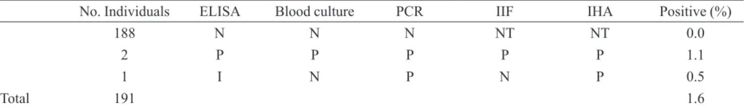
Of the 191 candidate blood donors who underwent serological screening for Chagas’ disease, 188 tested nega-tive on ELISA. Two individuals were posinega-tive on the ELI-SA, IIF, and the IHA whereas 1 person had inconclusive results on the ELISA, testing negative on IIF and positive on IHA. On the blood cultures, 189 individuals presented negative, and 2 positive results. For the PCR, a total of 188 individuals tested negative and 3 positive (Table I).
The results of the ELISA were conirmed by blood culture and PCR for 190 individuals (99.5%). In one case (0.5%), the ELISA was inconclusive, blood culture was negative, and PCR positive. Of the three cases in which the ELISA was positive or inconclusive, two were shown to be positive by blood culture, and three by PCR (Table I).
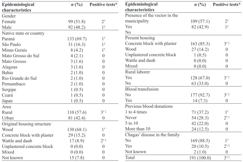
Table II shows that the epidemiological proile of
the population studied for risk of infection by T. cruzi was
similar for subjects with negative (188) and positive (3) tests. The majority of the individuals investigated were natives of states where Chagas’ disease is endemic and had lived in rural areas, both constituting risk factors associa-ted with this disease. Also, the sample was predominantly female and the majority reported no history of Chagas’ disease in the family. The three individuals with positive results on at least one of the tests were 48 years of age or older, presently residing in municipalities in Paraná (Co-lorado, Sarandi, and Floresta), were rural laborers, while two reported having had a case of Chagas’ disease in the family, also considered a risk factor for Chagas’ disease.
DISCUSSION
The HRM is a unit of intermediate complexity with macroregional responsibility, and provides support and assistance to the health network, supervises services, and performs serological examinations for all the collection units. It is a reference center for Northwestern Paraná, a region endemic for Chagas’ disease. After the resolution of the Ministry of Health came into effect in 2004 (Brasil, 2004), the HRM began to use only the ELISA test to screen blood donors for Chagas’ disease.
In the present study, the results of the ELISA were confirmed by blood culture and PCR in a high percentage of samples, afirming the use of ELISA as the sole test for screening prospective blood donors. Satisfactory results for different ELISA kits have been
previously reported (Oelemann et al., 1998; Leiby et
al., 2000; Blejer, Saguier, Salamone, 2001; Gadelha
et al., 2003; Pirard et al., 2005).However, the results
TABLE I - Results of tests performed on candidate blood donors at the Maringá Regional Blood Donation Center, Paraná, Brazil,
from March 2007 through February 2008
No. Individuals ELISA Blood culture PCR IIF IHA Positive (%)
188 N N N NT NT 0.0
2 P P P P P 1.1
1 I N P N P 0.5
Total 191 1.6
TABLE II - Epidemiological characteristics and positive tests for Trypanosoma cruzi among candidate blood donors at the Maringá
Regional Blood Donation Center, Paraná, Brazil, fromMarch 2007 through February 2008
Epidemiological
characteristics n (%) Positive tests*
Gender Female
Male 99 (51.8)92 (48.2) 2
†
1‡
Native state or country Paraná
São Paulo Minas Gerais Mato Grosso do Sul Mato Grosso Alagoas Bahia
Rio Grande do Sul Pernambuco Sergipe Ceará Japan 133 (69.7) 31 (16.3) 8 (4.2) 4 (2.1) 3 (1.6) 3 (1.6) 2 (1.0) 2 (1.0) 2 (1.0) 1 (0.5) 1 (0.5) 1 (0.5) 1† 1‡ 1† 0 0 0 0 0 0 0 0 0 Area Rural
Urban 110 (57.6)81 (42.4) 3
† ‡
0 Original housing structure
Wood
Concrete block with plaster Wattle and daub
Unplastered concrete block Mixed Not known 130 (68.1) 29 (15.2) 17 (8.9) 0 (0.0) 0 (0.0) 15 (7.8) 1† 0 2† ‡
0 0 0
Epidemiological
characteristics n (%) Positive tests*
Presence of the vector in the municipality
Yes No
109 (57.1)
82 (42.9) 2
†
1‡
Present housing
Concrete block with plaster Wood
Unplastered concrete block Wattle and daub
Mixed 163 (85.3) 27 (14.2) 1 (0.5) 0 (0.0) 0 (0.0)
3† ‡
0 0 0 0 Rural laborer Yes
No 128 (67.0)63 (33.0) 3
† ‡
0 Blood transfusion
No
Yes 177 (92.7)14 (7.3) 3
† ‡
0 Previous blood donations
1 to 4 times Never 5 to 10 More than 10
71 (37.2) 54 (28.3) 42 (22.0) 24 (12.5)
1†
2† ‡
0 0 Chagas’ disease in the family
No Yes Not known 169 (88.5) 20 (10.5) 2 (1.0) 1†
2† ‡
0
Total 191 (100.0) 3† ‡
n = Number; % = Percentage; * Candidate blood donors with positive results for Trypanosoma cruzi infection. † Elisa, Blood
culture, PCR, IIF, and IHA positive. ‡ Elisa inconclusive, Blood culture and IIF negative, PCR and IHA positive.
presented in this study must be interpreted cautiously by considering the samples analyzed, the serological tests and reagents, the technical procedures, quality control, and the protocols for extraction and ampliication of the DNA, and taking into account that the study area has a low prevalence of Chagas’ disease. Although high levels of agreement, principally between serological and mo-lecular tests, in diagnosing Chagas’ disease have been
reported (Avila et al., 1993; Britto et al., 1995;
Junquei-ra, Chiari, Wincker, 1996; Gomes et al., 1999; Castro et
al., 2002; Marcon et al., 2002; Salomone et al., 2003;
Vera-Cruz et al., 2003; Coronado et al., 2006),
positi-ve results on xenodiagnosis, blood culture or PCR, in conjunction with negative or inconclusive serological
tests, have also been demonstrated (Avila et al., 1993;
Gomes et al., 1999; Castro et al., 2002; Marcon et al.,
2002; Salomone et al., 2003; Vera-Cruz et al., 2003).
As a result of this lack of agreement, Salomone et al.
(2003) and Vera-Cruz et al. (2003) raised the question
as to whether a single serological test to screen blood bags for release is safe, and whether the ELISA test
underestimates the prevalence of T. cruzi infections.
In the present study, positivity for T. cruzi was 1.6%
(3/191) for all the tests used. This positivity rate is very similar to that reported by the regional blood donation
centers of Londrina/Paraná, Brazil (Oliveira-Marques et
al., 2005) and Iguatu/Ceará, Brazil (Sobreira et al., 2001),
relecting the success of the program to eliminate
Triato-ma infestans and of the regular and rigorous screening of donors at blood banks in place since the 1980s
(Franco-Paredes, Bottazzi, Hotez, 2009; Fernandes et al., 2009;
Villela et al., 2009). Monteon et al. (2005) and
Hernández-Becerril et al. (2005) also observed low seroprevalence
A.T. Kinoshita-Yanaga, M. Bértoli, V.A. Martins, A.S. Mizutani, M.J.O. Toledo, S.M. Araújo, M.L. Gomes
58
by Schmunis and Cruz (2005), Sabino et al. (2003), Silva
et al. (2007) and Araújo, Vianna, Berne (2008).
For two cases in which the ELISA was positive, the blood culture was also positive. In the one case which was inconclusive on ELISA, the blood culture was negative. These are very good results, because in the samples for which the ELISA was positive, the blood culture, a test
known for its low sensitivity (Chiari et al., 1989; Gomes
et al., 1998; Castro et al., 2002), also showed a positive result. This good performance of the blood culture may be related to the level of parasitemia of the individuals at the time of donation, and to the quality of execution of this technique in our laboratory, given it is laborious and requires aseptic procedures and good-quality reagents.
As mentioned, the ELISA was inconclusive in one individual, yet even in this case the protocol proposed by the Ministry of Health enabled elimination of the sample collected, since it was positive for IHA, despite testing negative on the IIF. In addition, the PCR result was posi-tive, indicating that the PCR technique could be a valid alternative if reference centers using this methodology are established. Other investigators have also shown that, in individuals with inconclusive serology, tests such as the PCR and the TESA-Blot can be used to conirm the results
(Umezawa et al., 1996; Gomes et al., 1999; Marcon et al.,
2002; Silveira-Lacerda et al., 2004; Batista et al., 2007;
Araújo, Vianna, Berne, 2008). Although Duarte et al.
(2006) observed sensitivity rates of 95.7 to 100% for IIF, IHA, and ELISA, and 1.2% for PCR, several investigators have reported good agreement of IIF and ELISA results
with those of the PCR (Britto et al., 1995; Junqueira,
Chiari, Wincker, 1996; Gomes et al., 1999;
Hernández-Becerril et al., 2005; Coronado et al., 2006), in-line with
the results of the present study.
Of the conventional serological tests used to diag-nose Chagas’ disease, the IHA has the lowest sensitivity (Oelemann et al., 1998; Leiby et al., 2000; Blejer, Saguier,
Salamone, 2001; Gadelha et al., 2003; Pirard et al., 2005).
However, in the present study, the IHA was able to con-irm the diagnosis of one individual with an inconclusive ELISA and a negative IIF result. This result corroborates the concerns of several investigators (Blejer, Saguier,
Salamone, 2001; Gadelha et al., 2003; Pirard et al., 2005;
Araújo, Vianna, Berne, 2008) who have emphasized the importance of combining several effective tests when screening for Chagas’ disease at the blood banks, thereby reducing false-negative or inconclusive results and
cross-reactions (Saéz-Alquezar et al.,1998).
In the present study, the inclusion of two other sero-logical tests (IIF and IHA) was only required to conirm the diagnosis in 3 individuals with positive results on at
least one of the tests applied (ELISA, blood culture or PCR), considering that the results of these three tests with different principles were concordant for most of the indivi-duals investigated. As recommended by Luquetti (1990), conirmation of the diagnosis of individuals suspected of
harboring T. cruzi infection screened in the blood-supply
network and/or that have positive results on other tests, requires high sensitivity and speciicity, made possible by combining two techniques (IIF and IHA).
According to Gontijo, Rocha, Oliveira (1996), risk factors for Chagas’ disease include being born in a rural area, living in states endemic for Chagas’ disease, and having a history of Chagas’ disease in the family. Using these criteria, candidate blood donors in the present study
all had a similar risk of infection by T. cruzi. Our results
reinforce the effectiveness of serological screening and the good performance of the ELISA test, which whilst not perfect, provide a satisfactory level of safety for blood screening. Other evidence that demonstrated the discrimi-natory power of the ELISA was the three individuals with positive or inconclusive results on this test who, in addition to exhibiting risk factors associated with Chagas’ disease, reported other important epidemiological characteristics such as presence of the vector in the municipalities of residence, and rural laborer status. With respect to age, all the individuals who were positive for infection were 48 years of age or older, a inding which may relect the
success of the Brazilian program to eliminate T. infestans,
the principal domestic vector of T. cruzi.
Determining the proile of individuals living in areas of low prevalence, as achieved in the present study, yields important information that can be applied in areas where Chagas’ disease is more prevalent and also in regions with active infection, thus allowing the groups at risk to be identiied. Consequently, appropriate health education such as that carried out under the ACHEI Program (Araújo
et al., 2000) can be developed. The ACHEI program has demonstrated, in practice, the beneits of psychological and psychosocial intervention in preventing aggravation of the disease, given that symptomatic patients were found to exhibit more physical and psychological stress, lower capacity for resistance (expressed as greater despair and emotional dificulties), and lower capacity for tenacity and
innovation (Mota et al., 2006).
CONCLUSION
blood donors at the HRM, since candidate blood donors with negative or positive tests showed a similar risk of
in-fection by T. cruzi. However, the possibility of conlicting
results from analyses performed in other blood-supply networks using different diagnostic kits cannot be ruled out. Taken together, these results demonstrate that the blood used in transfusions in Northwestern Paraná shows a satisfactory level of safety. Studies at multiple centers and including samples from regions with a high prevalence of Chagas’ disease should be conducted, in order to conirm the observations of the present study.
ACKNOWLEDGEMENTS
The authors would like to thank the staff of the HRM for their help collecting blood and executing the ELISA tests. This study was supported by the Araucária Foun-dation (Fundação Araucária) - Support for Scientiic and Technology Development of Paraná - Research Program for the SUS (PPSUS): Managing Shared Health 2006/2007 - Project Grant Agreement 191/2007, protocol 11,464.
REFERENCES
ARAÚJO, A.B.; VIANNA, E.E.S.; BERNE, M.E.A.
Anti-Trypanosoma cruzi antibody detection in blood donors in the southern Brazil. Braz. J. Infect. Dis., v.12, n.6, p.480-482, 2008.
ARAÚJO, S.M.; ANDO, M.H.; CASSAROTTI, D.J.; MOTA, D.C.G.A.; BORGES, S.; GOMES, M.L. Programa ACHEI: Atenção ao chagásico com educação integral no município de Maringá e região noroeste do Paraná, Brasil. Rev. Soc. Bras. Med. Trop., v.33, n.6, p.565-572, 2000.
AVILA, H.A.; PEREIRA, J.B.; THIEMANN, O.; PAIVA, E.; DEGRAVE, W.; MOREL, C.M.; SIMPSON, L. Detection of Trypanosoma cruzi in blood specimens of chronic chagasic patients by polymerase chain reaction ampliication of kinetoplast minicircle DNA: comparison with serology and xenodiagnosis. J. Clin. Microbiol., v.31, n.9, p.2421-2426, 1993.
BATISTA, A.M.; SILVA, C.A.; ALMEIDA, E.A.; GUARIENTO, M.E.; WANDERLEY, J.S.; BOTELHO COSTA, SC. Detecção de DNA de Trypanosoma cruzi em indivíduos com megaesôfago e sorologia não-reagente para doença de Chagas. Rev. Soc. Bras. Med. Trop., v.40, Supll. III, p.160, 2007.
BLEJER, J.L.; SAGUIER, M.C.; SALAMONE, H.J. Antibodies to Trypanosoma cruzi among blood donors in Buenos Aires, Argentina. Int.J. Infect. Dis., v.5, n.2, p.89-93, 2001.
BRASIL. Ministério da Saúde. Agência Nacional de Vigilância Sanitária. Resolução RDC n. 153, de 14 de Junho de 2004. Determina o Regulamento Técnico para os procedimentos hemoterápicos. Diário Oicial [da] República Federativa do Brasil. Brasília, DF, 24 jun. 2004. Available at: < http://e-legis.anvisa.gov.br/leisref/public/showAct.php?id=11662>. Accessed on: 08 jul. 2009.
BRITTO, C.; CARDOSO, M.A.; VANNI, C.; HASSLOCKER-MORENO, A.; XAVIER, S.; OELEMAN, W.; SANTORO, A.; PIMIMEZ, C.; MOREL, C.M.; WINCKER, P. Polymerase chain reaction detection of Trypanosoma cruzi in human blood samples for diagnosis and treatment evaluation. Parasitology, v.110, n.3, p.241-247, 1995.
CASTRO, A.M.; LUQUETTI, A.O.; RASSI, A.; RASSI, G.G.; CHIARI, E.; GALVÃO, L.M.C. Blood culture and polymerase chain reaction for the diagnosis of the chronic phase of human infection with Trypanosoma cruzi.
Parasitol. Res., v.88, n.10, p.894-900, 2002.
CHIARI, E.; DIAS, J.C.P.; LANA, M.; CHIARI, C.A. Hemocultures for the parasitological diagnosis of human chronic Chagas’ disease. Rev. Soc. Bras. Med. Trop., v.22, n.1, p.19-23, 1989.
CORONADO, X.; ZULANTAY, I.; REYES, E.; APT, W.; VENEGAS, J.; RODRIGUEZ, J.; SOLARI, A.; SANCHEZ, G. Comparison of Trypanosoma cruzi detection by PCR in blood and dejections of Triatoma infestans fed on patients with chronic Chagas disease. Acta Trop., v.98, n.3, p.314-317, 2006.
DUARTE, A.M.V.; ANDRADE, H.M.; MONTE, S.J.H.; TOLEDO, V.P.C.P.;GUIMARÃES, T.M.P.D. Assessment of chemiluminescence and PCR effectiveness in relation to conventional serological tests for the diagnosis of Chagas’ disease. Rev. Soc. Bras. Med. Trop., v.39, n.4, p.385-387, 2006.
A.T. Kinoshita-Yanaga, M. Bértoli, V.A. Martins, A.S. Mizutani, M.J.O. Toledo, S.M. Araújo, M.L. Gomes
60
FRANCO-PAREDES, C.; BOTTAZZI, M.E.; HOTEZ, P.J. The Uninished Public Health Agenda of Chagas Disease in the era of globalization. PLoS Negl. Trop. Dis., v.3, n.7, p.1-4, 2009.
GADELHA, A.A.M.; VERÇOSA, A.F.A.; LORENA, V.M.B.; NAKAZAWA, M.; CARVALHO, A.B.; FERREIRA, A.G.P.; SILVA, E.D.; KRIEGER, M.A.;GOMES, Y.M. Chagas’disease diagnosis: comparative analysis of recombinant ELISA with conventional ELISA and the haemaglutination test. Vox Sang., v.85, n.3, p.165-170, 2003.
GOMES, M.L.; MACEDO, A.M.; VAGO, A.R.; PENA, S.D.J.; GALVÃO, L.M.C.; CHIARI, E. Trypanosoma cruzi: Optimization of polymerase chain reaction for detection in human blood. Exp. Parasitol., v.88, n.1, p.28-33, 1998.
GOMES, M.L.; GALVÃO, L.M.C.; MACEDO, A.M.; PENA, S.D.J.; CHIARI, E. Comparative analysis among parasitologic, molecular and serologic tests in chronic Chagas disease diagnosis. Am. J. Trop. Med. Hyg., v.60, n.2, p.205-210, 1999.
GONTIJO, E.D.; ROCHA, M.O.C.; OLIVEIRA, U.T. Peril clínico-epidemiológico de chagásicos atendidos em laboratório de referência e proposição de modelo de atenção ao chagásico na perspectiva do SUS. Rev. Soc. Bras. Med. Trop., v.29, n.2, p. 101-108, 1996.
HERNÁNDEZ-BECERRIL, N.; MEJÍA, A.M.; BALLINAS-VERDUGO, M.A.; GARZA-MURILLO, V.; MANILLA-TOQUERO, E.; LÓPEZ, R.; TREVETHAN, S.; CARDENAS, M.; REYES, P.A.; HIRAYAMA, K.; MONTEÓN, V.M. Blood transfusion and iatrogenic risks in Mexico city. Anti-Trypanosoma cruzi seroprevalence in 43,048 blood donors, evaluation of parasitemia, and electrocardiogram findings in seropositive. Mem. Inst. Oswaldo Cruz, v.100, n.2, p.111-116, 2005.
JUNQUEIRA, A.C.; CHIARI, E.; WINCKER, P. Comparison of the polymerase chain reaction with two classical parasitological methods for the diagnosis of Chagas disease in a endemic region of northerneastern Brazil. Trans. R. Soc. Trop. Med. Hyg., v.90, n.2, p.129-132, 1996.
LEIBY, D.A.; WENDEL, S.; TAKAOKA, D.T.; FACHINI, R.M.; OLIVEIRA, L.C.; TIBBALS, M.A. Serologic testing for Trypanosoma cruzi: comparison of radioimmunoprecipitation assay with commercially available indirect imnmnofluorescence assay, indirect hemagglutination assay, and enzyme-linked immunosorbent assay kits. J. Clin. Microbiol., v.38, n.2, p.639-642, 2000.
LUQUETTI, A.O. Use of Trypanosoma cruzi deined proteins for diagnosis – multicentre trial serological and technical aspects. Mem. Inst. Oswaldo Cruz, v.85, n.4, p.497-505, 1990.
MARCON, G.E.B.; ANDRADE, P.D.; ALBUQUERQUE, D.M.; SILVA, J.; ALMEIDA, W.E.A., GUARIENTO, M.E.; COSTA, S.C.B. Use of a nested polymerase chain reaction (N-PCR) to detect Trypanosoma cruzi in blood samples from chronic chagasic patients and patients with doubtful serologies. Diagn. Microbiol. Infect. Dis., v.43, n.1, p.39-43, 2002.
MIYAMOTO, C.T.; GOMES, M.L.; MARANGON, A.V.; ARAÚJO, S.M.; BAHIA, M.T.; LANA, M.; TOLEDO, M.J.O. Trypanosoma cruzi: Sensitivity of the polymerase chain reaction for detecting the parasite in the blood of mice infected with different clonal genotypes. Exp. Parasitol., v.112, n.3, p.198-201, 2006.
MONTEÓN, V.M.; REYES-LÓPEZ, P.A.; SOSA-PALACIO, A.; LEÓN-TELLO, G.; MARTÍNEZ-MURGUÍA, J.; SOSA-JURADO, F. Heterogeneous distribution of the prevalence of anti-Trypanosoma cruzi antibodies among blood donors in the State of Puebla, Mexico. Salud Publica Mex., v.47, n.2, p.116-125, 2005.
MOTA, D.C.G.A.; BENEVIDES-PEREIRA, A.M.T.; GOMES, M.L.; ARAÚJO, S.M. Estresse e resiliência em doença de Chagas. Aletheia, v.24, n.2, p.57-68, 2006.
OELEMANN, W.M.; TEIXEIRA, M.D.; VERISSIMO da COSTA, G.C.; BORGES-PEREIRA, J.; CASTRO, J.A.F.; COURA, J.R.; PERALTA, J.M. Evaluation of three commercial enzyme-linked immunosorbent assays for diagnosis of Chagas diasease. J. Clin. Microbiol., v.36, n.9, p.2423-2427, 1998.
OLIVEIRA-MARQUES, D.S.; BONAMETTI, A.M.; MATSUO, T.; GREGORI JUNIOR, F.The epidemiologic proile and prevalence of cardiopathy in Trypanosoma cruzi
infected blood donor candidates, Londrina, Paraná, Brazil.
Rev. Inst. Med. Trop., v.47, n.6, p.321-326, 2005.
SABINO, E.C.; GONÇALEZ, T.T.; SALLES, N.A.; SILVA, G.R.; CHAMONE, D.F.Trends in the prevalence of Chagas’ disease among irst-time blood donors in São Paulo, Brazil.
Transfusion, v.43, n.7, p.853-856, 2003.
SAEZ-ALQUEZAR, A.; OTANI, M.M.; SABINO, E.C.; RIBEIRO-dos-SANTOS, G.; SALLES, N.; CHAMONE, D.F. Evaluation of the performance of Brazilian blood in test for Chagas’disease. Vox Sang., v.74, n.4, p.228-231, 1998.
SALLES, N.A.; SABINO, E.C.; CLIQUET, M.G.; ELUF-NETO, J.; MAYER, A.; ALMEIDA-NETO, C.; MENDONÇA, M.C.; ORLIACH-LLACER, P.; CHAMONE, D.F.; SAEZ-ALQUEZAR, A. Risk of exposure to Chagas’ disease among seroreactive Brazilian blood donors. Transfusion, v.36, n.11-12, p.969-973, 1996.
SALOMONE, O.A.; BASQUIERA, A.L.; SEMBAJ, A.; AGUERRI, A.M.; REYES, M.E.; OMELIANUK, M.; FERNÁNDEZ, R.A.; ENDERS, J.; PALMA, A.; BARRAL, J.M.; MADOERY, R.J. Trypanosoma cruzi in persons without serologic evidence of disease, Argentina. Emerg. Infect. Dis., v.9, n.12, p.1558-1562, 2003.
SANTOS, F.R.; PENA, S.D.J.; EPPLEN, J.T. Genetic and population study of a Y-linked tetranucleotide repeat DNA polymorphism with a simple non-isotopic technique. Hum. Genet., v.90, n.6, p.655-656, 1993.
SCHMUNIS, G.A.; CRUZ, J.R. Safety of the blood supply in Latin America. Clin. Microbiol. Rev., v.18, n.1, p.12-29, 2005.
SILVA, A. R.; VERAS, M. S. L.; CASTRO, J. A. F. Prevalência de sorologia positiva para infecção chagásica na Unidade Hemoterápica de Caxias, Estado do Maranhão. Rev. Soc. Bras. Med. Trop., v.40, Supll. III, p.140-141, 2007.
SILVEIRA, A.C.; ARIAS, A.R.; SEGURA, E.; GUILLÉN, G.E.V.; RUSSOMANO, G.; SCHENONE, H.; DIAS, J.C.P; PADILLA, J.V.; LORCA, M.; SALVATELLA, R.A. El control de la enfermedad de Chagas en los paises del Cono Sur de America: história de uma iniciativa internacional 1991/2001. Uberaba. Organização Pan-Americana da Saúde/Faculdade de Medicina do Triângulo Mineiro, 2002. 316 p.
SILVEIRA-LACERDA, E.P.; SILVA, A.G.; JUNIOR, S.F.; SOUZA, M.A.; KESPER, N.; BOTELHO-FILHO, A.; UMEZAWA, E.S. Chagas’ disease: application of TESA-blot in inconclusive sera from a Brazilian blood bank. Vox Sang., v.87, n.3, p.204-207, 2004.
SOBREIRA, A.C.M.; GOMES, F.V.B.A.F; SILVA, M.A.M.; OLIVEIRA, M.F. Chagasic infection prevalence in blood donors at the Regional Blood Donation Center of Iguatu.
Rev. Soc. Bras. Med. Trop., v.34, n.2, p. 193-196, 2001.
UMEZAWA, E.S.; NASCIMENTO, M.S.; KESPER JÚNIOR, N.; COURA, J.R.; BORGES-PEREIRA, J.; JUNQUEIRA, A.C.V.; CAMARGO, M.E. Immunoblot assay using excreted-secreted antigens of Trypanosoma cruzi in serodiagnosis of congenital, acute and chronic Chagas’ disease. J. Clin. Microbiol., v.34, n.9, p.2143-2147, 1996.
VERA-CRUZ, J.M.; MAGALLÓN-GASTELUM, E.; GRIJALVA, G.; RINCÓN, A.R.; RAMOS-GARCÍA, C.; ARMENDÁRIZ-BORUNDA, J. Molecular diagnosis of Chagas’ disease and use of an animal model to study parasite tropism. Parasitol. Res., v.89, n.6, p.480-486, 2003.
VILLELA, M.M.; SOUZA, J.M.B.; MELO, V.P.; DIAS, J.C.P. Avaliação do Programa de Controle da Doença de Chagas em relação à presença de Panstrongylus megistus na região centro-oeste do Estado de Minas Gerais, Brasil. Cad. Saúde Pública, v.25, n.4, p.907-917, 2009.
WINCKER, P.; BOSSENO, M.F.; BRITTO, C.; YAKSIC, N.; CARDOSO, M.A.; MOREL, C.M.; BRENIÈRE, S.F. High correlation between Chagas’ disease serology and PCR-based detection of Trypanosoma cruzi kinetoplast DNA in Bolivian children living in an endemic area. FEMS Microbiol. Lett.; v.124, n.3, p.419-423, 1994.
WORLD HEALTH ORGANIZATION. Control of Chagas disease. Second Report of the WHO Expert Committee. Series 905. Geneva, 2002. p1-109.
ZINGALES, B.; ANDRADE, S.G.; BRIONES, M.R.S.; CAMPBELL, D.A.; CHIARI, E.; FERNANDES, O.; GUHL, F.; LAGES-SILVA, E.; MACEDO, A.M.; MACHADO, C.R.; MILES, M.A.; ROMANHA, A.J.; STURM, N.R.; TIBAYRENC, M.; SCHIJMAN, A.G. A new consensus for Trypanosoma cruzi intraspecific nomenclature: second revision meeting recommends TcI to TcVI. Mem. Inst. Oswaldo Cruz, v.104, n.7, p. 1051-1054, 2009.
Received for publication on 26th March 2010