The Efficacy of Thoracic Ultrasonography in
Postoperative Newborn Patients after Cardiac
Surgery
Erkut Ozturk
1,2, MD; Ibrahim Cansaran Tanidir
1, MD; Okan Yildiz
3, MD; Yakup Ergul
1, MD; Alper Guzeltas
1, MD
Abstract
Objective: In this study, the efficacy of thoracic ultrasonography during echocardiography was evaluated in newborns.
Methods: Sixty newborns who had undergone pediatric cardiac surgery were successively evaluated between March 1, 2015, and September 1, 2015. Patients were evaluated for effusion, pulmonary atelectasis, and pneumothorax by ultrasonography, and results were compared with X-ray findings.
Results: Sixty percent (n=42) of the cases were male, the median age was 14 days (2-30 days), and the median body weight was 3.3 kg (2.8-4.5 kg). The median RACHS-1 score was 4 (2-6). Atelectasis was demonstrated in 66% (n=40) of the cases. Five of them were determined solely by X-ray, 10 of them only by ultrasonography, and 25 of them by both ultrasonography and X-ray. Pneumothorax was determined in 20% (n=12) of
the cases. Excluding one case determined by both methods, all of the 11 cases were diagnosed by X-ray. Pleural effusion was diagnosed in 26% (n=16) of the cases. Four of the cases were demonstrated solely by ultrasonography, three of them solely by X-ray, and nine of the cases by both methods. Pericardial effusion was demonstrated in 10% (n=6) of the cases. Except for one of the cases determined by both methods, five of the cases were diagnosed by ultrasonography. There was a moderate correlation when all pathologies evaluated together (k=0.51).
Conclusion: Thoracic ultrasonography might be a beneficial non-invasive method to evaluate postoperative respiratory problems in newborns who had congenital cardiac surgery.
Keywords: Pulmonary Atelectasis. Cardiac Surgical Procedures. Infant, Newborn. Postoperative Care. Ultrasonography.
1Department of Pediatric Cardiology, Istanbul Saglik Bilimleri University Istanbul Mehmet Akif Ersoy Thoracic and Cardiovascular Surgery Education and Research Hospital, Istanbul, Turkey.
2Istanbul Gelisim University, Istanbul, Turkey.
3Department of Cardiovascular Surgery, Istanbul Saglik Bilimleri University Istanbul Mehmet Akif Ersoy Thoracic and Cardiovascular Surgery Education and Research Hospital, Istanbul, Turkey.
This study was carried out at the Department of Pediatric Cardiology, Istanbul Saglik Bilimleri University Mehmet Akif Ersoy Thoracic and Cardiovascular Surgery Education and Research Hospital, Istanbul, Turkey.
No inancial support. No conlict of interest.
Correspondence Address: Erkut Ozturk
İstanbul Sağlık Bilimleri Universitesi Mehmet Akif Ersoy Eğitim Araştırma Hastanesi, İstasyon Mahallesi İstanbul Caddesi Bezirganbahçe Mevki 34303 Küçükçekmece- İstanbul
E-mail: [email protected]
Article received on January 24th, 2017. Article accepted on April 2nd, 2017. Abbreviations, acronyms & symbols
CT ICU NPV PPV RACHS-1 USG
= Computerized tomography = Intensive care unit
= Negative predictive value = Positive predictive value
= Risk Adjustment for Congenital Heart Surgery-1 = Ultrasonography
INTRODUCTION
The diagnosis of respiratory complications such as pleural efusion, pulmonary atelectasis, and pneumothorax are important in postoperative management of neonates after cardiac surgery. Imaging techniques are as well used veriication
DOI: 10.21470/1678-9741-2017-0017
of correct positions of chest tubes, central venous lines, and endotracheal tubes. X-ray is the most frequently used method in diferential diagnosis of these intrathoracic pathologies, but radiation exposure due to repetitive examination might have hazardous efect for neonates[1-3].
Radiation dose reduction is especially important for newborns, as they are more sensitive to harmful efects of ionizing radiation. Considering the long-life expectancy of newborns risk for development of immune dysfunction, cataract and malignancy increased after radiation exposure. Therefore, every efort should be made to minimize radiation exposure whenever possible, especially in neonatal period[4].
In this study, the eiciency of thorax USG during echocardiography was evaluated in postoperative neonates.
METHODS
Sixty neonates operated on between March 1, 2015, and September 1, 2015, were included in the study. The study was approved by the local ethics committee. A routine chest X-ray was performed on each of the patients every morning after the operation during their ICU stay, along with echocardiography and thorax USG for the evaluation of pleural efusion, pericardial efusion, atelectasis, and pneumothorax. USG and X-ray indings were compared.
Antero-posterior bedside chest radiographs were obtained using portable X-ray equipment. Atelectasis was identiied as an essentially homogenous opacity with loss of normal radiolucency. Pleural efusion in the supine position was considered as an increased homogenous density superimposed over lung ields. Pneumothorax was identiied by an increased radiolucency without lung markings in the costophrenic angle. Pericardial efusion was suspected when the X-ray showed an enlarged cardiac silhouette with or without an epicardial fat-pad sign and with lungs typically clear (Figure 1A).
Thorax USG was performed by a single pediatric cardiac intensivist (EO) skilled in echocardiographic examinations, using a Vivid S5 (GE, Vivid S5, Norway) with 7-MHz transducers. Examination was performed according to the literature (Figure 1B).
Exclusion criteria included congenital pulmonary problems, premature birth, and neurological problems causing respiratory distress. The data were collected from the medical records of the pediatric cardiovascular surgery ICU. The data reviewed included age, sex, weight, and the type and diagnosis of congenital
anomalies. The operative data included lesion and type of repair by Risk Adjustment for Congenital Heart Surgery-1 (RACHS-1) risk category[6].
Statistical analysis was performed using the Statistical Package for Social Science (SPSS, Chicago, Il, USA) version 15.0 for Windows. The descriptive analysis (frequency, median and range) was used to identify the general and speciic features of the studied sample. P<0.05 was considered statistically signiicant.
To assess the agreements of thorax ultrasound with chest X-ray, Cohen’s kappa coeicient (“k”) statistics were used, with k values ≤ 0 as indicating no agreement and 0.01–0.20 as none to slight, 0.21–0.40 as fair, 0.41–0.60 as moderate, 0.61–0.80 as substantial, and 0.81–1.00 as almost perfect agreement[7].
RESULTS
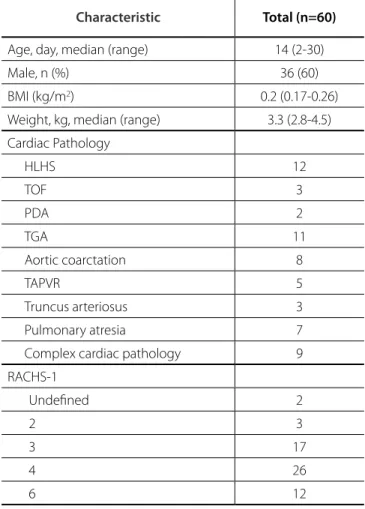
The majority, 60% (n=36) of the patients were male. Median age was 14 days (2-30 days), and median weight was 3.3 kg (2.8-4.5 kg). Median RACHS-1 score was 4 (2-6). The demographic features are listed in Table 1.
Atelectasis was demonstrated in 66% (n=40) of the cases. Five of the atelectasis cases were demonstrated only by X-ray, 10 of them by USG, and 25 of them both by X-ray and USG.
Pneumothorax was observed in 20% (n=12) of the cases. While 11 of them were demonstrated only by X-ray, one of them was discerned both by X-ray and USG.
Pleural efusion was demonstrated in 26% (n=16) of the cases. Four of these were demonstrated only by USG, three of them by X-ray, and nine of them both by X-ray and USG.
Pericardial efusion was demonstrated in 10% (n=6) of the cases. While one of the pericardial efusion cases was demonstrated both by X-ray and USG, the remaining ive were diagnosed by USG.
Fig. 1 -A 19-day old patient, operated for coarctation of aorta, on the 5th postoperative day. A) Chest X-ray revealed increased opacity on the
Three lung pathologic changes and pericardial efusion were found. There was a moderate correlation between abnormalities detected by thorax USG and X-ray (k=0.51). When thorax USG and X-ray were compared separately for diferent pathologies, the k was 0.64 for atelectasis, 0.22 for pneumothorax, 0.68 for pleural efusion and 0.28 for pericardial efusion. The evaluation of the presence of atelectasis, pneumothorax, pleural efusion, and pericardial efusion are listed in Table 2.
DISCUSSION
The beneits of utilizing thorax USG in emergency departments and ICU was supported by multiple studies[4,8-10].
In contrast, there is limited data about using thorax USG after congenital heart surgery in newborns. In this study, the eicacy of thorax USG in determining respiratory complications after congenital heart surgery in newborns was compared with that of a chest X-ray. This study’s results indicate that thorax USG is both eicient and useful in this condition. This is the irst comprehensive study about this topic in the literature to our knowledge.
Atelectasis is a clinical condition, particularly observed frequently in neonates and children who underwent congenital heart surgery. This morbidity can be associated with excessive and viscous pulmonary secretions, and it presents with associated respiratory symptoms. Accumulation of mucus causes susceptibility to pulmonary and systemic infections, causing prolonged need for mechanical ventilation and hospitalization[11,12].
The sensitivity and accuracy of USG in determining consolidation or atelectasis is reported as 80-100% and 90-100%, respectively[8,13]. Atelectasis was the most common respiratory
complication and was determined in 66% of the cases in the study. Atelectasis was shown in 88% of patients by USG and in 75% by X-ray. Although atelectasis was more frequently diagnosed by USG, imaging of right and left upper lobe atelectasis were
Table 2. The evaluation of chest X-ray and USG of the pathologies.
Observed problem Only X-ray Only USG USG and X-ray
Atelectasis 5 10 25
Right apical 3 0 2
Right basal 0 4 6
Left apical 2 0 3
Left basal 0 6 14
Pneumothorax 11 - 1
Right 8 - 0
Left 3 - 1
Pleural Efusion 3 4 9
Right 2 2 3
Left 1 2 6
Pericardial efusion - 5 1
Table 1. General clinical characteristics of the cases.
Characteristic Total (n=60)
Age, day, median (range) 14 (2-30) Male, n (%) 36 (60) BMI (kg/m2) 0.2 (0.17-0.26)
Weight, kg, median (range) 3.3 (2.8-4.5) Cardiac Pathology
HLHS 12
TOF 3
PDA 2
TGA 11
Aortic coarctation 8
TAPVR 5
Truncus arteriosus 3 Pulmonary atresia 7 Complex cardiac pathology 9 RACHS-1
Undeined 2
2 3
3 17
4 26
6 12
diicult. Atelectasis could not be demonstrated in 60% (n=5) of the cases with right upper lobe atelectasis and 40% (n=5) of the cases with left upper lobe atelectasis.
Pneumothorax is deined as the presence of air within the pleural space that prevents full expansion of the lung.
Pneumothorax may progress and cause hemodynamic instability, especially in patients receiving positive pressure ventilation[4,14].
In the literature, comparing the eiciency of X-ray and USG in determining pneumothorax was found 98% sensitivity, 99% speciicity, 98% positive predictive value (PPV), and 99% negative predictive value (NPV) for USG, whereas the results were 75%, 100%, 100%, and 90% for X-ray, respectively[13].
In a study including 126 patients with pneumothorax due to diferent etiologies (range 2 months-88 years) eiciency of USG in determining pneumothorax was compared with thorax computerized tomography (CT). The sensitivity, speciicity, accuracy, PPV, and NPV of chest USG was 89%, 88.5%, 88.9%,
96.7%,and 67.6%, respectively[15]. X-ray was more diagnostic than
USG for the demonstration of pneumothorax. Only 8% of the cases with pneumothorax (n=12) could be diagnosed by USG. The lack of experience in determining, localizing, and classifying pneumothorax might be the underlying reason in this study.
The other most common pathology after cardiac surgery is pleural efusion[3,16]. The compression efect of the efusion leads
to diferent degrees of aeration, even complete loss of alveolar aeration of that particular lung area. Out of the luid collection, the gradual restoration of the aeration in concordance with gradual manifestation of this consolidative process is often seen. This feature may lead to diferentiate the compressive atelectasis from pneumonia[17].
Vezzani et al.[13] demonstrated 100% sensitivity and 99%
diagnostic accuracy of chest ultrasound in their series. The false positive result was probably due to the presence of a small pleural efusion not identiied by chest X-ray. In this study, pleural efusion was demonstrated in 16 patients; 82% of them were diagnosed by USG and 75% by X-ray.
USG was better than X-ray for the diagnosis of pericardial efusion[1,4,17]. Only 16% of the cases (n=1) could be diagnosed
by X-ray. Results were similar for both methods for the diagnosis of pleural efusion.
Cost analysis of X-ray and ultrasonography also were compared, resulting in slightly lower costs for ultrasonography, but with a considerable advantage in terms of reduction of ionizing radiation exposure for both patients and staf[10,17]. X- ray
has certain limitations including risks of radiation exposure, high inter-observer and intra-observer variations.USG is a relatively smaller device that makes point of care more feasible. USG is easy, rapid, portable and repeatable.The USG has also shown less inter and intra-observer variations. Learning curve of techniques and interpretations of USG is simple and fast[18].
The sensitivity, speciicity, inter and intra-observer variability were found diferent in a small number of pediatric studies that compare USG and X-ray to determine the lung pathologies[5,18].
Yadav et al.[19] evaluated 118 community-acquired pneumonia
cases age between 2-59 months old with X-ray and USG. Abnormal X-ray were found in 101 (85.6%) and abnormal USG in 105 (89%) children. In diagnosing the speciic radiological
type of pneumonia, very good concordance (Kappa=0.7) was found between X-ray and USG. Kappa was found 0.9 especially in diagnosis of pleural efusion.
In the present study, there was a moderate relation (k=0.51) between USG and X-ray in evaluation of all of the pathologies. The concordance relation was much higher in pleural efusion (k=0.68) and atelectasis (k=0.64) than pneumothorax (k=0.22) and pericardial efusion (k=0.28).
Limitation
The current study was conducted at a single center with a limited number of cases. It would be more valuable to study this topic in a prospective randomized protocol with a larger patient population. Although computerized tomography (CT) is more accurate and almost a gold standard method in evaluation of thorax pathologies technical diiculties in practice, inancial cost and radiation exposure prevent its routine use. Thorax CT might be evaluated in addition to these two modalities.
CONCLUSION
Thorax USG might be a useful non-invasive method to evaluate the postoperative respiratory problems of neonates after congenital heart surgery. These indings should be supported by further studies.
REFERENCES
1. Bouhemad B, Zhang M, Lu Q, Rouby JJ. Clinical review: bedside lung ultrasound in critical care practice. Crit Care. 2007;11(1):205. 2. Lobo V, Weingrow D, Perera P, Williams SR, Gharahbaghian L. Thoracic
ultrasonography. Crit Care Clin. 2014;30(1):93-117.
3. Mayo P, Volpicelli G, Lerolle N, Schreiber A, Doelken P, Vieillard-Baron
Authors’ roles & responsibilities
EO
ICT
OY
YE
AG
Conception or design of the work; drafting the work; any part of the work appropriately investigated and resolved; final approval of the version to be published
Drafting the work; any part of the work appropriately investigated and resolved; final approval of the version to be published
Acquisition and analysis; any part of the work appropriately investigated and resolved; final approval of the version to be published
Revision of the work; any part of the work appropriately investigated and resolved; final approval of the version to be published
A. Ultrasonography evaluation during the weaning process: the heart, the diaphragm, the pleura and the lung. Intensive Care Med. 2016;42(7):1107-17.
4. Gargani L. Lung ultrasound: a new tool for the cardiologist. Cardiovasc Ultrasound. 2011;9:6.
5. Riu B, Ruiz J, Mari A, Silva S. Chest ultrasonography in pediatric critical care practice. Ann Fr Anesth Reanim. 2013;32(12):e219-23.
6. Jenkins KJ, Gauvreau K, Newburger JW, Spray TL, Moller JH, Iezzoni LI. Consensus-based method for risk adjustment for surgery for congenital heart disease. J Thorac Cardiovasc Surg. 2002;123(1):110-8.
7. Landis JR, Koch GG. The measurement of observer agreement for categorical data. Biometrics. 1977;33(1):159-74.
8. Tomà P, Owens CM. Chest ultrasound in children: critical appraisal. Pediatr Radiol. 2013;43(11):1427-34.
9. Trinavarat P, Riccabona M. Potential of ultrasound in the pediatric chest. Eur J Radiol. 2014;83(9):1507-18.
10. Vezzani A, Brusasco C, Palermo S, Launo C, Mergoni M, Corradi F. Ultrasound localization of central vein catheter and detection of postprocedural pneumothorax: an alternative to chest radiography. Crit Care Med. 2010;38(2):533-8.
11. Ozturk E, Tanidir IC, Haydin S, Onan IS, Odemis E, Bakir I. The use of dornase alpha for post-operative pulmonary atelectasis after congenital
heart surgery. Cardiol Young. 2014;24(5):807-12.
12. Peroni DG, Boner AL. Atelectasis: mechanisms, diagnosis and management. Paediatr Respir Rev. 2000;1(3):274-8.
13. Vezzani A, Manca T, Brusasco C, Santori G, Valentino M, Nicolini F, et al. Diagnostic value of chest ultrasound after cardiac surgery: a comparison with chest X-ray and auscultation. J Cardiothorac Vasc Anesth. 2014;28(6):1527-32.
14. Irwin Z, Cook JO. Advances in point-of-care thoracic ultrasound. Emerg Med Clin North Am. 2016;34(1):151-7.
15. Balesa J, Rathi V, Kumar S, Tandon A. Chest sonography in the diagnosis of pneumothorax. Indian J Chest Dis Allied Sci. 2015;57(1):7-11. 16. Grimberg A, Shigueoka DC, Atallah AN, Ajzen S, Iared W. Diagnostic
accuracy of sonography for pleural efusion: systematic review. Sao Paulo Med J. 2010;128(2):90-5.
17. Cantinotti M, Giordano R, Volpicelli G, Kutty S, Murzi B, Assanta N, et al. Lung ultrasound in adult and paediatric cardiac surgery: is it time for routine use? Interact Cardiovasc Thorac Surg. 2016;22(2):208-15. 18. Copetti R, Cattarossi L. Ultrasound diagnosis of pneumonia in children.
Radiol Med. 2008;113(2):190-8.