*Correspondence: R. B. Bazotte. Universidade Estadual de Maringá. Avenida Colombo 5790 - Bloco K68 – sala 101 – Maringá – PR – Brasil CEP: 87020-900. E-mail: [email protected]
A
vol. 52, n. 3, jul./sep., 2016 http://dx.doi.org/10.1590/S1984-82502016000300021
Glutamine dipeptide supplementation improves clinical responses
in patients with diabetic foot syndrome
Tuane Krupek
1, Maria Angélica Rafaini Covas Pereira da Silva
2, Débora de Mello Gonçalves
Sant´Ana
3, Márcia Regina Batista
4, Eliana Litsuko Tomimatsu Shimauti
4, Anacharis Babeto de
Sá-Nakanishi
5, Jurandir Fernando Comar
5, Christiano Rodrigues Schamber
1, Dorival Moreschi
Junior
6, Roberto Barbosa Bazotte
1,*1Pharmaceutical Sciences Postgraduate Program - State University of Maringá, Maringá, PR, Brazil, 2Department of Pharmacology and Therapeutics, State University of Maringá, Maringá, PR, Brazil, 3Department of Morphological Sciences,
State University of Maringá, Maringá, PR, Brazil, 4Department of Clinical Analysis and Biomedicine, State University of Maringá, Maringá, PR, Brazil, 5Department of Biochemistry - State University of Maringá, Maringá, PR, Brazil, 6Department
of Medicine - State University of Maringá, Maringá, PR, Brazil
The efect of glutamine dipeptide (GDP) supplementation in patients with diabetic foot syndrome was evaluated. A total of 22 patients took part in the study. GDP was supplied in 10 g sachets, and was dissolved in water immediately before use, with ingestion once a day, after lunch or after dinner (20 g/day) over a period of 30 days. Quantiication of foot insensitive areas, oxidative stress, blood cytokines, and biochemical, hematological and toxicological parameters was performed before and after GDP supplementation. We observed an increase in blood levels of interferon-α (P=0.023), interferon-γ (P=0.038), interleukin-4 (P=0.003), interleukin-6 (P=0.0025), interleukin-7 (P=0.028), interleukin-12 p40 (P=0.017), interleukin-13 (P=0.001), leukocytes (P=0.037), eosinophils (P=0.049), and typical lymphocytes (P<0.001) due to GDP administration. In addition, we observed a reduced number (P=0.048) of insensitive areas on the foot, and reduction (P=0.047) of fasting hyperglycemia. Patients also showed increased blood high density lipoprotein (P<0.01) and protein thiol groups (P=0.004). These favorable results were associated with the absence of renal and hepatic toxicity. These results are of clinical relevance, since supplementation with GDP over 30 days improved clinical responses in patients with diabetic foot syndrome.
Uniterms: Type 2 diabetes mellitus/treatment/study. Glutamine/efects. Nutraceuticals.
INTRODUCTION
Diabetic foot syndrome has been defined as a pathological condition in which peripheral vascular disease, peripheral neuropathy, and infection lead to tissue destruction, resulting in possible lower-extremity amputation in people with diabetes (Canavan et al., 2008).
Nerve damage in feet is characterized by increased oxidative stress, which leads to loss of neurons by apoptosis thereby reducing the regenerative capacity(Vicent et al.,
2004), associated with loss of foot sensitivity. Diabetic foot ulcers are very common in diabetic patients and may lead
to amputation(Schirmer, Ritter, Fansa, 2013). Moreover, following amputation, 45% of patients with neuropathic ulcers and 55% of patients with ischemic ulcers die within 5 years(Armstrong, Wrobel, Robbins, 2007).
The amino acid glutamine is involved in many processes that are vital to cell function. The molecular mechanisms of glutamine action remain to be elucidated but may involve changes in gene and protein expression, protein activity, and changes in oxidative status (Newsholme et al., 2003). For this reason, the enteral
and parenteral administration of glutamine has been recommended for critically ill patients(Newsholme et al., 2011; Vasconcelos, Tirapegui, 2002). In addition,
oral glutamine has been used by healthy individuals, in particular by athletes, to maintain immune function
that glutamine supplementation caused a reduction in systolic blood pressure, hyperglycemia, abdominal circumference(Mansour et al., 2015), and improved insulin secretion(Samocha-Bonet et al., 2015).
Although oral glutamine treatment is beneicial for human health, its low solubility and stability in aqueous solutions limits its availability in the blood. Furthermore, about 50% of orally administered glutamine is extracted by the splanchnic bed in healthy humans(Matthews, Marano, Campbell, 1993). However, this problem can be overcome with highly soluble stable L-alanyl-L-glutamine, a synthetic dipeptide composed of alanine and glutamine (Minguette-Camara et al., 2014; Rogero et al., 2002)
which is commonly known as glutamine dipeptide (GDP). Thus, based on the therapeutic potential of GDP, we evaluated the impact of supplementation with this dipeptide on the metabolic profile, oxidative stress, hematological parameters and blood levels of cytokines.
In this clinical investigation we used a well-established experimental approach in which each patient served as their own control(Sekhar et al., 2011;
Borges-Santos et al., 2012; Nguyen et al., 2014) eliminating the
interference of several factors such as age, duration of diabetes, and gender.
PATIENTS AND METHODS
Written consent to participate in this investigation was obtained from each patient and the study was conducted according to the ethical standards established by the Declaration of Helsinki and approved by Maringá State University Ethics Committee (COPEP - CAAE 204.758) and Clinical trial reg. n. DOI 10.1186/ISRCTN10878185, http://www.isrctn.com/.
Eligibility criteria included a diagnosis of type 2 diabetes and a confirmed diagnosis of diabetic foot syndrome. Exclusion criteria were severe hypertension, ischemic heart disease, pregnancy, and liver diseases.
Subjects
A total of 22 patients were selected to participate in the study after a medical consultation at the University Hospital of Maringá State University.
During the consultation, patients were interviewed using a questionnaire to obtain information about their socio-demographic and disease factors (age, sex, medical history, educational level, marital status, duration of diabetes, diabetes-related disorders etc.), therapeutic proile, and lifestyle.
Regarding diabetes, 33.3% of the patients had been
diagnosed with diabetes at least 10 years ago, 44.4% of the patients had been diagnosed for 11 to 20 years ago and 22.2% of the patients had been diagnosed over 21 years ago.
Most patients were female (83.3%), over 60 years of age (61.1%), sedentary (55.6), non-smokers (94.4%) and had at least 8 years of schooling (77.7%).
The majority of patients had one or more comorbidities associated with diabetes, namely hypertension (83.3%), retinopathy (66.7%), and/or nephropathy (27.8%).
Insulin, oral antidiabetic drugs, anti-hypertensive, and/or lipid-lowering drugs were used by 50%, 88.9%, 83.3% and 55.6% of patients, respectively. Regular insulin, NPH insulin, and regular insulin/NPH were used by 38.9%, 5.6%, and 5.6% of patients, respectively.
Despite the fact that most patients had a family history of type 2 diabetes (88.9%), the majority of patients showed an absence of knowledge about diabetic foot syndrome (55.6%).
Study Design
After the interview, a foot examination based on the National Hansen’s Disease Program developed by the University of Baton Rouge, USA was performed. This diabetic foot screening is not used to diagnose peripheral neuropathy, but to identify those patients who have lost protective sensation. The foot examination uses a 5.07 monoilament, which delivers 10 g of force to 12 locations on each foot, i.e., 24 points of sensation in total(Tan, 2010). During the physical examination, we found that many patients already had signs of peripheral vascular disease resulting from type 2 diabetes, such as intermittent
claudication (38.9%), absence of distal pulses (44.4%),
paresthesia (50.0%), calluses (83.3%), ungual mycosis (33.3%), and interdigital mycosis (22.2%). Some patients had already undergone surgical procedures in the lower limbs such as debridement (11.1%), revascularization (5.6%), or amputation (5.6%).
Venous blood was collected from each patient after an overnight fast as previously described (Zubioli et al.,
antioxidant activity was evaluated by means of total antioxidant capacity(Erel, 2004) and protein thiol groups (Faure, Lafond, 1995).
Serum eotaxin, interferon-α (IFN-α), interferon-γ (IFN-γ), interleukin-1 receptor antagonist (IL-1RA), interleukin-2 receptor (IL-2R), interleukin 4 (IL-4), interleukin 6 (IL-6), interleukin 7 (IL-7), interleukin 8 8), interleukin 10 10), interleukin 12 p40 (IL-12 p40), interleukin 13 (IL-13), interleukin 15 (IL-15), inducible protein 10 (IP-10), monocyte chemotactic protein 1 (MCP-1), monokine induced by IFN-γ (MIG), macrophage inflammatory protein-1α (MIP-1α), and
regulated on activation, normal T-cell expressed and secreted (RANTES) were quantified using the human cytokine magnetic plex panel produced by Invitrogen™ by means of the immunoassay Luminex - Magpix® platform.
Levels of erythrocytes, hemoglobin, hematocrit, platelets, leukocytes, basophils, eosinophils, immature neutrophils, segmented leukocytes, typical lymphocytes, atypical lymphocytes, and monocytes were evaluated, along with the amplitude of the distribution oferythrocyte size (ADES), mean corpuscular volume (MCV), mean corpuscular hemoglobin (MCH), and mean corpuscular hemoglobin concentration (MCHC) using BC Plus 3000 for Mindray Hematology Analyzers.
Patients were required to ingest GDP (Ajinomoto North America, NC, USA) which was supplied in sachets (10 g), and was dissolved in water immediately before using, once a day, after lunch or after dinner (20 g/day), for 30 days. After this period of treatment, all clinical procedures were repeated (blood collection, biochemical and hematological evaluation, quantiication of cytokines and foot examination).
The efect of the treatment with GDP was evaluated by comparing each patient before (day 1) and after treatment (day 30). In this way, each patient served as his or her own control.
Statistical analysis
For statistical analysis we used the software R.2.10.1. Results were analyzed using the Wilcoxon test
for comparing values before and after treatment. For quantitative variables, the Spearmanʼs correlation was used. Data were reported as the mean ± standard error (M ± SE). A p<0.05 level of probability was accepted as a statistically signiicant diference for all comparisons.
RESULTS AND DISCUSSION
A total of 18 patients with type 2 diabetes completed the study, while the remaining four patients were excluded because they did not take the GDP treatment as
recommended.
Supplementation with GDP reduced (P=0.048) the number of areas on the foot that lacked sensation from 5.9 ± 1.5 to 4.1 ± 1.3. Moreover, individual evaluation (Table I) showed that 10 patients (55.5%) experienced a reduction in the number of points without sensation after supplementation with GDP. In agreement with these results, supplementation with glutamine has been shown to reduce the loss of neurons in the duodenum of diabetic rats. This efect was attributed to the neuroprotective efect of glutamine which prevents oxidative stress by increasing the availability of reduced glutathione from glutamine (Zanoni et al., 2011).
Type 2 diabetic patients have been shown to have reduced antioxidant capacity(Kasznicki et al., 2012;
Oliveira et al., 2014; Rani, Mynthili, 2014), and, therefore,
the efect of oral GDP treatment on oxidative stress was evaluated. Treatment with GDP increased (P=0.004) protein free-thiol groups from 5.99 ± 0.17 nmol/mg protein to 6.73 ± 0.23 nmol/mg protein (M ± SE). However, the total antioxidant capacity remained unchanged (P=0.066), i.e., 0.73±0.02 µmol/mL (before supplementation) vs.
0.74 ± 0.02 µmol/mL (after supplementation). The
increased level of protein free thiol groups after oral GDP supplementation, suggests a contribution of the antioxidant properties of glutamine(Newsholme et al.,
2003, 2011).
It should be emphasized that partial recovery of sensation occurred in the presence of reduced (P=0.047) fasting hyperglycemia and increased (P<0.01) HDL after treatment with GDP. However, total cholesterol,
TABLE I - Individual evaluations of the number of areas on the foot without sensation (NAFWS) in type 2 diabetic patients before supplementation (BS) and after supplementation (AS) with glutamine dipeptide. The numbers 1-18 represent each patient included in the study
NAFWS 1 2 3 4 5 6 7 8 9 10 11 12 13 14 15 16 17 18
BS 16 1 11 7 2 1 16 9 16 7 4 15 1 0 0 1 0 0
triacylglycerol, total protein, and albumin remained unchanged (Table II).
The increased HDL-C, i.e., 2.9 mg/dL (Table II), is very important considering that: a) an elevation of 1 mg/ dL has been shown to reduce the risk of microvascular complications in type 2 diabetic patients(Toth et al.,
2012); b) patients with diabetic foot syndrome have a higher risk of cardiovascular disease(Pinto et al.,
2008); and c) hyperlipidemia is associated with diabetic neuropathy(Callaghan et al., 2012).
The increased urea values (P<0.001) after GDP supplementation (Table II) conirm the increased ingestion of this dipeptide.
The blood values of creatinine, AST, ALT, and GGT remained unaltered (Table II), suggesting the absence of renal and hepatic toxicity as consequence of supplementation with oral GDP. In agreement with these observations, it has been reported that glutamine (44-60 g/ day) does not cause any side efects (Bushen et al., 2004).
In agreement with previous studies (Weigelt et al.,
2009; Whitmont et al., 2013), we observed high blood levels
of C-reactive protein (a marker of acute inflammation) before GDP treatment. However, C-reactive protein levels were not inluenced by GDP treatment (Table II). This result could be partly explained by the fact that there is a simultaneous increase in the blood levels of pro-inflammatory (IFN-α, IFN-γ, IL-6, IL-7) and anti-inlammatory (IL-4, IL-13, IL-12 p40) cytokines (Table III). However, how can the synchronous increase of
pro-inlammatory and anti-inlammatory cytokines during GDP supplementation be accounted for?
We suggest that the simultaneous rise of pro-inflammatory and anti-pro-inflammatory cytokines during GDP supplementation is indicative of a pro-inlammatory and anti-inlammatory balance. In agreement with this suggestion, we previously reported a concurrent increase of blood pro-inlammatory and anti-inlammatory cytokines during an oral glucose tolerance test (Bazotte et al., 2016;
Eik Filho et al., 2016). Furthermore, other studies have
also demonstrated activation of pro-inlammatory and anti-inlammatory cytokines during sepsis (Mancilla-Ramírez et al., 1993), diabetes (Chatzigeorgiou et al., 2010), and
infections (Ng et al., 2003).
This balance of pro-inflammatory and
anti-inflammatory cytokines could represent an important negative feedback mechanism, which protects the body from excessive inlammation and its consequences.
Regarding the involvement of cytokines, it must be noted that these substances show pleiotropic efects in modulating immune responses and chronic inlammation (Akdis et al., 2011; Dinarello, 2007).
In this context, the elevation (P=0.003) of IL-4 induced by GDP supplementation (Table III) is very important, as IL-4 promotes activation of macrophages into reparative macrophagesin damaged tissues (Novak, Koh, 2013). Moreover, IL-13, whose efects on immune cells are similar to those of the closely-related cytokine IL-4(Novak, Koh, 2013) is also increased after oral GDP treatment (Table III). In addition, IFN-α and IFN-γ, which play a role in modulating immune responses by activating
immune cells(Kim, 2011; Aroor et al., 2013) are increased
(Table III) after oral treatment with GDP.
TABLE II - Biochemical and toxicological parameters (mean ± standard error) of diabetic patients before and after supplementation with glutamine dipeptide. Number of patients = 18
Biochemical parameters Before After P
Fasting glycemia (mg/dL) 155.6 ± 48.6 132.2 ± 50.5 0.047*
Creatinine (mg/dL) 1.3 ± 0.07 1.3 ± 0.09 0.396
Aspartate aminotransferase (U/L) 18.3 ± 1.77 24.8 ± 6.03 0.106
Alanine aminotransferase (U/L) 17.1 ± 2.45 17.3 ± 2.95 0.201
Gamma glutamyltransferase (U/L) 40.9 ± 8.61 44.4 ± 10.15 0.326
Total cholesterol (mg/dL) 190.2 ± 6.90 184.1 ± 7.63 0.289
High density lipoprotein (mg/dL) 54.3 ± 3.88 57.2 ± 3.84 <0.01*
Triacylglycerol (mg/dL) 158.0 ± 16.31 150.9 ± 15.00 0.472
Total protein (g/dL) 8.1 ± 0.08 8.1 ± 0.09 0.113
Albumin (g/dL) 4.5 ± 0.05 4.5 ± 0.05 0.777
Urea (mg/L) 43.3 ± 4.33 52.2 ± 4.39 <0.001*
C reactive protein (mg/L) 5.6 ± 2.6 6.5 ± 4.6 0.168
TABLE III - Serum cytokines levels (pg/mL) of diabetic patients before and after supplementation with glutamine dipeptide. N = number of patients
Cytokines N Before N After P
Eotaxin 18 118.39 ± 27.9 18 284.85 ± 28.4 0.492
IFN-α 17 22.41 ± 5.4 17 42.26 ± 6.5 0.023*
IFN-γ 15 4.52 ± 1.2 15 8.521 ± 1.2 0.038*
IL1-RA 17 168.68 ± 40.9 17 284.76 ± 40.2 0.638
IL-2R 18 75.36 ± 17.8 18 148.13 ± 16.1 0.084
IL-4 18 16.54 ± 3.9 18 35.89 ± 4.6 0.003*
IL-6 13 3.29 ± 0.9 13 5.41 ± 1.2 0.002*
IL-7 12 70.32 ± 20.3 12 156.33 ± 14.2 0.028*
IL-8 17 26.67 ± 6.5 17 47.84 ± 5.7 0.492
IL-10 16 1.37 ± 0.3 16 2.90 ± 0.4 0.286
IL-12p40 18 74.02 ± 17.4 18 253.49 ± 27.6 0.017*
IL-13 18 77.53 ± 18.3 18 239.10 ± 17.4 0.001*
IL-15 16 26.79 ± 6.7 16 49.92 ± 9.8 0.055
IP-10 18 31.13 ± 7.3 18 94.05 ± 8.8 0.184
MCP-1 18 98.50 ± 23.2 18 302.68 ± 30.9 0.113
MIG 16 40.65 ± 10.2 16 92.40 ± 13.0 0.071
MIP-1 α 16 18.11 ± 4.5 16 43.79 ± 5.4 0.105
RANTES 18 1880.4 ± 443.2 18 5148.6 ± 406.4 0.214
Interferon-α (IFN-α), interferon-γ (IFN-γ), interleukin-1 receptor antagonist (IL-1RA), interleukin-2 receptor (IL-2R), interleukin 4 (IL-4), interleukin 6 (IL-6), interleukin 7 (IL-7), interleukin 8 (IL-8), interleukin 10 (IL-10), interleukin-12 p40 (IL-12p40), interleukin 13 (IL-13), interleukin 15 (IL-15), IFN-γ inducible protein 10 (IP-10), monocyte chemotactic protein 1 (MCP1), monokine induced by IFN-γ (MIG), macrophage inlammatory protein-1α (MIP-1α), regulated on activation normally T-cell expressed and secreted (RANTES). *Non parametric Wilcoxon test. A P value of <0.05 was considered as statistically signiicant.
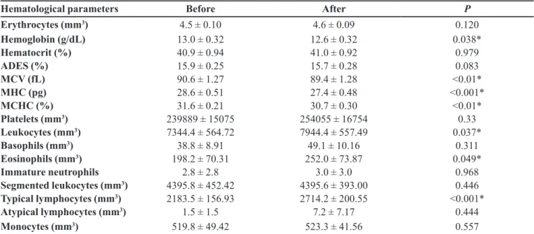
TABLE IV - Hematological parameters of diabetic patients before and after supplementation with glutamine dipeptide (mean ± standard error). Number of patients = 18
Hematological parameters Before After P
Erythrocytes (mm3) 4.5 ± 0.10 4.6 ± 0.09 0.120
Hemoglobin (g/dL) 13.0 ± 0.32 12.6 ± 0.32 0.038*
Hematocrit (%) 40.9 ± 0.94 41.0 ± 0.92 0.979
ADES (%) 15.9 ± 0.25 15.7 ± 0.28 0.083
MCV (fL) 90.6 ± 1.27 89.4 ± 1.28 <0.01*
MHC (pg) 28.6 ± 0.51 27.4 ± 0.48 <0.001*
MCHC (%) 31.6 ± 0.21 30.7 ± 0.30 <0.01*
Platelets (mm3) 239889 ± 15075 254055 ± 16754 0.33
Leukocytes (mm3) 7344.4 ± 564.72 7944.4 ± 557.49 0.037*
Basophils (mm3) 38.8 ± 8.91 49.1 ± 10.16 0.311
Eosinophils (mm3) 198.2 ± 70.31 252.0 ± 73.87 0.049*
Immature neutrophils 2.8 ± 2.8 3.0 ± 3.0 0.968
Segmented leukocytes (mm3) 4395.8 ± 452.42 4395.6 ± 393.00 0.446
Typical lymphocytes (mm3) 2183.5 ± 156.93 2714.2 ± 200.55 <0.001*
Atypical lymphocytes (mm3) 1.5 ± 1.5 7.2 ± 7.17 0.444
Monocytes (mm3) 519.8 ± 49.42 523.3 ± 41.56 0.557
Other cytokines that were shown to increase after oral treatment with GDP (Table III) include IL-6 (induces immune response, during infections, trauma, burns or other tissue damage), IL-7 (a hematopoietic growth factor secreted by stromal cells in the bone marrow and thymus), and IL-12 p40 (acts as an antagonist of IL-12, a pro-inlammatory cytokine).
In summary, the significant increases in IFN-α, IFN-γ, IL-4, IL-6, IL-7, IL-13, and IL-12 p40 may improve the immune responses after oral treatment with GDP. In agreement, with this proposition, as shown in Table IV, oral supplementation with GDP also increased the number of circulating leukocytes (P=0.037), eosinophils (P=0.049) and typical lymphocytes (P<0.001).
Finally, decreased hemoglobin (P=0.038), MCV (P<0.01), MHC (P<0.001), and MCHC (P<0.01) were detected (Table IV). This implies that glutamine is important for hematopoiesis, as previously reported (Rogero et al., 2008).
Our results are of clinical relevance, as treatment with oral GDP (20 g/day) over a period of 30 days improved clinical responses in patients with diabetic foot syndrome.
ACKNOWLEDGMENTS
This study was funded by the Research Program for the uniied health system (PPSUS) – Process 987/2013.
REFERENCES
AKDIS, M.; BURGLER, S.; CRAMERI, R.; EIWEGGER, T.; FUJITA, H.; GOMEZ, E.; KLUNKER, S.; MEYER, N.; O’MAHONY, L.; PALOMARES, O.; RHYNER, C.; OUAKED, N.; SCHAFFARTZIK, A.; VAN DE VEEN, W.; ZELLER, S.; ZIMMERMANN, M.; AKDIS, C. A. Interleukins, from 1 to 37, and interferon-gamma: receptors, functions, and roles in diseases. J. Allergy Clin. Immun.
v.127, n.3, p.701-21, 2011.
ARMSTRONG, D.G.; WROBEL, J.; ROBBINS, J.M. Guest editorial: are diabetes-related wounds and amputations worse than cancer? Int. Wound J., v.4, p.286-7, 2007.
AROOR, A.R.; MCKARNS, S.; DEMARCO, V.G.; JIA, G.; SOWERS, J.R. Maladaptive immune and inflammatory pathways lead to cardiovascular insulin. Metabolism., v.62, n.11, p.1543-52,2013.
BAZOTTE, R.B.; DE CASTRO, R.M.A.; DA SILVA, M.A.R.C.P.; KRUPEK, T.; EIK FILHO, W. Blood levels of pro-inlammatory and anti-inlammatory cytokines in a patient with a lat glucose curve. Acta Diabetol., v.53, n.4, p.1-3, 2016.
BORGES-SANTOS, M.D.; MORETO F.; PEREIRA P.C.; MING-YU, Y.; BURINI R.C. Plasma glutathione of HIV⁺ patients responded positively and differently to dietary supplementation with cysteine or glutamine. Nutrition., v.28, n.7-8, p.753-6, 2012.
BUSHEN, O.Y.; DAVENPORT, J.A.; LIMA, A.B.; PISCITELLI, S.C.; UZGIRIS, A.J.; SILVA, T.M.; LEITE, R.; KOSEK, M.; DILLINGHAM, R.A.; GIRAO, A.; LIMA, A.A.; GUERRANT, R.L. Diarrhea and reduced levels of antiretroviral drugs: improvement with glutamine or
alanyl-glutamine in a randomized controlled trial in northeast Brazil. Clin. Infect. Dis., v.38, n.12, p.1764-70, 2004.
CALLAGHAN, B.C.; CHENG, H.T.; STABLES, C.L.; SMITH, A.L.; FELDMAN, E.L. Diabetic neuropathy: clinical manifestations and current treatments. Lancet Neurol., v.11, n.6, p.521-34, 2012.
CANAVAN, R.J.; UNWIN, N.C.; KELLY, W.F.; CONNOLLY, V.M. Diabetes and nondiabetes related lower extremity amputation incidence before and after the introduction of better organized diabetes foot care: continuous longitudinal
monitoring using a standard method. Diabetes Care, v.31, n.3, p.459-63, 2008.
CHATZIGEORGIOU,A.;HAROKOPOS,V.; MYLONA-KARAGIANNI, C.; TSOUVALAS, E.; AIDINIS, V.; KAMPER, E.F. The pattern of inflammatory/anti-inlammatory cytokines and chemokines in type 1 diabetic patients over time. Ann. Med., v.42, n.6, p.426-38, 2010.
CURY-BOAVENTURA, M.F.; LEVADA-PIRES, A.C.; FOLADOR, A.; GORJAO, R.; ALBA-LOUREIRO, T.C.; HIRABARA, S.M.; PERES, F.P.; SILVA, P.R.; CURI, R.; PITHON-CURI, T.C. Efects of exercise on leukocyte death: prevention by hydrolyzed whey protein enriched with glutamine dipeptide: Eur. J. Appl. Physiol., v.103. n.3, p.289-94,2008.
EIK FILHO, W.; MARCON, S.S.; KRUPEK, T.; PREVIDELLI, I.T.; PEREIRA, O.C.; SILVA, M.A.R.C.P.; BAZOTTE R.B. Blood levels of pro-inlammatory and anti-inlammatory cytokines during an oral glucose tolerance test in patients with symptoms suggesting reactive hypoglycemia. Braz. J. Med. Biol. Res., v.49, p.pii:S0100-879X, 2016.
EREL, O. A novel automated direct measurement method for total antioxidant capacity using a new generation, more stable ABTS radical cation. Clin. Biochem., v.37, n.4, p.277-85, 2004.
FAURE, P.; LAFOND, J.L. Measurement of plasma sulfhidryl and carbonyl groups as a possible indicator of protein oxidation. In: FAVIER, A.E.; CADET, J.; KALNYANARAMAN, M.; FONTECAVE, M.; PIERRE, J.L. (Eds). Analysis of Free Radicals in Biological Systems. Boston, USA: Birkhäauser, 1995. p.238-47.
KASZNICKI, J.; KOSMALSKI, M.; SLIWINSKA, A.; MROWICKA, M.; STANCZYK, M.; MAJSTEREK, I.; DRZEWOSKI, J. Evaluation of oxidative stress markers in pathogenesis of diabetic neuropathy. Mol. Biol. Rep.,v.39, n.9, p.8669-78, 2012.
KIM, H. Glutamine as an immunonutrient. Yonsei Med. J., v.52, n.6, p.892-7, 2011.
MANCILLA-RAMÍREZ, J.; RAMÍREZ-HERRERA, M.; PORTILLO-GÓMEZ, L.; GAITÁN-MEZA, J.; DINARELLO, C.A. Antagonists in neonatal sepsis. Bol. Med. Hosp. Infant Mex., v.50, n.9, p.691-3, 1993.
MANSOUR, A.; MOHAJERI-TEHRANI, M.R.; QORBANI, M.; HESHMAT, R.; LARIJANI, B.; HOSSEINI, S. Efect of glutamine supplementation on cardiovascular risk factors in patients with type 2 diabetes. Nutrition, v.31, p.119-26,
2015.
MATTHEWS, D.E.; MARANO, M.A.; CAMPBELL, R.G. Splanchnic bed utilization of glutamine and glutamic acid
in humans. Am. J. Physiol., v.264, n.6, p.848-54, 1993.
MINGUETTE-CAMARA, V.C.; MARQUES, A.C.R.; VILELA, V.R.; SCHIAVON, F.P.M.; BRUSCHI, M.L.; BAZOTTE, R.B. A comparison of the effects of oral glutamine dipeptide, glutamine, and alanine on blood amino acid availability in rats submitted to insulin-induced hypoglycemia. Nutrients, v.6, n.10, p.4520-30,2014.
NEWSHOLME, P.; ABDULKADER, F.; REBELATO, E.; ROMANATTO, T.; PINHEIRO, C.H.; VITZEL, K.F.; SILVA, E.P.; BAZOTTE, R.B.; PROCOPIO, J.; CURI, R.; GORJAO, R.; PITHON-CURI, T.C. Amino acids and diabetes: implications for endocrine, metabolic and immune function. Front Biosci., v.16, p.315-39,2011.
NEWSHOLME, P.; LIMA, M.M.R.; PROCOPIO, J.; PITHON-CURI, T.C.; DOI, S.Q.; BAZOTTE, R.B.; PITHON-CURI, R. Glutamine and glutamate as vital metabolites. Braz. J. Med. Biol. Res., v.36, n.2, p.153-63, 2003.
NG, P.C.; LI, K.; WONG, R.P.; CHUI, K.; WONG, E.; LI, G.; FOK, T.F. Pro-inflammatory and anti-inflammatory cytokine responses in preterm infants with systemic infections. Arch. Dis. Child Fetal Neonatal Ed., v.88, n.3, p.209-13, 2003.
NGUYEN, D.; HSU, J.W.; JAHOOR, F.; SEKHAR, R.V. Efect of increasing glutathione with cysteine and glycine supplementation on mitochondrial fuel oxidation, insulin sensitivity, and body composition in older HIV-infected patients. J. Clin. Endocrinol. Metab., v.99, n.1, p.169-77, 2014.
NOVAK, M.L.; KOH, T.J. Macrophage phenotypes during tissue repair. J. Leukoc. Biol., v.93, n.6, p.875-81, 2013.
OLIVEIRA, A.C.P.; TEIXEIRA, C.J.; STEFANELLO, T.F.; CARRARA, M.A.; BAZOTTE, R.B.; SÁ-NAKANISHI, A.B.; COMAR, J.F.; BATISTA, M.R. Oxidative stress parameters as biomarkers of risk factor for diabetic foot among the patients with type 2 diabetes. Braz. Arch. Biol. Technol., v.57, n.2, p.223-27,2014.
PINTO, A.; TUTTOLOMONDO, A.; DI RAIMONDO, D.; FERNANDEZ, P.; LA PLACA, S.; DI GATI, M.; LICATA, G. Cardiovascular risk proile and morbidity in subjects affected by type 2 diabetes mellitus with and without diabetic foot. Metabolism, v.57, n.5, p.676-82, 2008.
ROGERO, M.M.; TIRAPEGUI, J.; PEDROSA, R.G.; CASTRO, I.A.; PIRES, I.S.O.; OLIVEIRA, A.A.M.; SALGADO, M.M.; PINTO, A.R.; UEDA, M. Effect of L-glutamine and L-alanyl-L-glutamine supplementation on the response to delayed-type hypersensitivity test (DTH) in
rats submitted to intense training. Rev. Bras. Ciênc. Farm., v.38, n.4, p.487-97, 2002.
ROGERO, M.M.; TIRAPEGUI, J.; VINOLO, M.A.; BORGES, M.C.; DE CASTRO, I.A.; PIRES, I.S.; BORELLI, P. Dietary glutamine supplementation increases the activity of peritoneal macrophages and hemopoiesis in early-weaned mice inoculated with Mycobacterium bovis bacillus Calmette-Guerin. J. Nutr., v.138, n.7, p.1343-8, 2008.
SAMOCHA-BONET, D.; CHISHOLM, D.J.; HOLST, J.J.; GREENFIELD, J.R. L-glutamine and whole protein restore first-phase insulin response and increase glucagon-like peptide-1 in type 2 diabetes patients. Nutrients, v.7, n.4, p.2101-8,2015.
SCHIRMER, S.; RITTER, R.G.; FANSA, H. Vascular surgery, microsurgery and supramicrosurgery for treatment of chronic diabetic footulcers to prevent amputations.PLoS One, v.13, p.1-14, 2013.
SEKHAR, R.V.; PATEL, S.G.; GUTHIKONDA, A.P.; REID, M.; BALASUBRAMANYAM, A.; TAFFET, G.E.; JAHOOR, F. Deicient synthesis of glutathione underlies oxidative stress in aging and can be corrected by dietary cysteine and glycine supplementation. Am. J. Clin. Nutr., v.94, n.3, p.847-53, 2011.
TAN, L.S. The clinical use of the 10g monofilament and its limitations: a review. Diabetes Res. Clin. Pract., v.90, n.1, p.1-7, 2010.
TOTH, P.P.; SIMKO, R.J.; PALLI, S.R.; KOSELLECK, D.; QUIMBO, R.A.; CZIRAKY, M.J. The impact of serum lipids on risk for microangiopathy in patients with type 2
diabetes mellitus. Cardiovasc. Diabetol., v.11, n.109, 2012.
VASCONCELOS, M.I. L; TIRAPEGUI, J. News aspects of nutritional support in the critically ill at intensive care unit (ICU).Rev. Bras. Ciênc. Farm., v.38, n.1, p. 23-32, 2002.
VINCENT, A.M.; RUSSELL, J.W.; LOW, P.; FELDMAN, E.L. Oxidative stress in the pathogenesis of diabetic neuropathy. Endocr. Rev., v.25, n.4, p.612-28, 2004.
WEIGELT, C.; ROSE, B.; POSCHEN, U.; ZIEGLER, D.; FRIESE, G.; KEMPF, K.; KOENIG, W.; MARTIN, S.; HERDER, C. Immune mediators in patients with acute diabetic foot syndrome. Diabetes Care,v.32, n.8, p.1491-96, 2009.
WHITMONT, K.; FULCHER, G.; REID, I.; XUE, M.; MCLKELVEY, K.; XIE, Y.; ABOUD, M.; WARD, C.; SMITH, M.M.; COOPER, A.; MARCH, L.; JACKSON, C.J. Low circulating protein C levels are associated with lower leg ulcers in patients with diabetes. Biomed. Res. Int., v.2013, p.719570, 2013.
ZANONI, J.N.; TRONCHINI, E.A.; MOURE, S.A.; SOUZA, I.D. Effects of L-glutamine supplementation on the myenteric neurons from the duodenum and cecum of
diabetic rats. Arq. Gastroenterol., v.48, n.1, p.66-71,2011.
ZUBIOLI, A.; SILVA, M.A.R.C.P.; TASCA, R.S.; CURI, R.; BAZOTTE, R.B. Pharmaceutical consultation as a tool to
improve health outcomes for patients with type 2 diabetes. Braz. J. Pharm. Sci., v.49, p.85-94, 2013.
Received for publication on 10th November 2015