*Correspondence: R.M. Sponchiado. Faculdade de Farmácia. Universidade Federal do Rio Grande do Sul – UFRGS. Av. Ipiranga, n.2752, 90610-000 - Porto Alegre - RS, Brazil. E-mail: [email protected]
A
vol. 52, n. 3, jul./sep., 2016 http://dx.doi.org/10.1590/S1984-82502016000300014
In vitro
evaluation of cutaneous penetration of acyclovir from
semisolid commercial formulations and relation with its effective
antiviral concentration
Rafaela Martins Sponchiado
*, Leticia Malgarim Cordenonsi, Nathalie Ribeiro Wingert,
Bibiana Verlindo de Araujo, Nadia Maria Volpato
Faculty of Pharmacy, Federal University of Rio Grande do Sul, Porto Alegre-RS, Brazil
The evaluation of drug permeation/penetration of semisolid formulations into animal skin can be useful to supplement the pharmaceutical equivalence. This paper describes the in vitro assessment of acyclovir (ACV) into porcine skin from commercial formulations with etermination of drug concentration
in diferent layers of cutaneous tissue to correlate with efective antiviral concentration in order to
improve the equivalence decision. Studies were conducted using Franz cells and porcine skin. Selected
pharmaceutical creams containing ACV had identical (reference and generic) and diferent (similar) excipients. A software program was employed for the simulation of antiviral efectiveness in the skin. Regarding ACV skin penetration, the irst batch of the generic product showed a signiicant diference
from reference and similar products, while in the second batch all products demonstrated equivalent drug penetration in the skin. Simulation studies suggest that formulations analysed exhibit a pharmacological
efect even when in contact with Herpes simplex strains of high IC50 (inhibitory concentration required
to reduce viral replication by 50%). According to results, it can be assumed that the in vitro cutaneous permeation/penetration study does not supply sensitivity information regarding small alterations of ACV semisolid formulations due to the variability inherent to the method, although it can be relevant to pharmaceutical equivalence studies in the development of semisolid products.
Uniterms: Acyclovir/skin penetration/in vitro studies. Acyclovir/semisolid formulations. Acyclovir/ pharmaceutical equivalence.
INTRODUCTION
A c y c l o v i r ( 2 a m i n o 1 , 9 d i h y d r o 9 ( ( 2 -hydroxyethoxy)methyl)-6H-purin-6-one) (ACV) (Figure 1) is a guanosine derivative drug with high specificity
for herpesviruses. Due to its high eicacy against herpes
simplex virus (HSV) and varicella-zoster virus, ACV has been the leading treatment for these pathologies until now (Field, Hodge, 2013). The action mechanism is based on competition with endogenous nucleosides for viral DNA incorporation, which disable DNA chain elongation and, therefore prevent viral replication (Barreca et al., 2003; Lupi, Silva, Pereira, 2000).
Regarding the topical dosage forms, currently
several brands from diferent manufacturers can be found
in the market, mainly as cream formulations. It is known that to obtain a better effect on skin virus affections, antiviral drugs must reach therapeutic concentrations in the basal cells of the epidermis, which are the earliest point of entry for virus propagation. Accordingly, penetration of an active pharmaceutical into the skin is a critical factor for successful treatment of oral herpes by ACV topical formulation (Hasler-Nguyen et al., 2009).
Implementation of in vitro methods to evaluate
the performance of topical pharmaceutical formulations is very important, not only to predict drug permeation/ penetration into the skin, but also as a valuable tool to compare generic and reference products. Some authors also point out its significance to monitor variations among batches manufactured on an industrial scale (De Paula, Martin, Bentley 2008; Shah et al., 1991). Undoubtedly, studies regarding cutaneous penetration/ permeation are important to optimize and develop semisolid pharmaceutical formulations. They enable a better understanding of what happens to the drug after it is applied to the skin, the release from the pharmaceutical formulation, penetration and percutaneous absorption.
According to the World Health Organization (WHO, 2006), studies regarding in vitro cutaneous absorption are increasingly being submitted for registration of topical products. Several published papers compare in vivo and in vitro results, and studies following OECD (Organization of Economic Co-operation and Development) directives demonstrate that in vitro tests provide a good prediction of cutaneous absorption (Alberti et al., 2001; Gannu et al, 2010; Tsai et al., 2010).
Currently, a release study with Franz vertical diffusion cells (VDC) is the in vitro method most employed and accepted by international regulation guidelines due to its good reproducibility (Sartorelli et al., 2000; OECD, 2004). FDA (Food and Drug Administration, USA) recommends VDC employing synthetic membranes to evaluate scale-up and post-approval alteration in topical formulations (FDA, 1997). However, cutaneous permeation studies employing skin specimens in this
difusion bi-compartment model usually provide more
variable results.
Therefore, the purpose of this paper was to verify whether in vitro determination of cutaneous permeation/ penetration of ACV from commercial formulations (reference – R, generic – G, and similar – S) can indicate the maintenance of drug activity and product equivalence,
relating the ACV concentration in different layers of
cutaneous tissue to efective antiviral concentration. A bioanalytical method for drug quantiication in skin layers
(epidermis and dermis) of pig ears was validated for this purpose.
MATERIAL AND METHODS
Generic and similar 5% ACV dermatologic creams were selected by analysing the composition of
excipients from diferent brands found in the Brazilian
market, compared to the reference product (Zovirax®, GlaxoSmithKline). Table I presents the qualitative composition of chosen formulations according to excipient function or action within the formulation. Two medicines with similar formulations and one with a different composition were selected. R and G products present excipients with equivalent characteristics for instance, including propylene glycol, whose presence may promote drug permeation. In contrast, medicine S did not contain that inactive ingredient.
All reagents were analytical or HPLC grade. Ultra-pure water was obtained by 3UV Millipore® Derect-Q (Molsheim, France). Pig ears were acquired from the Ouro do Sul slaughterhouse (Harmonia, Brazil).
Chromatographic system
The analytical method was developed in a Shimadzu 10A system (Kyoto, Japan) equipped with an LC-20AT pump, SPD-20AV UV-VIS variable wavelength detector (254 nm), DGU-20A5 degasser, CBM-20A controller system, and SIL-20A injection valve with 100
μL loop. Phenomenex C8 column (5 μm, 250 mm x 4.6 mm i.d.) and pre column C8 (4 x 3.0 mm) were kept at 25 °C. The mobile phase consisted of water and methanol
(95:5, v/v) in isocratic mode, with 1 mL/min low and 50
µL of sample injection was used.
TABLE I - Qualitative composition of 5 % ACV creams selected (water and ACV are elements present in all formulations and therefore not included in this table
Product Preservative Emulsiier Lipophilic vehicle/
Emollient
Absorption
promoter Others
Glaxo SmithKline
(Reference)
-Cetostearyl alcohol, polaxamer 407
Vaseline, liquid
parain. Propylene glycol Sodium lauril sulfate Prati-Donaduzzi
(Generic)
Propylparaben
Methylparaben Cetostearyl alcohol Liquid petrolatum Propylene glycol Sodium lauril sulfate
Cimed (Similar) Propylparaben
-Skin preparation
Pig ears, obtained before the slaughter line scalding step, were initially washed with water. The central portion of the external side was excised and cut with a scalpel and
moulded into a round shape to it Franz cells, wrapped with PVC ilm and aluminium foil and kept under -20 °C
for up to 30 days.
Acyclovir extraction procedure
In order to validate the extraction procedure, skin segments without blood vessels or scars were cut in 1.5 cm diameter circles (equivalent to the Franz cells contact area). For validation requirements, dermis and epidermis separation was performed with a scalpel after immersion into water at 60°C for 45 seconds. Dermis was additionally sectioned into small parts and, only then transferred to separate tubes. Dermis and epidermis were spiked with ACV aqueous solution and kept still for 2 hours. The cycle of ACV extraction from skin layers occurred by addition of 4 mL of water in the tubes and sonication for over 40 min
Bioanalytical method validation
Bioanalytical method validation followed national and international guidelines (ANVISA, 2012; ICH,
2005). Method speciicity was evaluated by comparing
chromatograms from samples with and without ACV after skin extraction.
Standard curves were analysed on three diferent
days with drug concentrations from 0.5 to 25 µg/mL. Samples were diluted with biological matrices in the same proportion as the extraction procedure (1.5 cm diameter skin per 8 mL of water). Results were submitted to linear regression analysis through minimum square method and ANOVA (analysis of variance) statistical evaluation. Limits of detection and quantification were estimated according to standard curve and confirmed through
experiments. Intercept signiicance was also veriied.
Method precision was assessed by ACV recovery from the main skin layers. After separation, dermis and epidermis were spiked with 50 µL of ACV solution at three known concentration levels (low, median, and high). The
inal concentration corresponded to 0.5, 12.5, and 25 µg/mL;
each level had n = 6 for both skin layers.
In vitro skin penetration studies
Skin samples were thawed at room temperature
and mounted in Franz difusion cells (Hanson Research,
Chatsworth, USA) with stratum corneum (SC) facing donor compartment and dermis in contact with receptor
compartment. The difusion area of Franz cells was 1.77
cm2, and the receptor container was filled with 7 mL phosphate bufer, pH 7.4. Pig skin was kept in contact with
the receptor solution at 32 °C (±1 °C) for up to 8 hours and aliquots of 1 mL were collected at predetermined times (1, 4, 6, and 8 h) to analyse ACV permeation through
the skin. The same volume of receptor luid withdrawn
was replaced to keep it constant. After 8 hours, skin was detached from the Franz cell and excess formulation carefully removed. Without water immersion, scalpel and scissor were applied to separate skin layers, epidermis and dermis, which were weighed and extraction proceeded as described previously.
Results are presented as µg of ACV per mg of tissue;
all data (n = 15) and mean values ± standard deviation (SD) were computed. Data were analysed by one-way ANOVA in order to compare the three pharmaceutical formulations.
Tukey test was applied to signiicant results (α = 0.05).
Pharmacodynamic study – simulation of ACV antiviral effect on skin layers
Scientific literature provides different in vitro
and in vivo values for inhibition of HSV-1 replication. Values of IC50 (inhibitory concentration for 50 % of viral
replication) found for plaque number reduction assay using Vero cells presented divergent patterns, which, for
the purpose of this study, were classiied as low, median,
and high potency, evidencing the characteristic of
drug-virus interaction (species-speciic) (Cavalli et al., 2009; Drugbank databese; Gong et al., 2004; Jalón et al., 2003; Suzuki, Okuda, Shiraki, 2006).
C o m p a r i s o n b e t w e e n e x p e c t e d e f f e c t s a t concentration ranges found for ACV in the in vitro
permeation assay from R, G, and S formulations was performed by means of Scientist® software program. employing a model of inhibitory efect for ACV and values
of IC50 described in the literature for diferent viral strains (Table II). The model is described by equation 1, where
E is the expected efect, E0 the efect on absence of drug
inhibition, Imax is the maximum inhibition promoted by
ACV (inhibition of replication), C is ACV concentration
evaluable in the tissue to produce an efect (in this situation
µg/g was employed), and IC50 (µg/mL) is the concentration
necessary to promote half of the maximum inhibitory
efect (Gabrielsson, Weiner, 2006).
To obtain a dose-response curve, low, median, and high IC50 (0.049, 0.265, and 0.850 µg/mL, respectively)
values from scientiic literature were used.
RESULTS AND DISCUSSION
Method validation
Analytical conditions allowed an adequate peak signal of ACV in the presence of biological matrix,
although with small interferences. According to oicial
regulation guidelines, the presence of a chromatographic response relative to blank sample along with analyte must
be lower than 20 % of the limit of quantiication (LoQ). On that basis, at the lower quantiication concentration
of ACV (0.5 µg/mL), the higher signal from biological matrices corresponded to 2.95 % and 11.75 % of the drug peak signal in epidermis and dermis, respectively (Figure 2).
The method developed presented an appropriate linearity at 0.5 – 25 µg/mL concentration range from 3 days analysis. The mean standard curve equation y =
153656x + 33034 had a determination coeicient (r2) equal
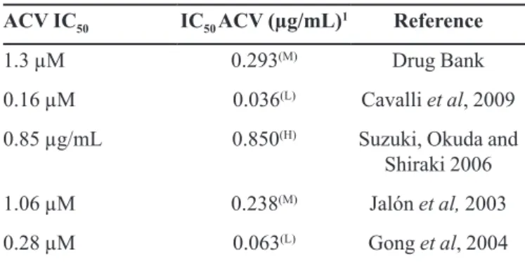
TABLE II - ACV IC50 values found in scientiic literature for distinct HSV-1 strains in plaque number reduction assay.
ACV IC50 IC50 ACV (µg/mL)1 Reference
1.3 µM 0.293(M) Drug Bank
0.16 µM 0.036(L) Cavalli et al, 2009
0.85 µg/mL 0.850(H) Suzuki, Okuda and Shiraki 2006
1.06 µM 0.238(M) Jalón et al, 2003
0.28 µM 0.063(L) Gong et al, 2004
(H): high value IC50; (M) median value of IC50; (L) low value IC50. 1: data were standardized in µg/mL
FIGURE 2 - Speciicity of chromatographic method for the analysis of ACV in skin layers. A) Epidermis; B) Dermis. Black line:
to 0.9999. According to data obtained from ANOVA it is
possible to ensure signiicant linear regression (Fcalculated
= 37278.2 >> Fcritical = 8.53) without deviation from linearity (Fcalculated = 0.35 < Fcritical = 4.20). Limits of
detection and quantiication were 0.11 and 0.36 µg/mL, respectively. Experimentally, ive injections at 0.3, 0.4, and
0.5 µg/mL presented an RSD of 0.77 %, 1.26 %, and 2.20 %, demonstrating that the proposed LoQ was precise and sensitive. During the study, every chromatographic run was performed with a new standard curve, which furnished an average slope of (n=18). The p-value for the intercept was always greater than 0.05.
Method precision and accuracy were evaluated in terms of ACV recovery from biological matrices. Table III presents data obtained for ACV recovery from skin layers (epidermis and dermis), and RSD for intra-day repeatability and intermediate precision at each concentration level of ACV added to matrices. Average data recovered were 92.74% for epidermis and 90.65% for dermis, values which are in agreement with
those recommended by scientiic guidelines. Regarding
precision assessment, RSD ranged from 4.36 % to 6.77 % which is considered acceptable for a bioanalytical method (<15 %) and also indicates suitability of the extraction
process and quantiication of ACV in the main skin layers.
Comparative cutaneous penetration study
A validated bioanalytical method was applied to the quantification of ACV in both skin layers (dermis and epidermis) after formulations were applied. ACV penetration was evaluated from three commercial creams (R, G, and S) in two different batches, corresponding to six samples. Results regarding accumulated amount of ACV in skin layers for R, G, and S samples in a cutaneous permeation study are shown in Figures 3
and 4. Evaluating Figure 3, it can be concluded that overall, a higher concentration of ACV is detected in epidermis than in dermis, a condition that is ideal for ACV
pharmacological efect against herpes virus. The amount
of drug accumulated in epidermis and dermis for R and S products was substantially lower than that obtained with
G, according to statistical evaluation. The main diference
between the formulations is the presence of propylene glycol in the R and G products. This excipient may have more than one function in a pharmaceutical formulation. Besides being a carrier, it can act as permeation promoter in amounts usually applied for dermatological products (Trottet et al., 2004). The action mechanism proposed for propylene glycol suggests that its input in SC increases penetrant solubility, with a consequent increment of the
low through skin tissue (Moser et al., 2001; Williams,
TABLE III - Accuracy and precision of the bioanalytical method for ACV quantiication
Skin Layers Level
Day 1 Day 2
Observed concentration ± SD
RSD (%) intra-day
Observed concentration ± SD
RSD (%) intra-day
RSD (%) inter-day
Epidermis
Low 0.42 ± 0.02 4.92 0.43 ± 0.02 4.65 4.36
Medium 11.82 ± 0.9 7.54 12.83 ± 0.05 0.36 6.41
High 22.41 ± 1.50 6.72 24.46 ± 0.24 0.99 6.32
Dermis
Low 0.47 ± 0.01 3.27 0.48 ± 0.04 7.94 5.6
Medium 10.95 ± 1.2 10.33 11.35 ± 0.16 1.38 6.77
High 22.15 ± 2.0 9.05 21.96 ± 0.70 3.17 6.10
FIGURE 3 - Retained amount of ACV in porcine skin layers
Barry, 2012). The manufacturers do not inform the amount of propylene glycol, but it is possible that one of those mechanisms occurred, explaining the higher concentration
of ACV from the irst batch of G product found in skin
layers.
Another difference that can be considered is the presence of sodium lauryl sulfate (SLS - anionic surfactant) in R and G formulations. The action mechanism of SLS has not yet been completely explained. However, it is well known that it interacts with keratin and intracellular lipids, and also promotes changes in the helix conformation of SC proteins, facilitating drug absorption (Nokhodchi et al., 2003; Rafeiro, 2013). Therefore, this characteristic will differ between formulations, according to the concentration of SLS.
I t sh o u l d b e h i g h l i g h t e d t h a t p h y si c a l a n d physicochemical characteristics of the drug and further elements in the formulation, as well as in the manufacturing process, may lead to variation of bioavailability that, for the generic product, may compromise bioequivalence and, therefore inter-changeability. Product R presented higher viscosity than products G and S, which supplied very
similar rheological proiles (data not shown).
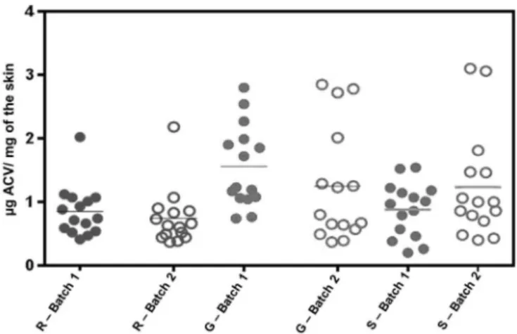
On the other hand, statistical evaluation of the second batch (Figure 4) has indicated the absence of a
signiicant diference (p>0.05) between formulations R,
G, and S. Dissimilar results regarding batches 1 and 2 for G product may indicate the poor homogeneity between manufactured batches. Semi solid pharmaceutical dosage forms are complex formulations and their physicochemical
release and penetration properties involve several factors. Initial separation of the system into two phases and the step of adding an active component are critical points in the semisolid fabrication process. Any change in one of these processes may alter the formulation properties, and the permeation and release characteristics of the active ingredient (OECD, 2004).
A statistical analysis performed between batches (for the same brand) pointed out the homogeneity of ACV
retained in the epidermis for formulation R, but a diferent
retention in the dermis. This also occurred for product G (epidermis: p = 0.539 and dermis: p = 0.009), while for product S, the retained amount in epidermis (p = 0.206) was considered equal comparing the two lots, as well as in dermis (p = 0.06).
As for the sum of ACV amounts retained in both
layers, statistical tests showed no signiicant diference
between the batches of pharmaceutical products (Figure 5). Statistical analysis of overall assessments (comparison
between two lots of three products) revealed a signiicant diference only between R product from the second batch and G product from the irst batch.
Whenever cutaneous permeation/penetration was evaluated (8 h), ACV was not detected in the receptor solution. According to Hasler-Nguyen et al.
(2009) ACV presents a hydrophobic region which can
interact with the hydrophobic structures in SC, afecting
drug permeation. On the other hands, dermis presents hydrophilic characteristics thereby representing a barrier against free transition of substances with high partition coefficient and limited water solubility. These results
conirm the release of ACV only in skin tissue (topically), which is the location required for efective treatment of
FIGURE 4 - Retained amount of ACV in porcine skin layers
regarding the second batch of reference (R), generic (G), and similar (S) products, expressed in µg/mg. Each symbol corresponds to an obtained value and the horizontal bar represents the mean concentration (n = 15).
cutaneous herpetic viruses, without achieving systemic
circulation and causing adverse efects.
It is worth noting that high values obtained for standard deviation during experiments performed can be explained by inherent variability by the inherent variability of studies with characteristics from cutaneous tissue (pig
skin) – interindividual diferences, growth, sex, and skin
thickness (Godin, Touitou, 2007). Those values might be related to skin layer weight which was fairly uniform: 22.2 ± 6.7 mg for epidermis and 92.0 ± 23.7 mg for dermis over ninety measurements (Table IV).
Recently, the national drug regulation agency of Brazil (ANVISA, 2014) declared that eleven topical brands of ACV were interchangeable, including the products employed in this study. In the USA, the FDA determines the optimal bioequivalence approach for each proposed generic topical formulation on a case-by-case basis. The approach may be pharmacokinetics, pharmacodynamics, clinical or in vitro considering drug mechanism and site of action, complexity of reference formulation, and feasibility and sensitivity of each approach. For ACV ointment 5%, generic and reference products must contain the same amounts of active and inactive ingredients and, as in the USA, the reference is considerably less complex than a cream, consisting of drug suspended in a polyethylene glycol (PEG) base. The
in vitro release approach (with inert membrane) can be used for equivalence purposes. Particle size, viscosity, morphic form and PEG molecular weight distribution are also required. No cutaneous permeation is mentioned in
the document. The products employed in this study are creams and ACV release through synthetic membrane was not evaluated (Davit, 2013).
Simulation of viral inhibitory effect on skin layers
When a pharmacodynamics study is performed, a direct relationship is expected between exposition and
efect, and thus higher drug exposition will result in an
increased viral inhibition response.
Results show that concentrations achieved for ACV in total skin, for all pharmaceutical specialities studied
and two batches analysed would present an antiviral efect
that is much superior to 50 % of inhibition. Therefore, studied products may be considered capable of providing ACV in cutaneous tissue in amounts that surpass IC50 viral
replication, even when there are strains with high IC50 values. Throughout the drug concentration range found in
the skin tissue, the efect calculated according to Equation
1 was greater than 98%. Therefore, medicines evaluable in the Brazilian market are believed to present an adequate
therapeutic efect against herpes virus, and are capable of
shortening the viral cycle and reducing the main symptoms (rash, burn, and redness).
CONCLUSION
The proposed analytical methodology is sensitive,
accurate and precise for the detection and quantiication of
ACV in skin layers of pig ears and therefore, in accordance
TABLE IV - Medium and Standard deviation (SD) from 15 determinations regarding the penetration study in porcine skin for the analyzed batch
Commercial Creams Batch Skin Layers* Medium (µg/mg)** Standard deviation – SD
(µg/mg)
R
1 E 0.60 0.34
D 0.25 0.10
2 E 0.62 0.42
D 0.12 0.06
G
1 E 1.09 0.49
D 0.47 0.20
2 E 0.94 0.76
D 0.30 0.36
S
1 E 0.70 0.37
D 0.18 0.08
2 E 0.97 0.73
D 0.26 0.15
with national and international standards. According to the studies performed the different composition with absence of propylene glycol in S product is not evidenced by means of an in vitro cutaneous penetration test and it can be assumed that the method possesses a limited
capability to distinguish some diferences in formulations
due to its inherent variability. The results obtained for the ACV amounts in the skin are in accordance with the interchangeability between products proposed by the national regulatory agency for medicines. Nevertheless, it may be important to evaluate drug skin absorption from semisolid formulations during the development phase, as well as to predict drug concentration in skin layers (dermis and epidermis).
REFERENCES
AGÊNCIA NACIONAL DE VIGILÂNCIA SANITÁRIA. ANVISA. Resolução RDC 58, de 10 de outubro de 2014. Dispõe sobre as medidas a serem adotadas junto à Anvisa pelos titulares de registro de medicamentos para a intercambialidade de medicamentos similares com o medicamento de referência. Diário Oficial da União, Brasília, Poder executivo, 2014.
AGÊNCIA NACIONAL DE VIGILÂNCIA SANITÁRIA. ANVISA. Resolução RE nº 27, de 17 de maio de 2012.
Diário Oicial da União, Brasília, 22 maio 2012.
ALBERTI, I.; KALIA, Y. N.; NAIK, A.; BONNY, J. D.; GUY, R. H. In vivo assessment of enhanced topical delivery of terbinaine to human stratum corneum. J. Control Rel., v.71, n.3, p.319-327, 2001.
BARRECA, M.L.; CHIMIRRI A., CLERCQ E.D., LUCA L.D., MONFORTE A.M.; MONFORTE P.; RAO A., ZAPPALÀ M. Anti-HIV agents: design and discovery of new potent RT inhibitors. Farmaco, v.58, n.3, p.259-263, 2003.
CAVALLI, R., DONALISIO, M., CIVRA, A., FERRUTI, P., RANUCCI, E., TROTTA, F., LEMBO, D. Enhanced antiviral activity of Acyclovir loaded into β-cyclodextrin-poly(4-cryloylmorpholine) conjugate nanoparticles. J. Control Rel., v.137, n.2, p.116-122, 2009.
DAVIT, B.M. Regulatory approaches for generic drugs: BE of topical drug products. In: PQRI workshop on the evaluation of new and generic topical drug products - current challenges in bioequivalence, quality and novel assessment technologies. Rockville, MD, 2013.
DE PAULA, D., MARTIN, C.A., BENTLEY, M.V.L.B. Development and validation of HPLC method for imiquimod determination in skin penetration studies. Biomed. Chromatogr., v.22, n.12, p.1416-1423, 2008.
DRUGBANK. Database. Acyclovir. Available at: <http://http:// www.drugbank.ca/drugs/DB00787>. Accessed at: 10 Oct. 2014.
FIELD, H. J.; HODGE, R. A. V. Recent developments in anti-herpesvirus drugs. Br. Med. Bull., v.106, n.1, p.213-249, 2013.
FOOD AND DRUG ADMINISTRATION. FDA. CENTER F O R D R U G E VA L U AT I O N A N D R E S E A R C H . CDER. Scale-Up and postapproval changes: chemistry, manufacturing, and Controls; in vitro release testing and in vivo bioequivalence documentation. Rockville, MD: FDA, Sep. 1997.
GABRIELSSON, J.; WEINER, D. Pharmacokinetic & pharmacodynamic data analysis: concepts and applications. 4. ed. Stockholm: Apotekarsocieteten, 2006.
GANNU, R.; PALEM, C.R.; YAMSANI, V.V.; YAMSANI, S.K.; YAMSANI, M.R. Enhanced bioavailability of lacidipine via microemulsion based transdermal gels: formulation optimization, ex vivo and in vivo characterization. Int. J. Pharm., v.388, n.1, p.231-241, 2010.
GODIN, B.; TOUITOU, E. Transdermal skin delivery: predictions for humans from in vivo, ex vivo and animal models. Adv. Drug Deliv. Rev., v.59, n.11, p.1152-1161, 2007.
GONG, Y.; RAJ, K.M.; LUSCOMBE, C.A.; GADAWSKI, I.; TAM, T.; CHU, J.; GIBSON, D; CARLSON, D; SACKS, S.L. The synergistic efects of betulin with acyclovir against herpes simplex viroses. Antivir. Res., v.64, n.2, p.127-130, 2004.
INTERNATIONAL CONFERENCE ON HARMONISATION. ICH. Harmonized tripartite guideline. Validation of analytical procedures: text and methodology Q2 (R1). Geneva, SW: ICH, 2005. v.1.
JALÓN, E.G.; BLANCO-PRÍETO, M.J.; YGARTUA, P.; SANTOYO, S. Increased efficacy of acyclovir-loaded microparticles against herpes simplex virus type 1 in cell culture. Eur. J. Pharm. Biopharm., v.56, n.2, p.183-187, 2003.
LUPI, O.; DA SILVA, A.G; PEREIRA Jr, A.C (Org). Herpes: clínica, diagnóstico e tratamento. Rio de Janeiro: Medsi, 2000.
MOSER, K.; KRIWET, K.; NAIK, A.; KALIA, Y.N.; GUY, R. H. Passive skin penetration enhancement and its quantiication in vitro. Eur. J. Pharm. Biopharm., v.52, n.2, p.103-112, 2001.
NOKHODCHI, A.; SHOKRI, J.; DASHBOLAGHI, A.; HASSAN-ZADEH, D.; GHAFOURIAN, T.; BARZEGAR-JALALI, M. The enhancement efect of surfactants on the penetration of lorazepam through rat skin. Int. J. Pharm., v. 250, p. 359-369, 2003.
ORGANIZATION FOR ECONOMIC COOPERATION AND DEVELOPMENT. OECD. Skin absorption: in vitro method, test guideline No 428. Guidelines for the testing of chemicals. Paris: OECD, v.1, n.4, p.1-8, 2004.
RAFEIRO, D.F.B. Novas estratégias de promoção de permeação transdérmica. 2013. Dissertação (Mestrado Integrado em Ciências Farmacêuticas) - Universidade Lusófona, Lisboa, 2013.
SARTORELLI, P.; ANDERSEN, H.R.; ANGERER, J.; CORISH, J.; DREXLER, H.; GÖEN, T.; GRIFFIN, P.; HOTCHKISS, S.A.M.; LARESE, F.; MONTOMOLI, L.; PERKINS, J.; SCHMELZ, M.; VAN DE SANDT, J.; WILLIAMS, F. Percutaneous penetration studies for risk assessment. Environ. Toxicol. Pharm., v.8, n.2, p.133-152, 2000.
SHAH, V.P., FLYNN, G.L., GUY, R.H., MAIBACH, H.I., SCHAEFER, H., SKELLY, J.P., WESTER, R. In vivo percutaneous penetration/absorption. Int. J. Pharm, v.74, n.8, p.1-8, 1991.
SUZUKI, M.; OKUDA, T.; SHIRAKI, K. Synergistic antiviral activity of acyclovir and vidarabine against herpes simplex virus types 1 and 2 and varicella-zoster vírus. Antivir. Res., v.72, p.157-161, 2006.
TROTTET, L., MERLY, C., MIRZA, M., HADGRAFT, J., DAVIS, A.F. Effect of finite doses of propylene glycol on enhancement of in vitro percutaneous permeation of loperamide hydrochloride. Int. J. Pharm, v.274, n.1, p.213-219, 2004.
TSAI, Y.H.; LEE, K.F.; HUANG, Y.B.; HUANG, C.T.; WU, P.C. In vitro permeation and in vivo whitening efect of topical hesperetin microemulsion delivery system. Int. J. Pharm., v.388, n.1, p.257-262, 2010.
WILLIAMS, A.C.; BARRY, B.W. Penetration enhancers. Adv. Drug Deliv. Rev., v.64. p.128-137, 2012.
WORLD HEALTH ORGANISATION. WHO. KIELHORN, J.; MELCHING-KOLLMUSS, S.; MANGELSDORF, I. Dermal Absorption. Environmental Health Criteria. Draft February 2006. Geneva, SW: WHO/IPCS, 2006. Available in: <http://www.inchem.org/documents/ehc/ehc/ehc235. pdf>. Accessed in: 8 mar. 2014.