Braz J Otorhinolaryngol. 2014;80(3):266-267
www.bjorl.org
Brazilian Journal of
OtOrhinOlaryngOlOgy
1808-8694/$ - see front matter © 2014 associação Brasileira de Otorrinolaringologia e Cirurgia Cérvico-Facial. Published by Elsevier Editora ltda. all rights reserved.
http://dx.doi.org/10.1016/j.bjorl.2013.01.001
CaSE rEPOrt
Mandibular mass as the primary manifestation of multiple myeloma
Tumor mandibular como manifestação primária do mieloma múltiplo
Kumar Pushpanshu
a,*, Silky Punyani
b, Rachna Kaushik
ca Department of Oral Medicine and Radiology, Dr BR Ambedkar Institute of Dental Sciences & Hospital, Patna, India b Department of Oral Medicine and Radiology, People’s Dental Academy, Bhopal, India
c Department of Periodontics, Dr BR Ambedkar Institute of Dental Sciences & Hospital, Patna, India
received 9 november 2012; accepted 3 January 2013
Please cite this article as: Pushpanshu K, Punyani S, Kaushik r. Mandibular mass as the primary manifestation of multiple myeloma. Braz J Otorhinolaryngol. 2014;80:266-7.
* Corresponding author.
E-mail: drpushpanshu@yahoo.co.in (K. Pushpanshu).
Introduction
Myelomas are plasma cell dyscrasias characterized by lym-phoid neoplastic proliferation of B cells, and the multiple myeloma (MM) is the most important symptomatic monoclo-nal gammopathy. it is characterized by numerous abnormal plasma cells permeating the bone marrow, and overproduc-tion of monoclonal light-chain or heavy-chain
immunoglobu-lins, that are identiiable in serum or urine.1 Because of its
tendency to widespread manifestations in multiple organs, this disease interests many medical specialists, including oral and maxillodental surgeons.2 MM manifestations in the
head and neck are common, but usually occur on the late stages of the disease; mandibular involvement as the initial presenting sign of the disease is extremely rare.3,4 the aim
of this article is to analyze this disorder, based on the pre-sentation of a 67-year-old woman with a painful mass in the mandible that prompted a MM diagnosis.
Case presentation
a 67-year-old woman presented a painful and progressively en-larging mass on the right posterior area of lower jaw, which had started approximately one month previously. She experienced dull and intermittent pain on the right side of lower jaw and
dificulty in eating. Physical examination showed swelling on
the same side of the mandible. her medical history indicated that she experienced fatigue for the last three months. intra-oral examination revealed a localized soft-tissue mass with central ulceration in right mandibular posterior region. Pa-noramic radiography showed a large radiolucent lesion of the right mandibular ramus together with multiple smaller
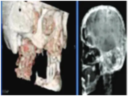
“pun-ched-out” lesions. Results of the contrast CT scan conirmed
the existence of a large osteolytic lesion in the right mandi-bular ramus (Fig. 1a). histopathological examination following incisional biopsy of the soft-tissue mass revealed sheets of plasma cells showing nuclear pleomorphism and lymphocytes, suggestive of plasmacytoma. a whole-body bone scan and a sys-temic skeletal radiographic survey revealed multiple “punche-d-out” osteolytic lesions involving the skull (Fig. 1B), clavicles
Mandibular mass as the primary manifestation of multiple myeloma 267
and pelvis. Urinalysis results were positive for Bence Jones
proteins. Multiple myeloma was conirmed, and the patient
was admitted to receive chemotherapy with proteasome-i-nhibitor bortezomib in combination with melphalan and prednisone, and to undergo local radiotherapy for the man-dibular mass.
Discussion
Oral lesions are seen with some frequency (30%) in patients with MM; however, when oral lesions are noted, the disease is usually in an advanced stage. Oral manifestations inclu-de jaw pain, tooth pain, paresthesia, swelling, soft tissue mass, mobility of teeth, migration of teeth, hemorrhage, and pathologic fracture.2 MM have a high potential to
pro-duce local and systemic perturbation of hemostasis and distinct radiographic bone alterations. Undiagnosed MM can impose a formidable emergency condition in dental practi-ce.5 thorough radiographic examination is critically impor-tant because it can be pathognomonic and may be the irst
sign of the disease.2 the presence of multiple punched-out
lesions on the jaw, and the skull bone is also observed in diseases such as langerhans’ cell histiocytosis, and metas-tatic malignant lesions.5 MM affecting the jaw bones has
a characteristic distribution, more frequently found in the mandible, especially in the posterior region, where hemato-poietic activity is greater.4
Final remarks
awareness of the maxillofacial manifestations of MM is important for early detection of the disease, which may
otherwise lead to delays and even errors in diagnosis and treatment. Being a rare disease, MM should not be kept at the forefront of the differential diagnosis of jaw lesions. rather, we hope the present report will serve to remind us that oral lesions may be the presenting sign of this rare pathology.
Conlicts of interest
The authors declare no conlicts of interest.
References
1. Pinto lS, Campagnoli EB, leon JE, lopes Ma, Jorge J.
Ma-xillary lesion presenting as a irst sign of multiple myeloma:
case report. Med Oral Patol Oral Cir Bucal. 2007;12:E344-7. 2. Pisano JJ, Coupland r, Chen Sy, Miller aS. Plasmacytoma
of the oral cavity and jaws: a clinicopathologic study of 13 cases. Oral Surg Oral Med Oral Pathol Oral radiol Endod. 1997;83:265-71.
3. Elias hg, Scott J, Metheny l, Quereshy Fa. Multiple myeloma
presenting as mandibular ill-deined radiolucent lesion with
numb chin syndrome: a case report. J Oral Maxillofac Surg. 2009;67:1991-6.
4. lee Sh, huang JJ, Pan Wl, Chan CP. gingival mass as the primary manifestation of multiple myeloma: report of two cases. Oral Surg Oral Med Oral Pathol Oral radiol Endod. 1996;82:75-9.