1879
M
EDICAL
J
OURNAL
SÃO PAULO
C
ASE
R
EPORT
Fonseca SF, Figueiredo MS, Cançado RD, Nakandakare F, Segreto R, Kerbauy J. Spinal cord compression in b-thalassemia: follow-up after radiotherapy.
Rev Paul Med 1998;116(6):1879-81
Spinal cord compression in
b
b
b
b
b
-thalassemia:
follow-up after radiotherapy
Silvana Fahel da Fonseca, Maria Stella Figueiredo, Rodolfo Delfini Cançado, Fernando Nakandakare, Roberto Segreto, José Kerbauy
Address for correspondence:
Maria Stella Figueiredo
Disciplina de Hematologia e Hemoterapia UNIFESP - EPM
Rua Botucatu, 740
São Paulo/SP - Brasil - CEP 04023-900 e-mail: [email protected]
Context: Spinal cord compression due to extramedullary hematopoiesis is a well-described but rare syndrome encountered in several clinical hematologic disorders, includingb-thalassemia. Case Report: We report the case of a patient with intermediate
b-thalassemia and crural paraparesis due to spinal cord compression by a paravertebral extramedullary mass. She was successfully treated with low-dose radiotherapy and transfusions. After splenectomy, she was regularly followed up for over four years without transfusion or recurrence of spinal cord compression. Discussion: Extramedullary hematopoiesis should be investigated in patients with hematologic disorders and spinal cord symptoms. The rapid recognition and treatment with radiotherapy can dramatically alleviate symptoms.
Uniterms: Extramedullary hematopoiesis. Spinal cord compression. Intermediate b-thalassemia.
INTRODUCTION
xtramedullary hematopoiesis (EMH) is a compensatory phenomenon that occurs in patients with hematological disorders when bone marrow function is not sufficient to maintain the circulatory demand. It has been seen in different types of severe anemia, such as polycytemia, leukemia and lymphoma, and after bone marrow irradiation, poisoning or neoplastic conditions.1
The most common sites of EMH are organs that have physiological hematopoiesis during embryonic life, especially the liver and spleen. Other sites of diffuse compensatory EMH include lymphonodes, adrenal glands, kidneys, breast, dura mater, adipose tissue and skin.2
Spinal EMH sufficiently severe to cause spinal cord compression rarely occurs in thalassemic patients and the
E
management of these patients remains controversial.3 We
report on a spinal cord compression due to EMH in a patient with intermediate b-thalassemia and the
therapeutic results obtained.
CASE REPORT
We report on a 20-year-old female with intermediate b-thalassemia that was asymptomatic and
had stable hemoglobin (Hb) at about 8.0 g/dl. The patient’s neurological manifestations had started some four months before this admission and her first symptom was intermittent leg pain, especially in the left leg, followed by a progressive decrease in sensitivity affecting both legs. During the three months prior to admission, she evolved progressive difficulty in walking and climbing stairs, and at admission, she was not able to walk or control sphincter.
1880
Fonseca SF, Figueiredo MS, Cançado RD, Nakandakare F, Segreto R, Kerbauy J. Spinal cord compression in b-thalassemia: follow-up after radiotherapy.
Rev Paul Med 1998;116(6):1879-81
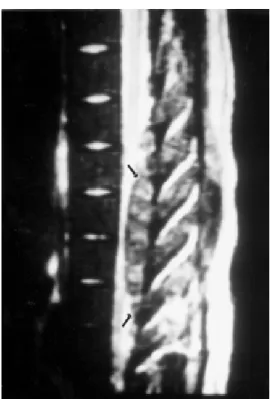
vibratory sense was diminished bilaterally to T9-T10, and there was hypoesthesia to touch and pinprick with a T9-T10 sensory level on both sides. Motor strength was decreased on the lower extremities, symmetrically. Tendon reflexes were hypoactive and symmetrical. Laboratory analysis showed Hb = 8.5 g/dl; hematocrit = 29% and reticulocyte count = 9%. Peripheral blood smear showed microcytosis, hypochromia, anisocytosis, poikilocytosis, and erythroblasts = 10%. Hemoglobin electrophoresis: Hb F = 81.5%; Hb A2 = 3.2% and Hb A = 15.3%. These were consistent with a diagnosis of intermediate b-thalassemia. Computerized tomography (CT) and magnetic resonance imaging (MRI) (Fig. 1) revealed a large posterior paravertebral lesion of soft tissue mass, with invasion of the epidural space of the spinal canal, extending from T4 through T8, with approximately 10 cm of longitudinal extension.
Radiation therapy (RT) was initiated immediately as an emergency measure. She received local RT totalling 1500 cGy between T4 and T8 using a linear accelerator. She was also transfused in order to mantain her Hb level greater than 9.5 g/dl. After RT, a significant clinical improvement was observed with no neurologic abnormalities, and this condition has been maintained until now. A follow-up MRI, two months after RT, demonstrated complete remission of cord compression, and marked decreased in the paraspinal mass (Fig. 2). There were no side effects during this treatment.
Splenectomy was performed one year after RT, and since then she has maintained a stable condition, with no need for blood transfusion. The patient has remained well for over four years without recurrence of spinal cord compression.
DISCUSSION
The first description of spinal cord compression by EMH dates from 1954.4 Since then, about 60 cases have
been reported, most of them in intermediate b-thalassemia
patients.5 This complication has mainly been observed in
the thoracic segment of the spinal cord but the reason for this predilection remains uncertain.6
The development of hematopoietic tissue in the vertebral canal is probably due to a bone marrow expansion leading to spinal cord compression. On CT and MRI, the EMH looks like a well-delimited lobular soft tissue with no erosion of the adjacent bone structures. Management strategies have included RT, laminectomy, and transfusion therapy.5,6 Spontaneous recovery with no therapeutic
intervention has also been reported, but may take several
months to occur and is subject to frequent recurrence.6
Patients treated only with transfusion have initially showed improvement, but with frequent recurrence.3,6
Figure 1 - Sagittal magnetic resonance imaging, at diagnosis, demonstrating posterior epidural mass with compression of the spinal cord from T4 through T8 (arrows).
1881
RT has been reported to yield excellent results with prompt neurological response since the hematopoietic tissue is radiosensitive and relatively small doses of radiation are needed. Complete recovery is achieved in as short a time as 3 to 7 days. Published reports and our own data support this conclusion.
Patients suffering from hematologic disorders with back pain and spinal cord symptoms should have EMH included in the differential diagnoses. MRI appears to be ideal for diagnosing and delineating the extent of the intraspinal masses. Prompt recognition is vital because RT can dramatically alleviate symptoms.2,6
R
ESUMO
Contexto: Compressão medular, secundária à hematopoese extramedular, é uma manifestação bastante descrita mas rara, encontrada em várias doenças hematológicas, incluindo a b-talassemia. Relato de caso: Nós relatamos o caso de uma
paciente com b-talassemia intermediária que apresentou paraparesia crural devido a compressão medular por uma massa extramedular paravertebral. Ela foi tratada com baixas doses de radioterapia e transfusões, com sucesso. Após esplenectomia, a paciente tem sido acompanhada por mais de quatro anos, sem apresentar necessidade de transfusão ou recurrência do quadro de compressão medular. Discussão: Hematopoese extramedular deve ser investigada em pacientes com doenças hematológicas e sintomas de compressão medular. O pronto reconhecimento e tratamento radioterápico pode aliviar a sintomatologia rapidamente.
REFERENCES
1. Gemenis T, Philippou A, Gouliamos A, et al. Atypical location of extramedullary hematopoietic masses in thalassemia. Radiologe 1989;29:295-6.
2. Konstantopoulos K, Vagiopoulos G, Kantouni R, et al. A case of spinal cord compression by extramedullary haematopoiesis in a thalassaemic patient: a putative role for hydroxyurea? Haematologica 1992;77:352-4.
3. Singounas EG, Sakas DE, Hadley DM, et al. Paraplegia in a pregnant thalassemic woman due to extramedullary hematopoiesis: successful management with transfusions. Surg Neurol 1991;36:210-5.
4. Gatto I, Terrana V, Biondi L. Compressione submidollo spinale da proliferazions di midollo osseo nello spazio epidurale in sogetts affeto da malattia di Colley splenectomizzato. Haematologica 1954;38:61-76. 5. Coraddu M, Floris F, Meleddu V, et al. Compression
médullaire et de la queue de cheval chez deux patients atteints de béta-thalassémie intermédiaire. Neurochirurgie 1991;37:58-60.
6. Kaufmann T, Coleman M, Giardina P, Nisce LZ. The role of radiation therapy in the management of hematopoietic neurologic complications in thalassemia. Acta Haematol. 1991;85:156-9.
Fonseca SF, Figueiredo MS, Cançado RD, Nakandakare F, Segreto R, Kerbauy J. Spinal cord compression in b-thalassemia: follow-up after radiotherapy.