406
CLINICS 2008;63:406-8
LETTER TO THE EDITOR
I Craniofacial Unit, Faculdade de Medicina da Universidade de São Paulo
– São Paulo/SP, Brazil.
II Neurosurgery Department, Faculdade de Medicina da Universidade de
São Paulo – São Paulo/SP, Brazil. [email protected]
ENCEPHALOCRANIOCUTANEOUS LIPOMATOSIS
(HABERLAND’S SYNDROME) - A CASE REPORT OF
A NEUROCUTANEOUS SYNDROME AND A REVIEW
OF THE LITERATURE
doi: 10.1590/S1807-59322008000300020
Giovanna Negrisoli Koishi,I Mauricio Yoshida,I Nivaldo Alonso,I Hamilton Matushita,II Dov GoldenbergI
INTRODUCTION
Encephalocraniocutaneous Lipomatosis (ECCL) is a rare neurocutaneous syndrome first described by Haberland and Perou in 1970.1 It is characterized by unilateral cutaneous, ocular, and neurologic malformations. There are 53 cases described in the literature, but only four of these are in Brazil.2-10
CASE REPORT
We present the case of a full-term girl born to non-consanguineous parents. Physical examination revealed a large (14 x 6 centimeters) hairless lesion on the right frontal-parietal scalp (Fig.1), multiple nodular lesions of the right upper eyelid and eyebrow, and a reddish bulbar conjunctival lesion on the right eye consistent with choriocystoma (Fig.2).
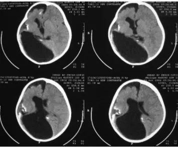
A CT scan of the brain showed cranial asymmetry, an arachnoid cyst of the right middle fossa, a right frontal subdural collection, a porencephalic cyst, hemiatrophy of the right hemisphere, and cortical calcifications of the parietal and occipital lobes (Fig.3).
At the age of two years, the patient underwent a complete excision of the scalp lesion after tissue expansion and excision of the nodules on the eyelid (Fig.4). The histopathological examination of the scalp lesion showed absence of hair follicles, a thinned dermis, and extended adipose tissue into the dermis; the cutaneous nodules were
Figure 1 - The hairless lesion (“naevus psiloliparus”)
407
CLINICS 2008;63:406-8 Encephalocraniocutaneous lipomatosis (Haberland’s Syndrome)
Koishi GN et al.
consistent with lipoma. In the four year follow-up period, the patient was noted to have mental retardation and seizures that were controlled with medication and neurological treatment (Fig.5).
DISCUSSION
ECCL is a rare, sporadic, neurocutaneous syndrome with no predominant gender, racial, or geographical association.11,12 The genetic mechanism has been hypothesized to involve lethal autosomal dominant genes
Figure 4 - Tissue expansion for surgical excision of the “naevus
psiloliparus”
Figure 5 - Post-operative results
Figure 3 - CT scan of the brain shows: porencephalic cyst, hemiatrophy of
the right hemisphere, subdural collection and parietal calcifications
that survive by mosaicism, and the pathogenesis is most likely a dysgenesis of the cephalic neural crest and anterior neural tube.3,11,13
Clinically, ECCL is characterized by unilateral abnormalities of the brain, eyes, and skin. The most common neurological findings are hemiatrophy, dilated ventricles, porencephalic cysts, abnormal calcifications, intracranial lipoma, and cranial asymmetry. Most patients present with seizures and mental retardation.3,9,11,12 The hairless lesion of the scalp (“naevus psiloliparus”) is pathognomonic, and the papular lesions of the eyelid, consistent with lipomas, are the most frequent feature in all cases reported. However, other lesions have also been reported, such as lipomas of the vertebral spine, odontom, and “café-au-lait” spots.3,14
408
CLINICS 2008;63:406-8 Encephalocraniocutaneous lipomatosis (Haberland’s Syndrome)
Koishi GN et al.
REFERENCES
1. Habeland C, Perou M. Encephalocraniocutaneous lipomatosis: a new example of ectodermal dysgenesis. Arch Neurol. 1970;22:144-55. 2. Hauber K, Warmuth-Metz M, Rose C, Bröcker E, Hamm H.
Encephalocraniocutaneous lipomatosis: a case with unilateral odontom as and review of the literature. Eur J Pediatr. 2003;162:589-93. 3. Gokhale NR, Mahajan PM, Belgaumkar VA, Pradhan SN, Uttarwar
NS. Encephalocraniocutaneous lipomatosis: a rare neurocutaneous syndrome. Indian J Dermatol Venereol Leprol. 2007;73:40-2. 4. Valladares MJ, Blanco MJ, Lopez-Lopez F, Gonzalez F. Bilateral ocular
involvement in encephalocraniocutaneous lipomatosis. Eur J Pediatr Neurol. 2007;11:108-10.
5. Rathoryia R, Shrivastava J. Encephalocraniocutaneous lipomatosis. Indian Pediatr 2006;43:262-3.
6. Sofiatti A, Cirto AG, Armone M, Romiti R, Santi C, Leite C, et al. Encephalocraniocutaneous lipomatosis: clinical spectrum of systemic involvement. Pediatr Dermatol 2006;23:27-30.
7. Donaire A, Carreno M, BargalloN, Setoain X, Agudo R, Martin G, et al. Presurgical evaluation and cognitive functional reorganization in Fishman syndrome. Epilepsy Behav. 2005;6:440-3.
8. Cultrera F, Guarnera F, Giardina MC. Overlap among neurocutaneous syndromes. Observations on encephalocraniocutaneous lipomatosis. Minerva Pediatr. 2004;56:219-22.
9. Lasierra R, Valencia I, Carapeto FJ, Ventura P, Samper MP, Rodriguez G, et al. Encephalocraniocutaneous lipomatosis: neurologic manifestation. J Child Neurol. 2003;18:725-9.
10. Guazzelli UF, Rufatto LA, Tavares AJ,Gehrke R. Encephalocraniocutaneous lipomatosis (Haberland syndrome). Rev Soc Bras Cir Craniomaxilofac. 2005;1:24.
11. Cruz AA, Schirmbeck T, Pina-Neto JM, Funayama CA. Cicatricial upper eyelid retraction in encephalocraniocutaneous lipomatosis: a rep[ort of two cases and review of literature. Ophthal Plast Reconstr Surg. 2002;18:151-5.
12. Almer Z, Vishnevskia-Dai V, Zadok D. Encephalocraniocutaneous lipomatosis: case report and review of the literature. Cornea. 2003;22(4): 389-90.
13. Happle R. Lethal gene surviving by mosaicism: a possible explanation for esporadic birth defects involving the skin. J Am Acad Dermatol. 1987;16:899-906.
14. Rubegni P, Risulo M, Sbano P, Buonocore G, Perrone S, Fimiani M. Encephalocraniocutaneous lipomatosis (Haberland syndrome) with bilateral cutaneous and viceral involvement. Clin Exp Dermatol. 2003;28:387-90.