www.bjorl.org
Brazilian
Journal
of
OTORHINOLARYNGOLOGY
ORIGINAL
ARTICLE
Intrasphenoid
septations
inserted
into
the
internal
carotid
arteries:
a
frequent
and
risky
relationship
in
transsphenoidal
surgeries
夽
Clauder
Oliveira
Ramalho
a,b,∗,
Horacio
Armando
Marenco
a,b,
Francisco
de
Assis
Vaz
Guimarães
Filho
c,
Marcos
Devanir
Silva
da
Costa
a,
Bruno
Fernandes
de
Oliveira
Santos
a,
Rodrigo
de
Paula
Santos
d,
Samuel
Tau
Zymberg
a,baUniversidadeFederaldeSãoPaulo(UNIFESP),DepartamentodeNeurocirurgia,SãoPaulo,SP,Brazil
bUniversidadeFederaldeSãoPaulo(UNIFESP),ProgramadePós-graduac¸ãodoDepartamentodeOtorrinolaringologiaeCirurgia
deCabec¸aePescoc¸o,SãoPaulo,SP,Brazil
cUniversidadeFederaldeSãoPaulo(UNIFESP),SãoPaulo,SP,Brazil
dUniversidadeFederaldeSãoPaulo(UNIFESP),DepartamentodeOtorrinolaringologia,SãoPaulo,SP,Brazil
Received7January2016;accepted19February2016 Availableonline22April2016
KEYWORDS
Sphenoidsinus; Sphenoidseptations; Skullbase;
Transsphenoidal surgery;
Expandedendonasal approach
Abstract
Introduction:Whenanexpandedendonasaltranssphenoidalsurgicalapproach isperformed, intrasphenoidseptations must be completely resected.If these structures areclose tothe internalcarotidartery(ICA),thentheirmanipulationmightcausevascularinjury.
Objective:Theobjectiveofthisstudyistodescribethefrequencyofintrasphenoidseptations intheinternalcarotidarteryprotuberance(ICAp).
Methods:Computedtomography(CT)scansof421patientswereanalysed.Intrasphenoid septa-tions(classifiedasintersphenoidoraccessory)andtheirrelationshiptotheICApweredescribed. Additionally,asphenoidsinusclassificationwasperformedbasedontheirdegreeof pneuma-tisationtodeterminewhetheradifferenceexistsinthefrequencyofintrasphenoidseptations insertedintoICApwithregardtosinustype.
Results:Thepatientmeanagewas39±21.4years.Overall,219patients(52%)hadseptations intheICAp;359patients(85.3%)hadintersphenoidseptations;ofthelatter,135(37.6%)had septationsintheICAp.Thisfrequencywashigheramongpatientswithsphenoidsinustype4or 5(44.7%and43.5%,respectively).Accessoryseptationswerefoundin255patients(60.6%);140 oftheseseptations(54.9%)wereintheICAp.Among351patientswithtypes3,4or5sphenoid
夽 Pleasecitethisarticleas:RamalhoCO,MarencoHA,GuimarãesFilhoFA,daCostaMD,deOliveiraSantosBF,dePaulaSantosR,etal.
Intrasphenoidseptationsinsertedintotheinternalcarotidarteries:afrequentandriskyrelationshipintranssphenoidalsurgeries.BrazJ Otorhinolaryngol.2017;83:162---7.
∗Correspondingauthor.
E-mail:[email protected](C.O.Ramalho).
PeerReviewundertheresponsibilityofAssociac¸ãoBrasileiradeOtorrinolaringologiaeCirurgiaCérvico-Facial.
http://dx.doi.org/10.1016/j.bjorl.2016.02.007
sinuses(i.e.,onlywell-pneumatisedsphenoidsinuses),219(62.4%)hadseptationsintheICAp. Thesefrequenciesarehigherthanthosereportedinmostpreviousstudies.
Conclusion: Thefrequency ofintrasphenoid septationsinthe ICApfoundis considerable.It ishigheramongpatientswithmorepneumatisedsinuses.Thisfindingjustifiesanappropriate pre-operativestudy,andcarefulattentionmustbepaidduringtranssphenoidalsurgery. © 2016 Associac¸˜ao Brasileira de Otorrinolaringologia e Cirurgia C´ervico-Facial. Published by Elsevier Editora Ltda. This is an open access article under the CC BY license (http:// creativecommons.org/licenses/by/4.0/).
PALAVRAS-CHAVE
Seioesfenoidal; Septac¸ões esfenoidais; Basedocrânio; Cirurgia transesfenoidal; Abordagemendonasal ampliada
Septac¸õesintraesfenoidaisinseridasnasartériascarotídeasinternas:umarelac¸ão frequenteearriscadanascirurgiastransesfenoidais
Resumo
Introduc¸ão: Quandoumaabordagemcirúrgicatransesfenoidalendonasalampliadaérealizada, septac¸õesintraesfenoidaisdevemsercompletamenteressecadas.Seestasestruturasestiverem próximasàartériacarótidainterna(ACI),amanipulac¸ãopodecausarlesãovascular.
Objetivo: Oobjetivodesteestudofoidescreverafrequênciadeseptac¸õesintraesfenoidaisna protuberânciadaartériacarótidainterna(pACI).
Método: Examesdetomografiacomputadorizada(TC)de421pacientesforamanalisados.As septac¸ões intraesfenoidais (classificadas como interesfenoidais ouacessórias) e suarelac¸ão comapACIforamdescritas.Alémdisso,umaclassificac¸ãodoseioesfenoidalfoirealizadacom basenoseugraudepneumatizac¸ãoparadeterminarseexisteumadiferenc¸anafrequênciade septac¸õesintraesfenoidaisinseridasempACIemrelac¸ãoaotipodeseio.
Resultados: Pacientes com idade média de 39±21,4 anos foram incluídos. No geral, 219 pacientes (52%) apresentavam septac¸ões na pACI; 359 (85,3%) tinham septac¸ões interes-fenoidais; 135(37,6%)comseptac¸õesnapACI. Estafrequênciafoimaiorentreospacientes comseioesfenoidaltipo4ou5(44,7e43,5%,respectivamente).Asseptac¸õesacessóriasforam encontradas em 255doentes (60,6%);140 dessasseptac¸ões(54,9%)estavam napACI. Entre 351 pacientescom seiosesfenoidaistipos 3, 4ou 5(isto é,apenas seios esfenoidais bem-pneumatizados),219 (62,4%)tinhamseptac¸ões napACI.Estasfrequências sãosuperiores às relatadasnamaioriadosestudos.
Conclusão:A frequência de septac¸ões intraesfenoidais na pACI encontrada é considerável, sendomaiorentrepacientescomseiosmaispneumatizados.Esteachadojustificaumestudo pré-operatório adequado euma atenc¸ãoespecial deve serdada duranteacirurgia transes-fenoidal.
© 2016 Associac¸˜ao Brasileira de Otorrinolaringologia e Cirurgia C´ervico-Facial. Publicado por Elsevier Editora Ltda. Este ´e um artigo Open Access sob uma licenc¸a CC BY (http:// creativecommons.org/licenses/by/4.0/).
Introduction
Transnasal transsphenoidal surgery has developed
sig-nificantly over recent decades. The cooperative work
betweenneurosurgeonsandear,neckandthroatsurgeons
has been essential for this development. The
introduc-tion of the endoscope was another landmark. Compared
with the microscope, the endoscope enabled additional
expansion of this surgical technique, thereby increasing
the possibility of resecting lesions not otherwise eligible
for transnasal transsphenoidal surgery.1 With the
emer-gence of the endoscopic expanded endonasal approach,
areas such as the clivus, the petrous bone, the
mid-dle cranial fossa and the infratemporal fossa became
accessible.2 An extensive sphenoidotomy with septation
resection is necessary to create an adequate surgical
corridor.3
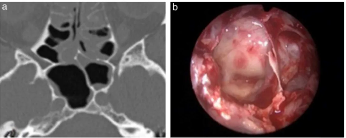
Intrasphenoid septations are bony structures found in
thesphenoidsinuswithseveralanatomicalconformations.
Because they are located in the sinus walls, they are
often adjacent to surrounding structures, especially the
internal carotid artery (ICA), which can increase the risk
of expanded transsphenoidal surgeries during septation
resection(Fig.1).
ICA injury is one of the most dramatic intraoperative
complications. This injury can lead to a challenging
sur-gical scenario featuring rapid blood loss that can result
in patient exsanguination.4 An appropriate pre-operative
radiologicevaluationofthesphenoidsinusanditsseptations
isnecessarytopreventthiscomplication.
Previousarticleshavedescribedthefrequencyof
intras-phenoidseptationsinthe ICAprotuberance(ICAp).1---5 The
majorityof thesearticleshave found fewerintrasphenoid
Figure1 (a)CTscanaxialview ofanintersphenoidseptationintheleft internalcarotid arteryprotuberance(ICAp)and(b) intraoperativeview(0◦endoscope)showingthesameseptationintheleftICAp.
currentarticledescribesthefrequencyofintrasphenoid
sep-tationsintheICApamongasampleof421patientsanalysed
usingcomputedtomography(CT)scansandcomparesthese
findingswiththoseofpreviousstudies.
Methods
Sampleandselectioncriteria
We searched the database of the department of
radiol-ogy of a hospital institution from January 2010 to April
2013 for patients who underwent CT scans of the skull
base. Individuals with a previous history of paranasal
sinusdisease orendonasal surgerywereexcluded. Atotal
of 421 patients were selected. Informed consent was
obtained from all individual participants included in the
study.
AllpatientsunderwentaCTscanwithskullbasesections
usingtheBrillianceCT64system(Philips,2004).The scan
wasperformedwith20×0.625collimation,apitchof0.348,
amatrixof512;200mmoffieldofview.Sectionthickness
ranged from0.6 to 1mm. The obtained data were
trans-ferredtotheExtendedBrillianceWorkspace(PhilipsMedical
System),wheretheimageswerereconstructedintheaxial,
coronal,andsagittalplanes.
Septationtypedefinitionanditsrelationshipwith ICAp
Intrasphenoid septations were classified as intersphenoid
whenthey(1) werelongitudinal andina medianor
para-median location and (2) separated the cavity into two
non-communicatingcompartmentsfromtheanteriortothe
posteriorsinuswall.Aseptationwasdefinedasanaccessory
whenitdidnotfollowalloftheintersphenoidsinuspatterns
(Fig.2).
Toconsiderthetruerelationshipbetweenseptationand
ICAp, aCTsection hadtoclearlyshowa septationin this
structure(Fig.2).
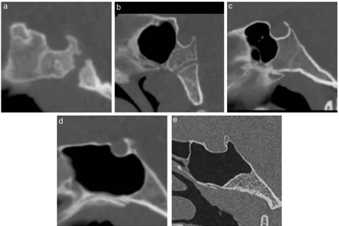
Sinusclassification
Sphenoidsinuses were classifiedbased ontheir degree of
pneumatisation,whichwasestablishedbythespatial
rela-tionship of the sinus posterior wall and the anterior and
posteriorwallsofthesellaturcica.Sinuseswereclassifiedas
follows:thosewithanabsenceofaerationorminimal
aera-tionwereclassifiedastype1;thosewiththeirposteriorwall
inapositionrostraltothesellaanteriorwallwereclassified
astype2;thosewiththeirposteriorwallbetweenthesella
anteriorandposteriorwallswereclassifiedastype3;those
withtheirposteriorwallreachingtheposteriorwallofthe
Figure3 Sinusclassification.MidsagittalreformattedimagesobtainedfromCTscans.(a---e)Types1through5sphenoidsinuses, respectively.
sellawereclassifiedastype4;andthosewithposterior
cli-noidaerationwereclassifiedastype5.Thepurposeofthis
analysiswastoassesswhetherthefrequencyofseptationin
theICApdifferedacrosspatientswithdisparatesinustypes
(Fig.3).
Statisticalanalyses
Categorical variableswere described bynumbers of cases
and percentages. Groups were comparedusing the z-test
for proportions and either the chi-square test or Fisher’s
exacttest,asappropriate.Continuousvariableswere
char-acterised as either the mean±standard deviation or the
median and interquartile range depending on normality;
between-group comparisonswere madeusingStudent’s t
-test or the Kruskal---Wallis test, respectively. A resultwas
considered significant when p<0.05. Statistical analyses
wereconductedwiththeSPSS17(Chicago,IL,USA).
Researchprotocol
Theethicscommitteeapprovedtheresearchprotocol
(doc-umentnumber186.717).
Results
Weidentified189maleand232femalepatients(meanage:
39±21.4years).Themostfrequenttypeofsinuswastype
4(61%ofcases)(Table1).
Atotalof359patients(85.3%)hadintersphenoid
septa-tions.Outoftheseseptations,135(37.6%)werefoundinthe
ICAp.Totalsof44.7%and43.5%ofpatientswithtype4and
5sinuseshadintersphenoidseptationsintheICAp,
respec-tively;only14.1%ofpatientswithtype3sinuseshadthese
septationsintheICAp.Patientswithtypes1or2sinusesdid
nothaveICAp-adjacentseptations.
Accessoryseptationswerefoundin255patients(60.6%).
These septations were present in only one-quarter of
patientswith type2 sinuses, whereasmost of those with
types 3, 4 or 5 sinuses had septations. Septations were
located in the ICAp of 140 patients (54.9% of those with
accessoryseptations;25.4%,42.8% and52.2% for types3,
4and5,respectively).Themaximumnumbersfoundwere
2,3,5,and4fortypes2,3,4,and5,respectively,whereas
themaximum numbersfoundintheICApwere2,3,and3
fortypes3,4,and5,respectively.
The number of patients with septation in the ICAp,
regardless of type, was 219 (52%). Those patients were
older than those without septations (43±18 vs. 34±23;
p<0.0001); no significant difference was observed with
regardtosex(femalescomprised 55%of allcasesin both
groups, p=0.963). Patients with types 4 and 5 sphenoid
sinusesweremorecommonthanthosewithtype3(Table2).
Ofthe351patientswithtype3,4or5sphenoidsinuses,
219 (62.4%) had septations in the ICAp. A total of 322
patients (91.7%) had intersphenoid septations; of these
patients,135(41.9%)hadseptationsintheICAp.Ofthe244
patientswithatleastoneaccessoryseptation,140(57.4%)
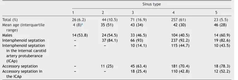
Table1 Intersphenoidandaccessoryseptationsbysinustype.
Sinustype
1 2 3 4 5
Total(%) 26(6.2) 44(10.5) 71(16.9) 257(61) 23(5.5)
Meanage(interquartile range)
4(8)a 35(51) 43(34) 42(30) 46(28)
Males 14(53.8) 24(54.5) 33(46.5) 104(40.5) 14(60.9)
Intersphenoidseptation --- 37(84.1) 66(93) 237(92.2) 19(82.6)
Intersphenoidseptation intheinternalcarotid arteryprotuberance (ICAp)
--- --- 10(14.1) 115(44.7) 10(43.5)
Accessoryseptation --- 11(25) 45(63.4) 181(70.4) 18(78.3)
Accessoryseptationin theICAp
--- --- 18(25.4) 110(42.8) 12(52.2)
aMediansignificantlydiffersfromtheothers(p<0.05).
Table2 Sphenoidseptationintheinternalcarotidartery protuberancebysinustype.
Total %
Type1 ---
---Type2 ---
---Type3 23 32.4% a
Type4 179 69.6% b
Type5 17 73.9% b
Proportionsidentifiedwithdifferentlettersaresignificantly dif-ferentaccordingtothez-testforproportions(p<0.05).
Discussion
Theexpandedendonasaltranssphenoidalapproachmarked a breakthrough in skull base surgery. With its develop-ment,lesionspreviouslyinaccessibleusingtheconventional endonasal route (e.g., those in the cavernous sinus, the planum sphenoidale, the middle cranial fossa, Meckel’s cave, the suprasellar region and the clivus) could be accessed.2 To obtain appropriate exposure and
accommo-datethesurgicalendoscopicinstruments,awidesphenoid
sinus opening is required with intrasphenoid septation
resection.3
Intrasphenoid septations are naturally occurring bony
structures inside the sphenoid sinus that divide it into
compartments. They are divided into intersphenoid and
accessory septations. The association with fusion lines
between ossification centres(synchondrosis)and the
sep-tationpositionsmightexplaintheirorigin.6,7Ingeneral,one
or more intersphenoidseptations are present. They show
greatvariability;therefore,theytypicallycreatetwo
asym-metriccompartments:rightandleft.Accessoryseptations
occur in different positions and are also common. Both
canbefound instructuresadjacent tothesphenoidsinus
increasingtheriskofneurovasculardamageduringsurgery,
especiallywhentheyarelocated intheICAp. Cope
previ-ouslydescribedthiscomplicationin1917.6
Inourstudy,CTscansrevealedthat 219patients(52%)
hadseptationsintheICAp.Amongpatientswithtype3,4,
or5sinuses(i.e.,well-pneumatisedsphenoidsinuses),this
prevalencewasevenhigher(62.4%).
Ourdatacontrastwiththoseofpreviouspapersshowinga
smallerprevalence.5,8---12However,Fernandez-Mirandaetal.
showedradiologicprevalenceof85%amongpatientswithat
leastoneseptationintheICAp.3
RennandRhotonfoundintersphenoidseptationsnextto
theICAchannelin32%ofcadavers.5Sethidescribed
inter-sphenoid septations in the ICAp in 40% of 30 cadavers in
an endoscopic study in 1995.8 Unal et al. and Abdullah
etal.reported30%and31% ofseptationsofthesphenoid
sinusattachedtothewalloftheICA,respectively,usingCT
scans.10,12
Elwany et al. found that 12.9% of patients had
septa-tions in the bone surrounding the ICAp in an endoscopic
studywith93cadavers.9Hamidetal.showedfrequenciesof
4.7%and6.75%forintersphenoidandaccessoryseptations
intheICAp,respectively.11 Bothofthesestudiesrepresent
thelowestfrequenciesintheliterature.
Thecurrentfindingssupporttheneedforapre-operative
radiologic study on intrasphenoid septations. Appropriate
knowledgeconcerningtheirpositionandtheirrelationship
tosurrounding structuresmight significantly decrease the
riskofsurgerycatastrophesduetovascularinjuries.
Pre-operativeCTscansaretheradiologicalevaluationof
choicebecausetheyadequatelyvisualise bonestructures.
Theanalysisoftheaxial,coronal,andsagittalplanesaswell
astheir3dimensionalreconstructionabilityallow
radiolo-giststoaccuratelydeterminewhetheraseptation isclose
tothestructuressurroundingthesphenoidsinus(e.g.,the
ICA).3,13---16
With regard to sinus classification, patients with type
4 or 5 sinuses weremore likelytohave septations in the
ICAp than patientswithtype 3sinuses. The aeration
pro-cessthatthesphenoidbodyundergoesexplainsthisresult.
WhentheICAispronounced,itcansubstantiallybulgeinto
thepneumatisedsinus,therebyincreasingtheareathatis
susceptibletoaseptationattachment.13
Thefactthatsinusesaremorepneumatisedinolder
peo-plemightexplainthehighermeanageofthepatientswho
The present study describes the anatomicalfindingsof
alarge,multiracialpopulation.Toourknowledge,itisthe
largestseriesregardingthisissue.
Conclusion
The high frequency of intrasphenoid septations in the
ICAprequiresanappropriatepre-operativeradiologicstudy.
Furthermore, careful attention should be paid during
transsphenoidal surgeryto reduce potentially serious
vas-cularinjuries.
Conflicts
of
interest
Theauthorsdeclarenoconflictsofinterest.
References
1.FelisatiG,Lenzi R,PipoloC,MaccariA, MessinaF,RevayM, etal.Endoscopicexpandedendonasalapproach---preliminary experiencewiththenew3Dendoscope.ActaOtorhinolaryngol Ital.2013;33:102---6.
2.Kassam AB, Gardner P, Snyderman C, Mintz A, Carrau R. Expanded endonasal approach fully endoscopic, completely transnasalapproachtothemiddlethirdoftheclivus,petrous bone,middlecranialfossa,andinfratemporalfossa.Neurosurg Focus.2005;19:E6.
3.Fernandez-Miranda JC, Prevedello DM, Madhok R, Morera V, Barges-Coll J, Reineman K, et al. Sphenoid septations and theirrelationshipwithinternalcarotidarteries:anatomicaland radiologicalstudy.Laryngoscope.2009;119:1893---6.
4.Valentine R, Wormald PJ. Controlling the surgical field during a large endoscopic vascular injury. Laryngoscope. 2011;121:562---6.
5.Renn W, Rhoton AL Jr. Microsurgical anatomy of the sellar region.JNeurosurg.1975;43:288---98.
6.CopeVZ.Theinternalstructureofthesphenoidalsinus.JAnat. 1917;51:127---36.
7.HaetingerRG,NavarroJA,LibertiEA.Basilarexpansionofthe humansphenoidalsinus:anintegratedanatomicaland comput-erizedtomographystudy.EurRadiol.2006;16:2092---9.
8.Sethi DS, Stanley RE, Pillay PK. Endoscopic anatomy of the sphenoidsinus and sellaturcica. JLaryngol Otol. 1995;109: 951---5.
9.ElwanyS,ElsaeidI,ThabetH.Endoscopicanatomyofthe sphe-noidsinus.JLaryngolOtol.1999;113:122---6.
10.UnalB,BademciG,BilgiliYK,BatayF,AvciE.Riskyanatomic variations of sphenoid sinus for surgery. Surg Radiol Anat. 2006;28:195---201.
11.HamidO,ElFikyL,HassanO,KotbA,ElFikyS.Anatomic varia-tionsofthesphenoidsinusandtheirimpactontrans-sphenoid pituitarysurgery.SkullBase.2008;18:9---15.
12.Abdullah BJ,Arasaratnam S,Kumar G, Gopala K. The sphe-noidsinuses:computedtomographicassessmentofseptation, relationshiptotheinternalcarotidarteries,andsidewall thick-nessintheMalaysianpopulation.HongKongJRadiol.2001;4: 185---8.
13.GuldnerC,PistoriusSM,DiogoI,BienS,SesterhennA,Werner JA.Analysisofpneumatizationandneurovascularstructuresof thesphenoidsinus usingcone-beam tomography(CBT).Acta Radiol.2012;253:214---9.
14.HewaidiG,OmamiG.Anatomicvariationofsphenoidsinusand relatedstructuresinLibyanpopulation:CTscanstudy.LibyanJ Med.2008;3:128---33.
15.Meloni F,Mini R, Rovasio S, Stomeo F,Teatini GP. Anatomic variationsofsurgicalimportanceinethmoidlabyrinthand sphe-noidsinus.Astudyofradiologicalanatomy.SurgRadiolAnat. 1992;14:65---70.
16.DavoodiM,Saki N,SakiG,RahimF.Anatomicalvariationsof neurovascularstructuresadjacentsphenoidsinusbyusingCT scan.PakJBiolSci.2009;12:522---5.