Short Report
Printed in Brazil - ©2017 Sociedade Brasileira de Química0103 - 5053 $6.00+0.00*e-mail: [email protected]
Phenylethylpyranone and Aristolochic Acid Derivatives from
Aristolochia urupaensis
Juliana C. Holzbach, Isabele R. Nascimento* and Lucia M. X. Lopes
Instituto de Química, Universidade Estadual Paulista (Unesp), CP 355, 14800-060 Araraquara-SP, Brazil
A new pyranone, (S)-2-(4-hydroxyphenylethyl)-6-methyl-2,3-dihydro-4H-pyran-4-one, with unusual carbon skeleton, and three new aristolochic acid derivatives (7-O-methylaristolochic acid F, sodium 7-O-methylaristolochate F and sodium aristolochate F) were isolated from Aristolochia urupaensis (Aristolochiaceae) stems together with 31 known compounds. The structures of the compounds were determined by spectroscopic analyses, including Fourier transform infrared (FTIR) and 1D and 2D nuclear magnetic resonance (NMR) techniques, and high-resolution mass spectrometry (HRMS).
Keywords: Aristolochia urupaensis, Aristolochiaceae, sodium aristolochate, phenylethylpyranone
Introduction
Aristolochia is the largest genus of the family
Aristolochiaceae with about 500 species worldwide and 92 native species to Brazil.1,2 The interest in phytochemical
studies on Aristolochia is due to the extensive use of
its species in traditional and homeopathic medicine.3
According to Heirinch et al.,4 99 species of Aristolochia
have been reported for medicinal uses, including for treatment of sexually transmitted diseases (STDs), gastrointestinal complaints, snakebites and poisoning, eczema and fungal skin diseases, as well as abortifacient. Many studies on Aristolochia species are linked to
aristolochic acid nephropathy (AAN), a disease associated with kidney failure and upper urothelial carcinoma (UUC). Although aristolochic acids I and II (AAs) are considered to be responsible for these nephrotoxic and carcinogenic effects,5 other aristolochic acids and aristolactams, also
present in these species, may be considered as nephrotoxic agents.6,7
In continuation to previous chemical studies on plants belonging to the Aristolochiaceae family, we report the isolation and structural elucidation of 35 compounds (Figure S1 in the Supplementary Information (SI) section) from the stems of Aristolochia urupaensis Hoehne. Among
them, a pyranone, an aristolochic acid and two sodium aristolochates were isolated and identified for the first time. The known compounds were identified by comparing their
physical and spectroscopic data with those of authentic samples and/or data reported in the literature.
Experimental
General experimental procedures
One-dimensional (1H, 13C and TOCSY (total correlation
spectroscopy)) and two-dimensional (1H-1H COSY
(homonuclear correlation), HSQC (heteronuclear single-quantum correlation) and HMBC (heteronuclear multiple bond correlation) NMR (nuclear magnetic resonance) experiments were performed on a Bruker Avance III 600 spectrometer (14.1 T) at 600 MHz (1H) and 151 MHz
(13C), using deuterated solvents (CDCl
3 and DMSO-d6)
(99.98% D) as internal standards for 13C NMR chemical
shifts and residual solvent as an internal standard for 1H NMR. d values are reported relative to TMS
(tetramethylsilane). High-resolution mass spectra (HRMS) were obtained on a Q-TOF Bruker MaXis ImpactTM
(MD-2018 Plus) and CD (2095 Plus) detectors. A Zorbax RX C18 (5 µm, 250 × 9.4 mm, Agilent) and Microsorb 100 Å Phenyl (5 µm, 250 × 4.6 mm, Agilent) columns were used for semi-preparative analysis. Solvents were HPLC grade from Mallinckrodt. Ultrapure water was obtained from Direct-Q3 UV System from Millipore.
Plant material
The plant materials (stems and leaves) were collected in the city of Porto Nacional (Tocantins State, Brazil) in December 2014, and identified as Aristolochia urupaensis
Hoehne by Dr Vinicius Castro Souza and MSc Joelcio Freitas. A voucher specimen (MBML 50517, 28/07/2016) was deposited at the herbarium of Museu de Biologia Prof Mello Leitão (MBML) in the city of Santa Teresa (Espírito Santo State, Brazil). The materials were separated according to the plant parts and dried (ca. 45 °C).
Extraction and isolation
The stems (194.7 g) were ground and exhaustively extracted by maceration at room temperature with hexanes, acetone and ethanol (3 × ca. 200 mL, 48 h, and shaken manually every 12 h for 2 min for each extraction), successively. Then, the residue was extracted with ethanol in a Soxhlet apparatus and extracts were individually concentrated.
The crude ethanol extract (3.5 g) was washed with methanol. The insoluble fraction gave 33 (530.0 mg). The
soluble fraction was concentrated (3.0 g) and subjected to the column chromatography (CC) (C18, 18.7 × 3.0 cm, H2O-MeOH gradient, 9:1 to 100% MeOH) to give
11 fractions (ca. 100 mL each; Fr1-Fr11). Fr6 gave 2
(21.7 mg). Fr2, Fr4, Fr5, Fr9 and Fr10 were subjected to C18 prep-HPLC by using different H2O-MeOH gradients for
further separation. Fr2 gave 25 + 34 (2.0 mg), 26 (0.9 mg)
and 34 (1.0 mg); Fr4 gave 10 (0.3 mg), 21 (0.5 mg), 27 + 30
(0.5 mg) and 28 + 31 (0.4 mg); Fr5 gave 18 + 20 (0.4 mg), 19 (3.9 mg), 22 (0.7 mg) and 23 (0.5 mg); Fr9 gave 3
(1.4 mg), 4 + 5 (1.2 mg), 7 (0.7 mg), 8 (0.8 mg), 9 (1.1 mg)
and 15 (2.5 mg); and Fr10 gave 3 (0.3 mg) and 11 (0.2 mg).
The mixture 4 + 5 was subjected to HPLC by using phenyl
column and eluted with H2O-ACN 11:9 to give 5 (0.4 mg).
The crude ethanolic Soxhlet extract (7.7 g) was fractioned on Amberlite XAD-16 column eluting with H2O (600 mL), MeOH (300 mL) and EtOAc (200 mL),
successively. The MeOH portion was concentrated (1.5 g) and subjected to CC (C18, 13.0 × 1.0 cm, H2O-MeOH gradient, 9:1 to 100% MeOH) to give
12 fractions (ca. 100 mL each; Fr1-Fr12). Fr1 and Fr9
gave 33 (26.3 mg) and 3 (87.2 mg), respectively. Fr7,
Fr8 and Fr10 were subjected to C18 prep-HPLC by using different H2O-MeOH gradients for further separation. Fr7
gave 1 (1.0 mg), 2 (1.0 mg), 6 (1.1 mg), 6 + 9 (1.0 mg), 12 + 13 (1.0 mg), 16 (0.7 mg), 24 (1.2 mg) and 29 + 32
(0.3 mg). Fr8 gave 2 (1.0 mg), 9 (0.7 mg), 14 (0.2 mg)
and 16 (0.5 mg), and Fr10 gave 3 (1.0 mg), 11 (0.6 mg), 17 (0.2 mg) and 35 (0.9 mg).
(−)-(S )-2-(4-Hydroxyphenylethyl)-6-methyl-2,3-dihydro-4H-pyran-4-one (1)
Yellow amorphous powder; [α]D28 –12.9 (c 0.001, MeOH);
CD (c 0.0107, MeOH) [Θ]270−131826, [Θ]318−202997;
UV (MeOH) λ / nm 268, 324; ATR-FTIR ν / cm-1
3386, 1647; 1H NMR (600 MHz, DMSO-d
6) d 1.85 (dddd,
1H, J 13.1, 9.0, 5.6, 4.4 Hz, H-1’a), 1.97 (dddd, 1H, J 13.1, 9.6, 8.1, 5.9Hz, H-1’b), 1.98 (br s, 3H, H-7), 2.31
(ddd, 1H, J 16.8, 3.7, 0.7 Hz, H-3a), 2.40 (dd, 1H, J 16.8,
13.2 Hz, H-3b), 2.58 (ddd, 1H, J 13.1, 9.0, 5.9Hz, H-2’a),
2.64 (ddd, 1H, J 13.1, 9.6, 4.4Hz, H-2’b), 4.32 (dddd, 1H, J 13.2, 8.1, 5.6, 3.7 Hz, H-2), 5.26 (br s, 1H, H-5), 6.67
(d, 2H, J 8.4, H-3’’/5’’), 7.00 (d, 2H, J 8.4 Hz, H-2’’/6’’); 13C NMR (151 MHz, DMSO-d
6) d 20.8 (C-7), 29.7 (C-2’),
35.8 (C-1’), 40.3 (C-3), 78.4 (C-2), 104.4 (C-5), 115.3 (C-3’’/5’’), 129.4 (C-2’’/6’’), 131.3 (C-1’’), 155.6 (C-4’’), 174.2 (C-6), 192.1 (C-4); HRMS (ESI QTOF, positive mode) m/z (rel. int.): 233.1175 [M + H]+ (100) (calcd. for
C14H17O3, 233.1178).
7-O-Methylaristolochic acid F (5)
Yellow oil; UV (MeOH) λ / nm 264, 308, 380; ATR-FTIR ν / cm-1 1340, 1521, 1699, 3145; 1H and 13C NMR data, see Table 1; HRMS (ESI QTOF, positive
mode) m/z (rel. int.): 342.0610 [M + H]+ (45), 298.0707
[M + H − CO2]+ (100) (calcd. for C17H12NO7, 342.0613).
Sodium 7-O-methylaristolochate F (8)
Yellow amorphous powder; UV (MeOH) λ / nm 260, 308, 376; ATR-FTIR ν / cm-1 1350, 1590; 1H and 13C NMR
data, see Table 1; HRMS (ESI QTOF, positive mode) m/z
(rel. int.): 364.0428 [M + H]+ (20), 342.0608 [M + H − Na]+
(35) (calcd. for C17H11NO7Na, 364.0428).
Sodium aristolochate F (9)
Yellow amorphous powder; UV (MeOH) λ / nm 268, 308, 376; ATR-FTIR ν / cm-1 1360, 1542, 1591, 3240; 1H
and 13C NMR data, see Table 1; HRMS (ESI QTOF, positive
mode) m/z (rel. int.): 350.0268 [M + H]+ (40) (calcd. for
C16H9NO7Na, 350.0271); HRMS (ESI QTOF, negative
mode) m/z (rel. int.): 326.0315 [M − Na]− (100) (calcd. for
Results and Discussion
Compounds 1-35 (Figure S1, in the SI section) were
isolated from the ethanolic and ethanolic Soxhlet extracts of the stems by column chromatography followed by semipreparative HPLC. The structures of the known compounds were determined by comparison of their physical and spectroscopic data with those of authentic samples and/ or data reported in the literature. The known compounds were identified as aristolochic acid IIIa (2), aristolochic
acid II (3), aristolochic acid I (4),8 sodium aristolochate
IIIa (6), sodium aristolochate II (7), aristolactam IIIa
N-β-glucoside (10),9 aristolactam II (11),10 cepharanone A
N-β-glucoside (12),11 aristolactam IIIa (13),12 aristolactam
AII (14),13 cepharadione A (15),14 tuberosinone (16),15
magnoflorine (17), trans-N-feruloyltyramine (18),
t r a n s - N- f e r u l o y l - 3 -O- m e t h y l d o p a m i n e (1 9) ,
c i s-N- f e r u l o y l t y r a m i n e (2 0) ,1 6 q u e r c e t i n
-3-O-β-glucopyranosyl-(1→6)-β-glucopyranoside (21),17
quercetin-3-O-β-glucopyranoside (22),18
kaempferol-3-O-β-glucopyranosyl-(1→6)-β-glucopyranoside (23),19
tiliroside (2 4),20 icariside D
2 (2 5),21
tyrosol-1-O-β-xylopyranosyl-(1→6)-O-β-glucopyranoside (26),22
trans-ferulic acid (27),23 trans-6-O-(p
-coumaroyl)-glucopyranoside (2 8),24 (E)-ethyl p-coumarate
(29),25 cis-ferulic acid (30),23 cis-6-O-(p
-coumaroyl)-glucopyranoside (31),24 (Z)-ethyl p-coumarate (32),25
(R)-allantoin (33),9 adenosine (34)26 and (−)-9,9’-di-[O-(E
)-feruloyl]secoisolariciresinol (35).27 The flavonoids 21-23, the
glycosidic phenylpropanoid 26 and the lignan 35 are being
reported for the first time in the Aristolochiaceae family. Compound 1 showed UV absorption bands at 268 and
324 nm, and IR absorption bands at 1647 and 3386 cm-1
characteristics of α,β-unsaturated ketone and hydroxyl group, respectively. The HRMS spectrum of 1 showed
peak at m/z 233.1175 [M + H]+ for protonated molecule,
indicating the molecular formula C14H16O3 (calcd. for
C14H17O3, 233.1178). The 1H NMR and HSQC spectra
showed signals for an aromatic ring 1,4-substituted
(dH 7.00 d, J 8.4 Hz, 2H, dC 129.4 and dH 6.67 d, J 8.4Hz,
2H, dC 115.3), one olephinic CH (dH 5.26 br s; dC 104.4),
one carbinolic CH (dH 4.32 dddd, J 13.2, 8.1, 5.6, 3.7 Hz; dC 78.4) and one methyl (dH 1.98 br s; dC 20.8) groups. In
addition, three non-equivalent methylenes were observed (dH 2.40 dd, J 16.8, 13.2 Hz and dH 2.31 ddd, J 16.8, 3.7,
0.7Hz, dC 40.3, CH2-3; dH 1.97 dddd, J 13.1, 9.6, 8.1, 5.9Hz
and dH 1.85 dddd, J 13.1, 9.0, 5.6, 4.4Hz, dC 35.8, CH2-1’;
and dH 2.64 ddd, J 13.1, 9.6, 4.4Hz and dH 2.58 ddd, J 13.1,
9.0, 5.9 Hz, dC 29.7, CH2-2’). The multiplicities of the
methylene hydrogens were determined with the help of spectral simulations using the WINDNMR-Pro program28
(Figure S3, in the SI section). 1H-1H COSY experiment
showed correlations between H-3 and H-2, as well as between H-1’ and H-2’ and H-2 (Figure 1). The correlations observed by HMBC experiment between C-2’ (dC 29.7)
and H-2’’,6’’ (dH 7.00); C-3 (dC 40.3) and H-5 (dH 5.26);
C-5 (dC 104.4) and H-7 (dH 1.98), as well as the molecular
formula determined for this compound led to establishing of a 2-(4-hydroxyphenylethyl)-6-methyl-2,3-dihydro-4H-pyran-4-one structure for 1 (Figure 1), which was
confirmed by correlations between H-2 and 2H-3, 2H-1’, and 2H-2’ observed by 1D-TOCSY experiments.
Based on the magnitude of the coupling constant between H-2 and H-3 (J 13.2 Hz), a pseudo-axial conformation was
assigned to H-2. Similar synthetic (S
)-2-(phenylethyl)-2,3-dihydro-4H-pyran-4-ones showed negative optical
rotation,29-31 while the (R
)-2-ethyl-6-methyl-2,3-dihydro-4H-pyran-4-one hepialone showed [α]D20 +106.4 (c 1.09,
EtOH) and positive Cotton effects at 261 and 312 nm in its CD curve.32 Since compound 1 showed [α]
D28 –12.9 and
negative Cotton effects at 270 and 318 nm, the structure could be established as (S
)-2-(4-hydroxyphenylethyl)-6-methyl-2,3-dihydro-4H-pyran-4-one. This is the first
time that a pyranone, isolated from a natural source, with this carbon skeleton is being described in the literature.
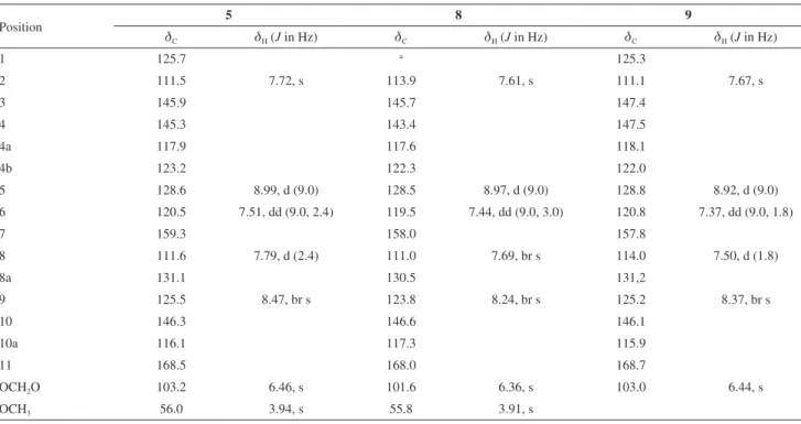
A molecular formula C17H11NO7 was determined
for 5 based on the HRMS spectra, which showed
peak at m/z 342.0610 [M + H]+ (calcd. for C
17H12NO7,
342.0613). The FTIR spectrum of this compound showed characteristic absorption bands to carboxylic acid at 1699 and 2800-3500 cm-1, and to nitro group at 1340 and 1521 cm-1.
The 1H and 13C NMR, HMBC and HSQC spectra (Table 1)
of a mixture comprising compounds 5 + 4 (2:1) showed
signals for aristolochic acid I (4) which were identified with
those of authentic sample. In addition, these spectra showed signal for 14 aromatic carbons, one acyl (dC 168.5), one
methylenedioxyl (dH 6.46 s, 2H, dC 103.2) and one methoxyl
(dH 3.94 s, 3H, dC 56.0) groups. The 1H NMR spectrum of
5 showed, in the aromatic region, a trisubstituted system
with three mutual coupled ABX pattern signals at d 8.99 (d, 1H, J 9.0 Hz), 7.51 (dd, 1H, J 9.0, 2.4 Hz), and 7.79 (d,
1H, J 2.4 Hz), which were assigned to H-5, H-6 and H-8,
respectively. In addition, two aromatic CH were observed in these spectra (dH-2 7.72 s, dC-2 111.5 and dH-9 8.47 br s, dC-9
125.5). These data and the UV absorption at 264, 308 and 380 nm are in accordance with a nitrophenanthrene structure, such as shown by aristolochic acids.33 The substituent
positions on the AA structure were assigned with the help of HMBC experiments (Figure 1). These experiments showed correlations between H-2 (dH 7.72) and C-11 (dC 168.5)
confirming the acyl group position in the structure, between H-9 (dH 8.47) and C-8 (dC 111.6), as well as H-5 (dH 8.99) and
OCH3 (dH 3.94) and C-7 (dC 159.3). These latter correlations
are also in accordance with a methoxyl group linked to C-7 on the C ring. Thus, this new compound was determined as 7-O-methylaristolochic acid F.
The 1H and 13C NMR spectra of compounds 8 and 9 are
very similar to those of 5. However, compounds 8 and 9
showed in their FTIR spectra absorption bands at ca. 1590 cm-1, characteristic of carboxylate instead of
carboxylic acid of AAs (1660-1710 cm-1).34 The spectra
revealed also absorption bands that indicated the presence of NO2 group (ca. 1350 and 1540 cm-1), and for 9 showed
absorption band for hydroxyl group at 3240 cm-1. The
HRMS spectra of compounds 8 and 9 showed peaks for
protonated molecules at m/z 364.0428 and 350.0268, in
accordance with the molecular formulae C17H10NO7Na
and C16H8NO7Na, respectively, and suggest 8 had a
methoxyl substituent, whereas 9 a hydroxyl in the
nitrophenanthrene structure. In addition, the protonated molecule of compound 8 is 22 Da higher than 5. Thus,
a sodium aristolochate derivative could be proposed for 8 and 9. The correlations observed by HMBC
experiments between the oxygenated carbons C-4, C-11 and H-2, C-7 and H-5, as well as C-8 and H-9 corroborate with this suggestion (Figure 1). Comparison of 1H NMR
data of 5, 8 and 9 confirmed the lower values for dH-9
observed for sodium aristolochates than aristolochic acids (5: dH-9 8.47, 8: dH-9 8.24, 9: dH-9 8.37).34 Thus,
compounds 8 and 9 were determined as 7-methoxy and
7-hydroxy sodium aristolochates, respectively. Moreover, an analogous acid of 9, which is known as aristolochic acid
F,33 showed NMR data considerably different from those
observed for 9.
Table 1. NMR spectroscopic data for compounds 5, 8 and 9 (14.1 T, DMSO-d6)
Position 5 8 9
dC dH (J in Hz) dC dH (J in Hz) dC dH (J in Hz)
1 125.7 a 125.3
2 111.5 7.72, s 113.9 7.61, s 111.1 7.67, s
3 145.9 145.7 147.4
4 145.3 143.4 147.5
4a 117.9 117.6 118.1
4b 123.2 122.3 122.0
5 128.6 8.99, d (9.0) 128.5 8.97, d (9.0) 128.8 8.92, d (9.0)
6 120.5 7.51, dd (9.0, 2.4) 119.5 7.44, dd (9.0, 3.0) 120.8 7.37, dd (9.0, 1.8)
7 159.3 158.0 157.8
8 111.6 7.79, d (2.4) 111.0 7.69, br s 114.0 7.50, d (1.8)
8a 131.1 130.5 131,2
9 125.5 8.47, br s 123.8 8.24, br s 125.2 8.37, br s
10 146.3 146.6 146.1
10a 116.1 117.3 115.9
11 168.5 168.0 168.7
OCH2O 103.2 6.46, s 101.6 6.36, s 103.0 6.44, s
OCH3 56.0 3.94, s 55.8 3.91, s
Conclusions
To date, 35 different compounds have been isolated from A. urupaensis, including the new
2,3-dihydro-4H-pyran-4-one (1). Compounds with this pyranone
carbon skeleton have not been isolated from natural sources yet. The new compounds 5, 8 and 9 are aristolochic acid
derivatives with unusual C-7 oxygenated substituents, and the compounds 21-23, 26 and 35 are being reported for the
first time in the Aristolochiaceae family.
Supplementary Information
Supplementary information (1D and 2D NMR, MS and FTIR spectroscopic data of compounds 1, 5, 8 and 9) is
available free of charge at http://jbcs.sbq.org.br as PDF file.
Acknowledgments
The authors thank Dr Vinicius C. Souza and MSc Joelcio Freitas for plant identification, and FAPESP, CAPES and CNPq for financial support and fellowships to L. M. X. L. and J. C. H.
References
1. Freitas, J.; Lírio, E. J.; González, F.; Phytotaxa2013, 124, 55. 2. http://floradobrasil.jbrj.gov.br/reflora/floradobrasil/FB15749,
accessed in January 2017.
3. Lopes, L. M. X.; Nascimento, I. R.; Silva, T. D.; Res. Adv. Phytochem.2001, 2, 19.
4. Heinrich, M.; Chan, J.; Wanke, S.; Neinhuis, C.; Simmonds, M. S. J.; J. Ethnopharmacol.2009, 125, 108.
5. Chen, C.-H.; Dickman, K. G.; Moriya, M.; Zavadil, J.; Sidorenko, V. S.; Edwards, K. L.; Gnatenko, D. V.; Wu, L.; Turesky, R. J.; Wu, X.-R.; Pu, Y.-S.; Grollman, A. P.; Proc. Natl. Acad. Sci. U. S. A.2012, 109, 8241.
6. Michl, J.; Kite, G. C.; Wanke, S.; Zierau, O.; Vollmer, G.; Neinhuis, C.; Simmonds, M. S.; Heinrich, M.; J. Nat. Prod.
2016, 79, 30.
7. Michl, J.; Ingrouille, M. J.; Simmonds, M. S. J.; Heinrich, M.;
Nat. Prod. Rep.2014, 31, 676.
8. Zhang, Y.-T.; Jiang, J.-Q.; Helv. Chim. Acta2006, 89, 2665. 9. Nascimento, I. R.; Lopes, L. M. X.; Phytochemistry2003, 63,
953.
10. Akasu, M.; Itokawa, H.; Fujita, M.; Tetrahedron Lett.1974, 15,
3609.
11. Wu, T.-S.; Leu, Y.-L.; Chan, Y.-Y.; J. Chin. Chem. Soc.2000,
47, 221.
12. Priestap, H. A.; Phytochemistry1985, 24, 849.
13. Tsuruta, A. Y.; Bomm, M. D.; Lopes, M. N.; Lopes, L. M. X.;
Eclet. Quim.2002, 27, 1.
14. Ma, J.; Jones, S. H.; Marshall, R.; Johnson, R. K.; Hecht, S. M.; J. Nat. Prod.2004, 67, 1162.
15. Dayun, Z.; Baode, W.; Baoshan, H.; Rensheng, X.; Yunping, Q.; Xiuzhen, C.; Dejian, Q.; Acta Chim. Sin.1983, 41, 74. 16. Holzbach, J. C.; Lopes, L. M. X.; Molecules2010, 15, 9462.
17. Byun, E.; Jeong, G.-S.; An, R.-B.; Min, T. S.; Kim, Y.-C.; Arch. Pharmacal. Res.2010, 33, 67.
18. He, D.; Huang, Y.; Ayupbek, A.; Gu, D.; Yang, Y.; Aisa, H. A.; Ito, Y.; J. Liq. Chromatogr. Relat. Technol.2010, 33, 615.
19. Moriyama, H.; Iizuka, T.; Nagai, M.; Yakugaku Zasshi2001,
121, 817.
20. Refaat, J.; Samy, M. N.; Desoukey, S. Y.; Ramadan, M. A.; Sugimoto, S.; Matsunami, K.; Kamel, M. S.; Med. Chem. Res.
2015, 24, 2939.
21. Wu, T.; Kong, D. Y.; Li, H. T.; Acta Pharm. Sin.2004, 39, 534.
22. Sawasdee, K.; Chaowasku, T.; Likhitwitayawuid, K.; Molecules
2010, 15, 639.
23. Salum, M. L.; Robles, C. J.; Erra-Balsells, R.; Org. Lett.2010,
12, 4808.
24. Huang, S.-X.; Liao, X.; Nie, Q.-J.; Ding, L.-S.; Peng, S.-L.;
Helv. Chim. Acta2004, 87, 598.
25. Carta, F.; Vullo, D.; Maresca, A.; Scozzafava, A.; Supuran, C. T.;
Bioorg. Med. Chem.2013, 21, 1564.
26. Ciuffreda, P.; Casati, S.; Manzocchi, A.; Magn. Reson. Chem.
2007, 45, 781.
27. Chen, J.-J.; Yang, C.-S.; Peng, C.-F.; Chen, I.-S.; Miaw, C.-L.;
J. Nat. Prod.2008, 71, 1016.
28. Reich, H. J.;Simulating NMR Spectra with WINDNMR-Pro; University of Wisconsin; http://www.chem.wisc.edu/areas/ reich/plt/windnmr.htm, accessed in March 2017.
29. Reiter, M.; Ropp, S.; Gouverneur, V.; Org. Lett.2004, 6, 91.
30. Zipp, G. G.; Hilfiker, M. A.; Nelson, S. G.; Org. Lett.2002, 4, 1823.
31. Denmark, S. E.; Heemstra, J. R.; J. Org. Chem.2007, 72, 5668. 32. Kubo, I.; Matsumoto, T.; Wagner, D. L.; Shoolery, J. N.;
Tetrahedron Lett.1985, 26, 563.
33. Cai, Y.; Cai, T.-G.; Chem. Pharm. Bull.2010, 58, 1093.
34. Leu, Y.-L.; Chan, Y.-Y.; Wu, T.-S.; Phytochemistry1998, 48, 743.