Rev Bras Cardiol Invasiva. 2014;22(3):271-4
© 2014 Sociedade Brasileira de Hemodinâmica e Cardiologia Intervencionista. Published by Elsevier Editora Ltda. All rights reserved.
Aortic Coarctation in Children Weighing Less than 25 kg:
Percutaneous Axillary Artery Approach
Germana Coimbra, Elio Vitor Duarte, Luiz J. Kajita, Pedro Lemos, Raul Arrieta
ABSTRACT
Background: Percutaneous treatment of aortic coarctation is the method of choice in children over 6 months of age and without aortic arch hypoplasia. However in patients less than 25 kg the classical access route (femoral) may pose a problem, especially in cases of stenting, due to the size of the size of the introducers. The objective of this study was to report our experience with the axillary artery approach for the percutaneous treatmentof patients with aortic coarctation weighing less than 25 kg. Methods: The arterial puncture was performed with a 21 G needle, with the arm abducted at 90°, and a 0.014 inch guidewire was positioned in the descending aorta. A 5 F × 7 cm pediatric introducer was initially used for the procedure and whenever required, it was replaced in a larger one. Manual-compression hemostasis was performed after the intervention. Results: Ten children were treated, eight with residual and two with native coarctation, mean age was 51.1 ± 30.8 months and weight 15.8 ± 5.8 kg. Puncture was performed in all cases without technical dificulty and the median introducer size was 7 F. Eight stents were implanted in eight patients and two patients were only treated by balloon angioplasty. Technical success was observed in all patients. After removal of the introducer, there was no permanent pulse loss and one patient had a small local hematoma. Conclusions: In our experience the axillary artery approach for percutaneous treatment of patients with aortic coarctation proved to be a safe and effective alternative in patients weighing less than 25 kg.
DESCRIPTORS: Aortic coarctation. Child. Axillary artery. Stents. Heart defects, congenital.
Instituto do Coração, Escola de Medicina, Universidade de São Paulo, São Paulo, SP, Brazil.
Correspondence to: Germana Coimbra. Avenida Dr. Enéas de Carvalho Aguiar, 44 – Cerceira César – CEP: 05403-900 – São Paulo, SP, Brazil
E-mail: [email protected]
Received on: 06/01/2014 • Accepted on: 08/09/2014
Original Article
RESUMO
Coarctação da Aorta em Crianças com Menos de 25 kg: Tratamento Percutâneo por Punção da Artéria
Axilar
Introdução: O tratamento percutâneo da coarctação da aorta é método de escolha em crianças acima de 6 meses de idade e sem hipoplasia do arco aórtico. No entanto, nos pacientes com menos de 25 kg, a via de acesso clássica (femoral) pode representar um problema, principalmente nos implantes de stents, devido ao tamanho dos introdutores. O objetivo deste estudo foi relatar a experiência com a punção da artéria axilar como via de acesso para o tratamento percutâneo de pacientes com coarctação da aorta e peso < 25 kg. Métodos: A punção foi realizada com agulha 21 G, com o braço abduzido em 90°, sendo introduzido io-guia 0,014 polegada, posicionado na aorta descendente. Um introdutor 5 F pediátrico de 7 cm foi inicialmente utilizado para realização do procedimento, sendo substituído, quando necessário, por um introdutor maior. Após a intervenção, foi realizada compressão hemostática manual. Resultados: Foram tratadas dez crianças, sendo oito com recoarctação pós-cirúrgica e duas com coactação nativa, com idades de 51,1 ± 30,8 meses e peso de 15,8 ± 5,8 kg. A punção foi realizada em todos os casos sem diiculdade técnica, e a mediana do calibre do introdutor foi de 7 F. Em oito pacientes, foram implantados oito stents e, em dois, foi realizada apenas angioplastia com balão. Houve sucesso técnico em todos os casos. Após a retirada do introdutor, não houve perda de pulso deinitiva e um paciente apresentou pequeno hematoma local. Conclusões: Na nossa experiência, o acesso axilar por meio de punção mostrou ser uma alternativa segura e eicaz neste grupo de pacientes.
DESCRITORES: Coartação aórtica. Criança. Artéria axilar. Stents. Cardiopatias congênitas.
I
n the percutaneous treatment of aortic coarctation, the choice of the approach in children is a fun-damental step in intervention planning, especially in small patients weighing < 25 kg. Most of these procedures can be performed by an arterial approach, both via femoral artery or by direct puncture into thecarotid artery, the latter being the most widely used approach today.1,2 This vascular access has proven safe
and effective; but not without complications.3,4 The
Coimbra et al.
Percutaneous Axillary Artery Approach of the CoAo
Rev Bras Cardiol Invasiva. 2014;22(3):271-4
272
The aim of this study was to report an initial ex-perience with the axillary-artery-puncture procedure as the main access route in the treatment of children under 25 kg with aortic coarctation.
METHODS
This was an observational cohort study of children with aortic coarctation undergoing interventional pro-cedures performed by axillary-artery puncture. Data were collected retrospectively by an analysis of clinical charts. The procedures were performed after an informed consent was obtained from parents or guardians.
Between June 2012 and May 2014, 53 interven-tions in patients with aortic coarctation, including ten performed by axillary approach in children weighing less than 25 kg, were performed.
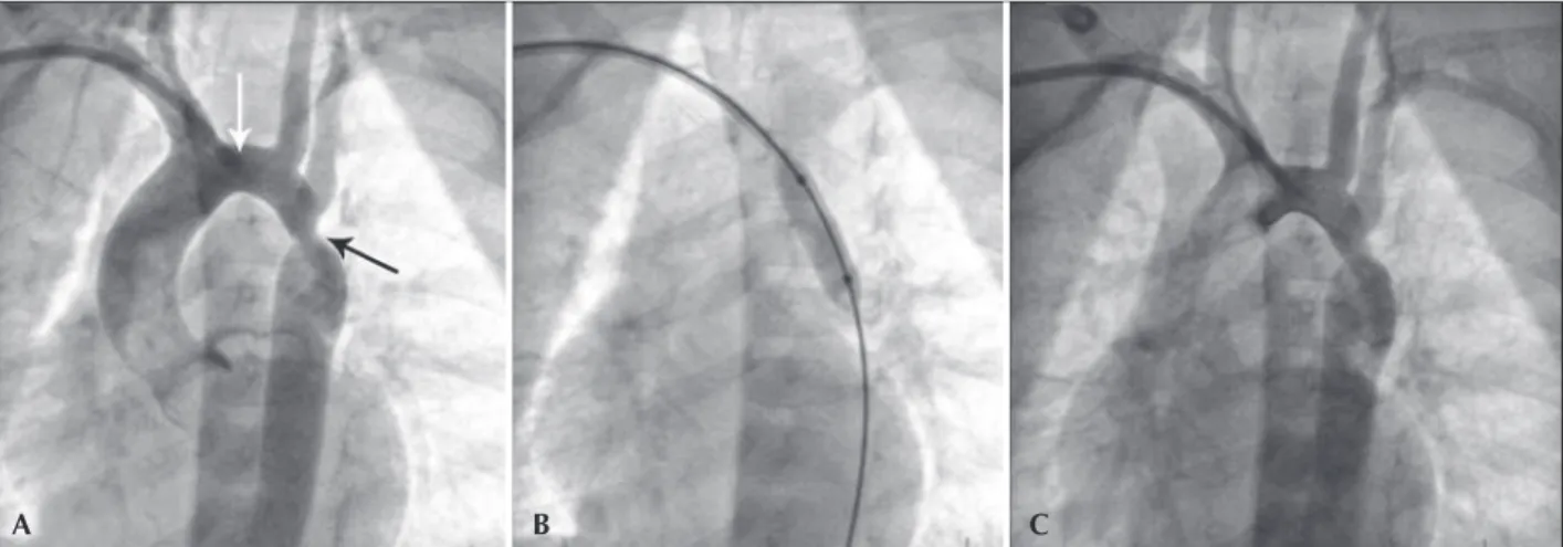
The axillary-artery puncture was conducted with the child under general anesthesia, with the arm abducted at 90°, and the head slightly tilted to the side opposite of the puncture site. After arterial pulse palpation in the axillary fossa, the arterial puncture was performed with a 21 G needle, and subsequently a guidewire of 0.014” (Boston Scientiic, Natick, United States) was introduced and positioned in the descending aorta. A pediatric 5 F sheath of 7 cm (Terumo Corporation, Tokyo, Japan or Cordis Corporation, Warren, United States) was initially used to perform the procedure, and was replaced, when necessary, by a larger sheath, according to the proile of catheters and balloons to be used. After the puncture, heparin was administered at a dose of 150 U/kg. The position regarded as ideal was obtained when the dis-tal end of the sheath was positioned in the aortic arch (Figure 1A). For most interventions, contrast injections were performed through the sheath sidearm, without requiring the use of additional angiographic catheters.
The decision to perform only balloon angioplasty was based on the following criteria: localized injury, without isthmus or aortic arch hypoplasia (local co-arctation) in children weighing < 10 kg, and without clinical signs of heart failure or of echocardiographic heart cavity enlargement. After the procedure, the sheath was removed, and a local manual compression was held for 15 to 20 minutes, with the low through the limb controlled by the pulse oximeter sensor. In all patients, heparin was neutralized with protamine.
In the recovery unit, the limb was kept warm us-ing orthopedic cotton for six hours. The mobility and sensitivity of the limb were clinically assessed before and immediately after surgery and during hospitaliza-tion. In all patients, the choice of this approach was planned before the procedure.
RESULTS
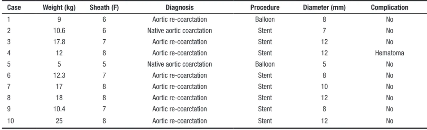
Ten children with mean age of 51.1 ± 30.8 months, and mean weigh of 14.8 ± 5.5 kg were included in the study. The demographic characteristics of these patients and procedures are described in the table. In all cases, the axillary artery puncture was performed without technical dificulty and the right axillary artery was used for the entire group. The median diameter of the sheath was 7 F. Postsurgical recoarctation was diagnosed in eight patients; in the remaining child, native coarctation was diagnosed.
Balloon angioplasty was performed in two patients weighing between 5 and 9 kg, and PowerFlex Cordis
balloons with diameters of 5 and 8 mm (Figure 1B and 1C) were used. In eight patients, eight stents were implanted; seven were XD Genesis
Cordis mounted on PowerFlex Cordis, with varying diameters (Figure
2). In one patient, a pre-mounted Express
SD (Boston
B C
A
Coimbra et al. Percutaneous Axillary Artery Approach of the CoAo
Rev Bras Cardiol Invasiva. 2014;22(3):271-4
273
Scientiic, Natick, United States) stent of 7 × 15 mm was used. Technical success was observed in all pa-tients. After sheath removal, no pulse loss was noted, and one patient had a small hematoma, with no need of transfusion.
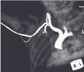
During an outpatient follow-up of 18.3 ± 2.2 months, one patient was referred for surgery because of aneurysm formation at the site of stenting. The re-maining children showed neither signs of re-coarctation nor pulse disorders or pressure difference between their upper limbs. In four children, a control-computed tomography (CT) scan for coarctation and a punctured axillary artery study were performed, showing no change in the access site (Figure 3).
DISCUSSION
Percutaneous treatment of aortic coarctation is currently considered the main therapeutic alternative in children over 1 year of age with aortic arch hypo-plasia. Angioplasty with balloon catheter is the irst choice for children weighing < 25 kg. The majority of patients progress favorably, but a non-negligible num-ber of aortic wall aneurysm formations still occur at the site of dilation, a fact of great concern every time this technique is used. Recently, stents have been used for treatment of aortic coarctation with less formation of aneurysms and excellent long-term results. Stents considered ideal are those that can be redilated up to adulthood; however, for implantation of these devices,
TABLE
Procedural and demographic characteristics
Case Weight (kg) Sheath (F) Diagnosis Procedure Diameter (mm) Complication
1 9 6 Aortic re-coarctation Balloon 8 No
2 10.6 6 Native aortic coarctation Stent 7 No
3 17.8 7 Aortic re-coarctation Stent 12 No
4 12 8 Aortic re-coarctation Stent 12 Hematoma
5 5 5 Native aortic coarctation Balloon 5 No
6 12.3 7 Aortic re-coarctation Stent 8 No
7 17 8 Aortic re-coarctation Stent 10 No
8 18 8 Aortic re-coarctation Stent 12 No
9 10.4 7 Aortic re-coarctation Stent 8 No
10 25 8 Aortic re-coarctation Stent 12 No
A B
Coimbra et al.
Percutaneous Axillary Artery Approach of the CoAo
Rev Bras Cardiol Invasiva. 2014;22(3):271-4
274
sheaths of calibers larger than 7 F are required, which may limit their use in patients of lower weight.
The actual incidence of vascular complications (stenosis) after percutaneous treatment of aortic coarcta-tion is dificult to estimate, since there can be appar-ent pulse symmetry with arterial stenosis presappar-ent, and imaging studies like ultrasound not always are ordered for diagnosis. Vascular complications, when using the femoral approach, range from 3 to 12%, depending on the experience of the group, and are more frequent in smaller children.7
The use of other vascular access options, such as carotid or even axillary artery dissection, has been de-scribed in percutaneous treatment of various congenital heart diseases with good results; however, until now their use in pediatric aortic coarctation treatment has not been reported. The present report details an initial experience using axillary artery puncture to treat this group of patients weighing < 25 kg. As demonstrated, the axillary-artery puncture technique was possible in all cases, and it should be emphasized that axillary pulse was universally present in the group and that the anatomy of the great vessels had been deined by echocardiogram or CT before the procedure. These facts are extremely important, since in patients with no palpable pulse or with an anomalous origin of the right subclavian artery, as shown by supplementary methods, this technique cannot be used.
Although the puncture was not guided by ultrasound, which would allow for measuring the vessel diameter, it is likely that the axillary artery has a larger caliber than the femoral artery one in this kind of defect, al-lowing for the use of sheaths of relatively higher caliber
compared to the weight of the child. Thus, it was possible to use the ideal stent without the occurrence of vascular complications. The absence of ischemic complications in the limb used can also be explained by the judicious puncture technique and by the fact that the axillary artery is not a branch of terminal irriga-tion. The upper member remains perfused throughout the intervention through the second intercostal artery and the acromial artery, minimizing the occurrence of distal ischemic complications.
During the follow-up, an anatomical evaluation of the punctured vessel was possible in 40% of patients, indicating preservation of the anatomy in all cases. In these patients, CT was performed as part of the clinical follow-up protocol at this institution, with the aim of evaluating the possible presence of vascular abnormali-ties in the coarctation site.
CONCLUSIONS
The axillary access obtained by direct vessel punc-ture was, in this initial experience, a good alternative for the treatment of children weighing less than 25 kg with aortic coarctation, with excellent results and a low complication rate.
CONFLICTS OF INTEREST
The authors declare no conlicts of interest.
FUNDING SOURCES
None.
REFERENCES
1. McCrindle BW, Jones TK, Morrow WR, Hagler DJ, Lloyd TR, Nouri S, et al. Acute results of balloon angioplasty of native coarctation versus recurrent aortic obstruction are equivalent. Valvuloplasty and Angioplasty of Congenital Anomalies (VACA) Registry Investigators. J Am Coll Cardiol. 1996;28(7):1810-7. 2. Fletcher SE, Nihill MR, Grifka RG, O’Laughlin MP, Mullins
CE. Balloon angioplasty of native coarctation of the aorta: midterm follow-up and prognostic factors. J Am Coll Cardiol. 1995;25(3):730-4.
3. Fawzy ME, Fathala A, Osman A, Badr A, Mostafa MA, Mo-hamed G, et al. Twenty-two years of follow-up results of balloon angioplasty for discreet native coarctation of the aorta in adolescents and adults. Am Heart J. 2008;156(5):910-7. 4. Dua JS, Osborne NJ, Tometzki AJ, Martin RP. Axillary artery
approach for balloon valvoplasty in young infants with severe aortic valve stenosis: medium-term results. Catheter Cardiovasc Interv. 2006;68(6):929-35.
5. Schranz D, Michel-Behnke I. Axillary artery access for cardiac interventions in newborns. Ann Pediatr Cardiol. 2008;1(2):126-30. 6. Lawless S, Orr R. Axillary artery monitoring of pediatric
pa-tients. Pediatrics. 1989;84(2):273-5.
7. Ammar RI. Balloon angioplasty for native aortic coarctation in children and infants younger than 12 months: immediate and medium-term follow-up. J Invasive Cardiol. 2012;24(12):662-6.