© 2014 Sociedade Brasileira de Hemodinâmica e Cardiologia Intervencionista. Published by Elsevier Editora Ltda. All rights reserved.
Balloon/Artery Ratio and Neointimal Hyperplasia
Volume Obstruction After Zotarolimus-Eluting
Stent Implantation
Gustavo do Prado Monteiro, J. Ribamar Costa Jr., Carlos Collet, Juliano Slhessarenko, Fausto Feres,
Ricardo Costa, Áurea J. Chaves, Marinella Centemero, Amanda G.M.R. Sousa, Alexandre Abizaid
ABSTRACT
Background: Intimal hyperplasia volume is correlated to arterial injury after bare-metal stenting. However, little is known about the impact of arterial injury on the inflam-matory/proliferative response with drug-eluting stents. We investigated the impact of arterial injury, evaluated by the balloon/artery ratio, on neointimal hyperplasia volume ob-struction, evaluated by intravascular ultrasound, 12 months after zotarolimus-eluting stent implantation. Methods: Balloon/ artery ratio was defined as the ratio of the maximum balloon diameter, using the maximal implantation or post-dilatation pressure, and the reference diameter of the vessel obtained before the procedure. Patients were divided into two groups: high balloon/artery ratio (≥ 1.15) and low balloon/artery ratio (< 1.15). Results: A total of 86 patients were included in the low balloon/artery ratio group (n = 47/48 lesions) or high balloon/artery ratio (n = 39/48 lesions). The clinical, angiographic and procedure related characteristics were not different between groups, except for the vessel reference diameter (2.73 ± 0.45 mm vs. 2.97 ± 0.40 mm; p = 0.01). At 12 months, similar in-stent late loss was observed (0.59 ± 0.32 mm vs. 0.62 ± 0.42 mm; p = 0.92), as well as binary restenosis (4.2% in both cohorts; p > 0.99). In stent neointi-mal hyperplasia volume obstruction (15.2 ± 14.3% vs. 12.5 ± 10.1%; p = 0.62) showed no difference between groups. No correlation was observed between balloon/artery ratio and neointimal hyperplasia obstruction volume at 12 months (R2 = 0.0025; p = 0.88). Conclusions: In the present study arterial injury was not correlated with the amount of intimal hyperplasia after zotarolimus-eluting stent implantation.
Descriptors: Coronary restenosis. Percutaneous coronary in-tervention. Drug-eluting stents.
Instituto Dante Pazzanese de Cardiologia, São Paulo, SP, Brazil. Correspondence to: Gustavo do Prado Monteiro. Avenida Dr. Dante Pazzanese, 500 − Vila Mariana − CEP: 04012 180 − São Paulo, SP, Brazil E mail: [email protected]
Received on: 06/04/ 2014 • Accepted on: 08/18/2014
Original Article
RESUMO
Relação Balão/Artéria e Volume de Obstrução Neontimal Após Implante de Stent Eluidor de
Zotarolimus
Introdução: O volume de hiperplasia intimal correlaciona-se com a injúria arterial após implante de stents não farma-cológicos. No entanto, pouco se sabe a respeito do impacto da injúria arterial na resposta inlamatória/proliferativa com os stents farmacológicos. Investigamos o impacto da injúria arterial, avaliada pela relação balão/artéria, no volume de obstrução neontimal, avaliado pelo ultrassom intracoronário 12 meses após o implante de stents farmacológicos com eluição de zotarolimus. Métodos: A relação balão/artéria foi deinida como a razão do diâmetro máximo do balão, obtido no implante ou na pós-dilatação, e o diâmetro de referência do vaso pré-procedimento. Os pacientes foram categorizados em dois grupos: relação balão/artéria alta (≥ 1,15) e relação balão/artéria baixa (< 1,15). Resultados: Foram incluídos 86 pacientes, nos grupos relação balão/artéria baixa (n = 47/48 lesões) ou relação balão/artéria alta (n = 39/48 lesões). As características clínicas, angiográicas e do procedimento não diferiram entre os grupos, à exceção do diâmetro de referência dos vasos (2,73 ± 0,45 mm vs. 2,97 ± 0,40 mm; p = 0,01). Aos 12 meses, observou-se semelhante perda tardia intra-stent (0,59 ± 0,32 mm vs. 0,62 ± 0,42 mm; p = 0,92), bem como reestenose binária (4,2% em ambas as coortes; p > 0,99). O volume de obstrução neointimal intra stent (15,2 ± 14,3% vs. 12,5 ± 10,1%; p = 0,62) não mostrou diferença entre os grupos. Não observamos correlação entre a relação balão/ artéria e o volume de obstrução neointimal aos 12 meses (R2 = 0,0025; p = 0,88). Conclusões: A injúria arterial não se correlacionou com a quantidade de hiperplasia intimal após implante de stents farmacológicos com eluição de zotarolimus.
Monteiro et al. Balloon/Artery Ratio and Neointimal Hyperplasia Volume Obstruction Rev Bras Cardiol Invasiva.
2014;22(3):252-7
253
C
oronary restenosis is the result of exacerbated heal-ing response secondary to vascular injury caused to the arterial wall by the balloon1 and comprises two basic mechanisms: neointimal hyperproliferation and vascular remodeling.The advent of bare-metal stents (BMS) virtually abolished negative vascular remodeling, as their metal structure prevents retraction of the treated vascular segment.2 However, the presence of a foreign metallic body in the vessel, in association with the barotrauma resulting from the pressure employed to release the stent, triggers a local inlammatory response that initiates the healing cascade, which in some cases can result in neointimal hyperproliferation and lead to further vessel obstruction.3
Studies with intravascular ultrasound (IVUS) in pa-tients treated with BMS suggest that stent overexpansion might result in more traumas to the vascular wall and thereby exacerbate the repair response, resulting in a greater amount of neointimal tissue.4
A little over a decade ago, drug-eluting stents (DES) were introduced into clinical practice. Due to their capacity to inhibit the neointimal proliferative response, these devices greatly reduced rates of restenosis and made percutaneous coronary intervention (PCI) the treatment of choice for coronary artery disease.
However, little is known about the impact of arte-rial injury on inlammatory/proliferative response after implantation of DES. The aim of the present study was to investigate, through IVUS, the impact of arte-rial injury on the neointimal obstruction volume after zotarolimus-eluting stent implantation.
METHODS
Study design and target population
This was a retrospective, single-center, single-arm study conducted in a tertiary hospital in the state of São Paulo. All patients treated with the Endeavor stent
(Medtronic, Santa Rosa, United States) from January to July 2011 were evaluated, submitted to angiographic reassessment and IVUS at 12 months. These patients were from different research protocols, thus having dif-ferent inclusion and exclusion criteria. In common, the lesions evaluated were de novo in native coronaries, with diameter between 2.5 and 3.5 mm, treated with a single DES (up to 30 mm). This analysis excluded patients treated in the acute phase of acute myocardial infarction (AMI) with ST-segment elevation or those with renal failure (creatinine clearance < 60 mL/minute). Patients with multivessel disease were treated according to the same procedure or in staged procedures.
The different study protocols were approved by the Research Ethics Committee of the institution and
all patients signed the informed consent prior to the procedure.
Procedure
PCI procedures were performed according to the routine of the institution and in accordance with current recommendations. The choice of access route (femo-ral or radial) was made by each surgeon, and direct stenting without pre-dilation was allowed. The use of post-dilation was at the surgeons’ discretion.
Antiplatelet protocol consisted in the administra-tion of acetylsalicylic acid (ASA) and clopidogrel. Pre-treatment included ASA at a dose of 100 to 200 mg per day in case of chronic use (> 7 days) or loading dose of 200 to 300 mg given > 24 hours before PCI. For clopidogrel, the loading dose of 300 mg was employed > 24 hours before the intervention in elective cases, or 600 mg > 2 hours before of the procedure, in cases of acute coronary syndrome. After the procedure, the use of ASA (100 mg/day) was recommended indeinitely and clopidogrel (75 mg/day) was administered for a minimum period of 12 months. Regarding anticoagula-tion therapy during the procedure, intravenous heparin was administered at a dose of 70 to 100 U/kg body weight to maintain the activated clotting time > 250 seconds (or > 200 seconds in case of concomitant use of glycoprotein IIb/IIIa inhibitor).
Outcome and definitions
The primary study endpoint was in-stent neointimal obstruction volume obtained by IVUS in the angiographic reevaluation 12 months after the procedure.
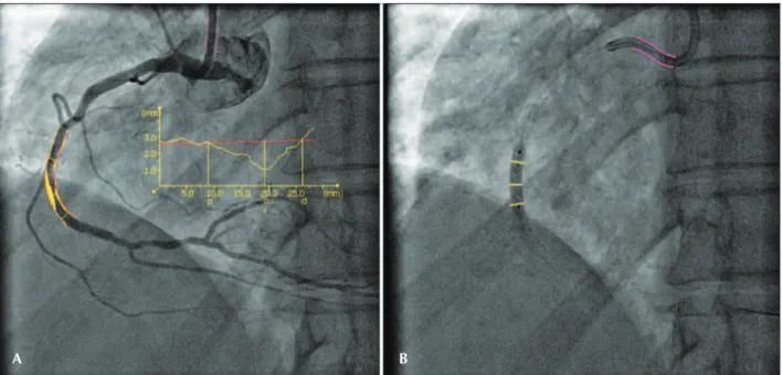
Arterial injury was defined by the balloon/artery ratio (BAR) during stenting or post-dilation, calcu-lated as the ratio of the maximum balloon diameter by reference vessel diameter prior to the procedure. This definition has been previously validated in stud-ies with bare-metal stents.5 Figure 1 illustrates the calculation of the BAR.
For this analysis, patients were categorized into two groups: low BAR (< 1.15) or high BAR (≥ 1.15). Angiographic analysis was performed using a validated software program (QAngioXA, version 7.3; Medis,
Leiden, Netherlands).
Image acquisition and intravascular ultrasound analysis
Image acquisition was performed using a single, rotational transducer with a frequency of 40 MHz, using a 2.6 F sheath, with motorized retraction in an automatic pullback system at a speed of 0.5 mm/sec, and commercial scanners (Galaxy 2; Boston Scientiic
For the volumetric analysis, the three-dimensional image reconstruction was performed using a com-mercially available computerized planimetry program (EchoPlaque 3.0; INDEC Systems Inc., Mountain View,
United States). The lumen, stent, and vessel (external elastic membrane) areas in the analyzed segment were determined by computerized planimetry at every mil-limeter. The neointimal hyperplasia area was calculated as the stent area minus the lumen area. Volumes (lumen, stent, and vessel) were calculated by Simpson’s rule. In-stent neointimal obstruction volume was calculated as the ratio between the neointimal hyperplasia volume and the stent volume × 100.
Statistical analysis
Continuous variables were expressed as mean and standard deviation, and categorical variables as per-centage and absolute value. Continuous variables were analyzed using Student’s t-test or the Mann-Whitney test. Categorical variables were compared using the chi-squared test or Fisher’s exact test. Linear regression using the least square method was used to analyze the association between in-stent neointimal obstruction volume and BAR at 12 months.
RESULTS
A total of 86 patients were evaluated, with 96 le-sions submitted to PCI with Endeavor stent
implanta-tion, of which 48 lesions were treated with low BAR and 48 with high BAR.
The baseline clinical and angiographic characteristics of the treated population are summarized in table 1.
As observed, the mean age of the population in the two groups was around 60 years, and most patients were males. There was a slight numerical predominance, but not significant, of diabetic pa-tients in high BAR group (30% vs. 38%; p = 0.39), while the majority of the population in both cohorts comprised patients with stable coronary disease. Overall, the left anterior descending artery was the most often treated vessel, and most lesions were of moderate complexity (B1/B2) in both groups, accord-ing to the classification of the American College of Cardiology/American Heart Association (ACC/AHA). During the procedure, no differences were observed between the groups regarding pre- or post-dilation rates, and both were performed in less than half the assessed population.
Table 2 depicts the quantitative coronary angiog-raphy data during the procedure and evaluation at 12 months. In the high BAR group, the treated vessels had smaller diameters (2.73 ± 0.45 mm vs. 2.97 ± 0.40 mm; p < 0.01), but with similar pre-intervention lesion extension and stenosis diameter. The mean BAR in the group with more aggressive implantation was 1.35 ± 0.13, whereas in the low BAR group, it was 1.03 ± 0.08. At 12 months, the observed in-stent late loss was similar (0.59 ± 0.32 mm vs. 0.62 ± 0.42 mm; p = 0.92), as well as binary restenosis (4.2% in both cohorts, p > 0.99) between the groups.
A B
Monteiro et al. Balloon/Artery Ratio and Neointimal Hyperplasia Volume Obstruction Rev Bras Cardiol Invasiva.
2014;22(3):252-7
255
TABLE 1
Clinical, angiographic, and procedural characteristics.
BAR < 1.15 (n = 47 patients/48 lesions)
BAR ≥ 1.15 (n = 39
patients/48 lesions) p-value
Age, years 59.9 ± 10.9 60.5 ± 9.3 0.93
Male gender, n (%) 37 (78) 30 (77) 0.89
Arterial hypertension, n (%) 38 (81) 31 (79.5) 0.87
Diabetes mellitus, n (%) 14 (30) 15 (38.5) 0.39
Dyslipidemia, n (%) 30 (63.8) 26 (66.7) 0.78
Previous AMI, n (%) 23 (49) 21 (53.8) 0.65
Smoking, n (%) 7 (15) 2 (5.1) 0.14
Renal failure, n (%) 1 (2.1) 2 (5.1) 0.45
Clinical presentation 0.87
Stable angina 41 (87) 35(89.7)
Acute coronary syndrome 6 (13) 4 (10.3)
Target vessel, n (%) 0.29
Left anterior descending artery 19 (39.6) 18 (37.5)
Left circumlex artery 10 (20.8) 14 (29.2)
Right coronary artery 19 (39.6) 16 (33.3)
Lesion classiication (ACC/AHA), n (%) 0.34
A 0 2 (4.2)
B1/B2 40 (83.3) 35 (72.9)
C 8 (16.7) 11 (22.9)
Pre-dilation, n (%) 21 (43.7) 21 (43.7) > 0.99
Post-dilation, n (%) 18 (37.5) 17 (35.4) 0.83
Final dilation pressure, atm 12.2 ± 5.1 14.0 ± 5.4 0.35
BAR: balloon-artery ratio; AMI: acute myocardial infarction; ACC/AHA: American College of Cardiology/American Heart Association.
TABLE 2
Quantitative coronary angiography at baseline and after 12 months.
BAR < 1.15 (n = 48 lesions)
BAR ≥ 1.15
(n = 48 lesions) p-value
Basal
RVD, mm 2.97 ± 0.40 2.73 ± 0.45 < 0.01
Lesion length, mm 16.6 ± 11.7 14.3 ± 8.7 0.61
Stenosis diameter, % 67.5 ± 11.0 68.0 ± 10.6 0.18
12 months
Late intrastent luminal loss, mm 0.59 ± 0.32 0.62 ± 0.42 0.92
Stenosis diameter, % 18.2 ± 11.9 19.6 ± 11.6 0.92
Binary restenosis, n (%) 2 (4.2) 2 (4.2) > 0.99
BAR: balloon-artery ratio; RVD: reference vessel diameter.
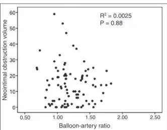
In the IVUS analysis at 12 months, signiicant dif-ferences were found regarding volume of the vessel (249.2 ± 207.5 mm3 vs. 325 ± 161.8 mm3; p < 0.01), lumen (110.7 ± 91 9 mm3 vs. 154.2 ± 91.3 mm3; p < 0.01), and stent (134 ± 113.8 mm3 vs. 174.9 ± 98
was no signiicant correlation between the BAR and the in-stent neointimal obstruction volume at 12 months (R2 = 0.0025; p = 0.88; Figure 2).
DISCUSSION
The main inding of this study concerns the lack of correlation between the degrees of arterial injury, quantiied by the BAR, with the formation of neointimal tissue within the zotarolimus-eluting stents, quantiied by the neointimal obstruction volume.
The main mechanism of restenosis after bare-metal stent implantation is exacerbated formation of neointi-mal tissue within the stent. Balloon inlation causes a fracture of atherosclerotic plaque. Activated platelets release mitogens, including thromboxane A2, serotonin, and platelet-derived growth factor, promoting smooth muscle cellproliferation.6,7 Consequently, activated smooth muscle cells migrate to the intimal layer. Endothelial dysfunction can contribute to the proliferation and migration of smooth muscle cells through the reduced synthesis of nitric oxide, which inhibits their growth.
Hoffmann et al.5 documented a signiicant correlation between neointimal hyperplasia assessed by IVUS and the product of BAR by the maximum inlation pressure, suggesting that BMS restenosis may be related to the degree of arterial overstretch. In contrast, Kuriayama et al.8 reported that overstretch in arteries with diameter ≥ 3 mm resulted in a lumen with larger inal dimen-sions. Thus, the risk of restenosis can be reduced with higher-pressure dilation in large vessels.
Recently, Eshtehardi et al.9, evaluating 196 patients treated with irst-generation drug-eluting stents (Cypher
and Taxus) showed no correlation between arterial
injury in the implant and neointimal tissue formation with these stents.
Study limitations
The main limitations of this study were the relatively small number of patients evaluated with IVUS and the fact it was not a randomized sample, which does not preclude the existence of other variables that might affect the neointimal hyperplasia volume, in addition to stent overexpansion. Additionally, the retrospective analysis of patients submitted to IVUS at 12 months might have caused a selection bias.
CONCLUSIONS
This study found no correlation between arterial injury assessed by the balloon/artery ratio and the neointimal obstruction volume after zotarolimus-eluting stent implantation.
CONFLICTS OF INTEREST
The authors declare no conlicts of interest.
FUNDING SOURCES
None. TABLE 3
Results of intravascular ultrasound at 12 months.
BAR < 1.15 (n = 48 lesions)
BAR ≥ 1.15
(n = 48 lesions) p-value
Vessel volume, mm3 249.2 ± 207.5 325.0 ± 161.8 < 0.01
Luminal volume, mm3 110.7 ± 91.9 154.2 ± 91.3 < 0.01
Stent volume, mm3 134.0 ± 113.8 174.9 ± 98.0 < 0.01
Intimal-hyperplasia volume, mm3 26.2 ± 36.7 20.5 ± 18.3 0.62
Intimal in-stent-obstruction volume, % 15.2 ± 14.3 12.5 ± 10.1 0.62
BAR: balloon-artery ratio.
Neointimal obstr
uction v
olume
Balloon-artery ratio R2 = 0.0025 P = 0.88
0.50 1.00 1.50 2.00 2.50
Monteiro et al. Balloon/Artery Ratio and Neointimal Hyperplasia Volume Obstruction Rev Bras Cardiol Invasiva.
2014;22(3):252-7
257
REFERENCES
1. Forrester JS, Fishbein M, Helfant R, Fagin J. A paradigm for restenosis based on cell biology: clues for the development of new preventive therapies. J Am Coll Cardiol. 1991;17(3):758-69. 2. Costa MA, Sabate M, Kay IP, de Feyter PJ, Kozuma K, Serrano
P, et al. Three dimensional intravascular ultrasonic volumetric quantiication of stent recoil and neointimal formation of two new generation tubular stents. Am J Cardiol. 2000;85(2):135-9. 3. Wilcox JN, Okamoto EI, Nakahara KI, Vinten-Johansen J. Peri-vascular responses after angioplasty which may contribute to postangioplasty restenosis: a role for circulating myoibroblast precursors? Ann N Y Acad Sci. 2001;947:68-90.
4. Hoffmann R, Mintz GS, Mehran R, Pichard AD, Kent KM, Satler LF, et al. Intravascular ultrassound predictors of angiographic restenosis in lesions treated with Palmaz-Schatz stents. J Am Coll Cardiol. 1998;31(1):43-9.
5. Hoffmann R, Mintz GS, Mehran R, Kent KM, Pichard AD, Satler LF, et al. Tissue proliferation within and surrounding Palmaz-Schatz stent is dependent on the aggressiveness of stent implantation technique. Am J Cardiol. 1999;83(8): 1170-4.
6. Welt FG, Rogers C. Inlammation and restenosis in the stent era. Arterioscler Thromb Vasc Biol. 2002;22(11):1769-76. 7. Costa MA, Simon DI. Molecular basis of restenosis and
dru-geluting stents. Circulation. 2005;111(17):2257-73.
8. Kuriyama N, Kobayashi Y, Kuroda N, Desai K, Yamamoto Y, Komiyama N, et al. Effect of coronary stent overexpansion on lumen size and intimal hyperplasia at follow-up. Am J Cardiol. 2002;89(11):1297-9.