The mitochondrial genome is highly conserved and com-pact, usually including genes that code for 13 proteins, 22 tRNAs and two rRNA, as well as an important noncoding sequence (con-trol region, CR) in vertebrates. Particularly, the CR in vertebrate mitochondrial genomes contains two promoters (LSP and HSP) for the transcription, heavy-strand replication origin (OH), and the displacement loop (D-loop) (CLAYTON 1982, CHANG & CLAYTON
1986). However, based on the distribution of the variable nucle-otide positions and different nuclenucle-otide frequencies, the mito-chondrial CR is divided into three domains (BROWNet al. 1986, SACCONEet al. 1991): termination associated sequence (TAS) do-main, central conserved domain (CD), and conserved sequence block (CSB) domain. The TAS domain has been shown to con-tain sequences associated with termination of newly synthesized H-strands during replication (DODAet al. 1981, BROWNet al. 1986, SBISÀet al. 1997). The CD, which contains several areas of highly
conserved sequences, is more conserved with respect to the TAS and CSB domains. However, the nature of these conserved se-quences varies among vertebrate classes. For example, ANDERSON
et al. (1981) indentified several conserved sequence boxes (CSB), B, C, D, E, and F in the CD of the human mitochondrial CR. Of those, only three (CSB B, CSB D, and CSB F) can be found in the avian mitochondrial CD (RUKONEN & KVIST 2002). The CSB
do-main usually contains the origin of the H-strand transcription (WALBERG & CLAYTON 1981, BROWNet al. 1986, KING & LOW 1987, FORANet al. 1988). In many taxa, the TAS and CSB domains, which are more variable than the CD, have variable numbers of tandem repeats (VNTRs) (BROUGHTON & DOWLING 1994, SBISÀ et al.
1997, ZARDOYA & MEYER 1998a, DELPORTet al. 2002, FUet al. 2006, ZHANG et al. 2009, XIONGet al. 2010).
Extant turtles have been divided into two monophyletic clades, Pleurodira and Cryptodira. However, virtually all
stud-Organization and variation of mitochondrial DNA control region in
pleurodiran turtles
Ling Wang
1; Xuming Zhou
2& Liuwang Nie
1, 31 Life Science College, Anhui Normal University, The Provincial Key Lab of the Conservation and Exploitation Research of
Biological Resources in Anhui, 1 East Beijing Road, Wuhu, Anhui, 241000, China.
2 Key laboratory of Animal Ecology and Conservation Biology, Institute of Zoology, Chinese Academy of Sciences, Beijing
100101, China.
3 Corresponding author: E-mail: [email protected]
ABSTRACT. Three complete mitochondrial DNA (mtDNA) control regions (CRs) of Chelodina rugosa (Ogilby, 1890), Chelusfimbriata (Schneider, 1783), and Podocnemisunifilis (Troschel, 1848) were firstly determined using Long-PCR method and the length were 1,016 bp, 1,149 bp, and 985bp, respectively. Together with CRs of Pelomedusa subrufa (Bonnaterre, 1789) and nearly complete CR of Podocnemis expansa (Schweigger, 1812) obtained from GenBank, the structural and evolutionary characteristics of mtDNA CRs in pleurodiran turtle were analyzed in this study. We identified three functional domains (TAS, CD, and CSB domains) as well as their conservation sequences (TAS, CSB-F, and CSB-1) according to their homology to those of other turtles. Within the TAS domain, an interrupted poly-C stretch was found in C. rugosa, C. fimbriata, and P. subrufa, which also exists in the published mt DNA CRs of Chrysemys picta (Schneider, 1783), Trachemys scripta (Thunberg in Schoepff, 1792), and Trionyx triunguis (Forskål, 1775). The analysis of the origin for the poly-C sequences in TAS domain from six turtles suggested that the poly-C sequences are more related to “goose hairpin” in birds rather than CSB2 in CSB domain. In the CSB domain, CSB2 and CSB3, which were determined in CRs of Cryptodira, were absent in Pleurodira CRs, indicating the regulative mechanisms of transcription may be varied in both two suborders and the lack of CSB2 and CSB3 could be proposed as one of diagnostic characters between Pleurodira and Cryptodira at molecular level. As for CR of other cryptodiran turtles, variable number of tandem repeats (VNTRs) in the 3’ end of the CRs was found in the five pleurodiran turtles. Interestingly, the long repeated motifs from each species could form stable stem-loop secondary structures, suggesting that the repeated sequences may play an important role in regulating replication of the mitochondrial genome in Pleurodiran, and the secondary structures of VNTRs may provide some potential information in phylogenetic inference.
ies on turtle mitochondrial genomes have focused on Cryptodira. Studies comparing the mitochondrial CRs of pleurodiran turtles have been limited because there is only one published complete CR from one species – Pelomedusa subrufa (Bonnaterre, 1789). In the present study, three complete CR of three pleurodiran turtles – Chelodina rugosa (Ogilby, 1890), Chelus fimbriata (Schneider, 1783), and Podocnemis unifilis (Troschel, 1848) – representing two families (Chelidae and Podocnemididae) are characterized. Additionally, together with the complete CR of P. subrufa and a nearly complete CR of Podocnemis expansa (Schweigger, 1812) obtained from GenBank, we have compared the CR sequences of pleurodiran turtles with the CRs of other vertebrates. Additionally, the features which are shared or not among the pleurodiran turtles and other verte-brates have been identified and are discussed in detail.
MATERIAL AND METHODS
Sample and sequencing
Specimens of C. rugosa, C. fimbriata, and P. unifilis were stored at the Anhui Normal University. Total genomic DNA was extracted from their muscles with the proteinase K method (SAMBROOK & RUSSELL 2001) and kept at -20°C for PCR
amplifica-tion.
The mitochondrial CR was amplified by long-PCR. The entire CR and 3 tRNAs (tRNAThr, tRNAPr°, and tRNAPhe) as well as partial Cyt b and 12S rRNA gene sequences were amplified in one single step using a pair of long-PCR primers:
LCR-F: 5’-CTTCCTATTTGCCTATGCTATC-3’ LCR-R: 5’-TATTTTGGGCTCCTGGTGTA-3’
Long-PCR conditions were: one minute at 95°C, then 30 cycles of 10 seconds at 98°C, five minutes at 55°C, followed by a final extension for 10 minutes at 72°C. PCR products were iso-lated using a Gel Extract Purification Kit (TaKaRa Co., Ltd, Dalian, China) after 1% agarose gel electrophoresis. The purified Prod-ucts were sequenced with an ABI3730 automated sequencer.
Sequence analysis
In order to determine the complete CR sequence of the three turtles, the sequences obtained for C. rugosa, C. fimbriata, and P. unifilis were compared with the complete mtDNA of P. subrufa (ZARDOYA & MEYER 1998b), and the tRNAs were
identi-fied using tRNAscan-SE 1.21(LOWE & EDDY 1997). The sequence containing almost complete CRs of P. expansa was retrieved from GenBank. Subsequently, five pleurodiran turtle sequences were aligned using ClustalX 1.8 software (THOMPSONet al. 1997)
and then checked manually in order to define the conserved sequence blocks.
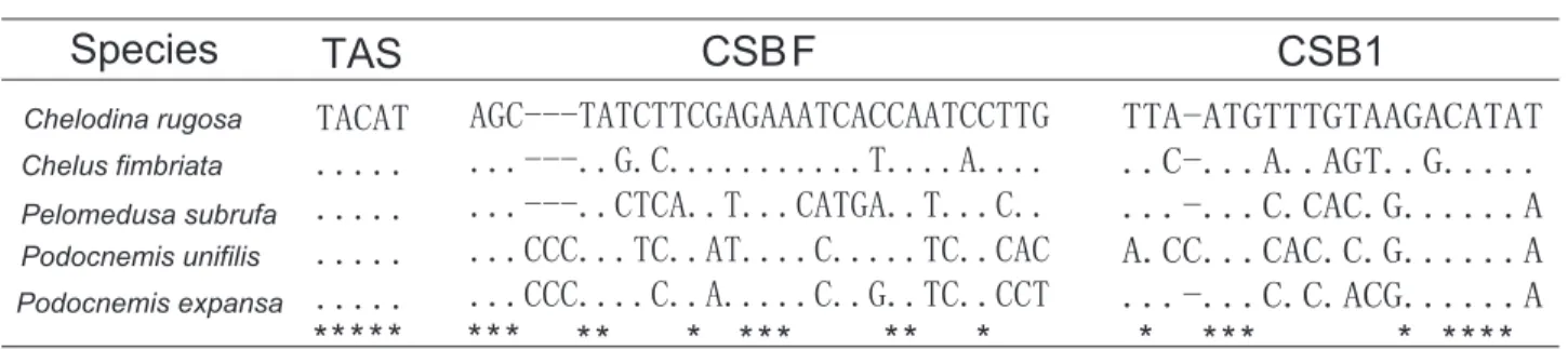
After comparison with published data from other taxa (the sequences used are listed in the Appendix), the conserved box F (CSB F) was delimited in the aligned sequences. The con-served block 1 (CSB1) with the characteristic motif GACATA was also delimited. The boundary of the TAS domains and the Central conserved domain was the starting point of the CSB F.
The CSB domain was always set to start with the conserved sequence box 1 (CSB1) (ZHANGet al. 2009, XIONGet al. 2010). The program ‘Tandem Repeats Finder’ (BENSON 1999) was used
in order to identify the VNTRs in the CRs. Furthermore, puta-tive secondary structures in the CRs were determined using the software RNA structure (MATHEWSet al. 1999). Subsequently,
the computer program RNAdraw (HOFACKERet al. 1995) was employed to prepare secondary structures for publication.
RESULTS AND DISCUSSION
The length and base composition of pleurodiran
turtle CRs
The CRs of C. rugosa, C. fimbriata, and P. unifilis have 1,016 bp, 1,149 bp, and 985bp, respectively. They are rich in adenine and thymine and lack L strand guanines, which is evi-dent in each domain, particularly in the CSB domain (Tab. I). However, the composition of the CR among the three domains is not uniform. The Central conserved domain is poor in ad-enine and rich in L strand guanines compared to the flanking TAS and CSB domains. Interestingly, among the five turtles compared, the TAS domain was found to be richer in adenine and thymine in three species: C. rugosa, C. fimbriata, and P. subrufa. This TAS domain composition is consistent with that of cryptodiran turtles (ZHANG et al. 2009, XIONG et al. 2010).
However, in the other two pleurodiran turtles (P. unifilis and P. expansa), the TAS domain was rich in cytosine and thymine.
The organization of turtle CRs
Like in most cryptodiran turtles, the CRs in the mito-chondrial genomes of pleurodiran turtles are located between the tRNAPro and tRNAPhe genes. The CSB F and CSB-1 were easy to define in the five turtles (Fig. 1). In the CSB F, 12 of 29 (41.4%) nucleotide positions were fixed among the five turtles. In the CSB1, 9 of 20 (45%) nucleotide positions were fixed. Compar-ing with the cryptodiran turtles (ZHANGet al. 2009, XIONGet al. 2010), the conserved blocks of pleurodiran turtles varied greatly. This variation may due to the age of the group. The fossil record has shown that most pleurodiran turtles diverged about 100-150 mya (million years ago) (GAFFNEY 1990). During this long
evolutionary history, the conserved blocks sequences of the mtDNA CRs might have changed independently, accumulat-ing more variation than the CRs of cryptodiran turtles, in which most species have diverged after 100 mya (NEARet al. 2005).
TAS domain
The TAS domain was located between the 5’end of the CR and the beginning of the CSB F. The length of the TAS do-main varied from 243 bp to 357 bp, and the sequences were heterogeneous and could not be unambiguously aligned. How-ever, the TAS sequences were easily determined and were iden-tical among the pleurodiran and cryptodiran turtles (Fig. 1).
occurs in four other cryptodiran turtles: Chrysemys picta (Schneider, 1783), Trachemys scripta (Thunberg in Schoepff, 1792), Trionyx triunguis (Forskål, 1775), and P. subrufa (ZARDOYA
& MEYER 1998a). It is one remarkable feature of the TAS do-main sequence in these turtles. The interrupted poly-C stretch is repeated once in C. picta and twice in T. scripta. The similar poly-C stretch sequence in many birds (QUINN & WILSON 1993,
RANDI & LUCCHINI 1998, RITCHIE & LAMBERT 2000) and crocodiles
(RAY & DENSMORE 2002) is referred to as “goose hairpin”. In birds, it could potentially form a stable hairpin structure, which is usually characterized by several consecutive cytosine residues separated from the complementary guanine by a putative stop sequence (TCCC). The poly-C stretch in crocodiles (Crocodylus) was reported as having a region with high cytosine content that could form a similar stem-loop structure. However, in the present study, the only poly-C stretches that have the capacity to form a similar secondary structure are those of C. fimbriata
and P. subrufa (data not shown). ZARDOYA & MEYER (1998a)
re-ported that the interrupted poly-C of the TAS domain in P. subrufa was remarkably similar to the CSB2. In order to discuss the origin of these similar poly-C sequences, we performed clus-tering analyses using the Neighbor-joining (NJ) method on the poly-C sequences of the TAS domain of the six turtles, the CSB2s from 31 turtles, and the “goose hairpin” sequence of Gallus gallus (Linnaeus, 1758). Interestingly, the sequences grouped into two major clades, A and B (Fig. 2). All CSB2s sequences grouped within Clade A. Though the relationships among these CSB2 may not represent the “species tree” of turtles, the fact that they clustered indicates that the CSB2 of these turtles is conserved. Clade B consists of the poly-C sequences in the TAS domain from the six turtles and the “goose hairpin” sequence from G. gallus. Clades A and B indicate that the origin of the poly-C sequences in the TAS domain were closer to “goose hair-pin” in the TAS domain rather than the CSB2.
Table I. The base composition (%) and length of the five pleurodiran turtle CRs.
Species Complete CR TAS domain
T C A G Total T C A G Total
Chelodina rugosa 32.00 19.9 35.00 13.10 1016.0 32.4 22.50 33.80 11.3 275.0
Chelus fimbriata 31.80 19.8 37.60 10.90 1149.0 28.6 24.90 32.70 13.9 245.0
Pelomedusa subrufa 34.80 18.7 37.90 8.60 1194.0 32.9 25.90 27.20 14.0 243.0
Podocnemis unifilis 32.40 26.0 28.90 12.70 985.0 31.4 32.50 24.60 11.5 357.0
Podocnemis expansa 32.30 25.1 29.80 12.80 1113.0 28.2 33.30 27.10 11.3 354.0
Average 32.66 21.9 33.84 11.62 1091.4 30.7 27.82 29.08 12.4 294.8
Species Central conserved domain CSB domain
T C A G Total T C A G Total
Chelodina rugosa 37.00 19.20 26.90 16.90 349.0 27.30 18.60 43.10 11.0 392
Chelus fimbriata 37.70 19.00 27.50 15.90 353.0 29.40 18.00 46.30 6.4 551
Pelomedusa subrufa 34.70 24.80 25.30 15.20 363.0 35.70 11.90 50.00 2.4 588
Podocnemis unifilis 32.80 25.30 24.20 17.60 363.0 33.20 18.10 41.10 7.5 265
Podocnemis expansa 32.40 24.80 24.80 18.10 420.0 36.60 16.80 38.90 7.7 339
Average 34.92 22.62 25.74 16.74 369.6 32.44 16.68 43.88 7.0 427 Note: In the CR of P.expansa, the TAS and Central conserved domain are complete, and CSB domain is almost complete.
Central conserved domain
The Central conserved domain (CD) is more conserved than the peripheral domains (TAS and CSB domains). It is 353-420bp long in the five turtles. Our resulting alignment had102 conserved sites, 273 variable sites, and 127 parsimony-informa-tive sites. Although the extreme conservation of this region in vertebrates may suggest that it has played an important role in the evolutionary history of the mitochondrial genome, its func-tion remains completely unknown. On other hand, the high degree of similarity makes this domain suitable for phylogenetic inference (SACCONEet al. 1991). However, we failed to reconstruct the phylogenetic relationships based on it. We believe that the phylogenetic distances in this domain are subject to high statis-tical fluctuations among family-level taxa of turtles because of the reduced number of sites in this region (353-420bp). Thus, the phylogenetic relationships of the five pleurodiran turtles reconstructed based on this domain were not well resolved.
CSB domain
The CSB domain, ranging from 265bp (P. unifilis) to 588bp (P. subrufa), has several conserved blocks involved in the regula-tion of replicaregula-tion and transcripregula-tion. These short conserved se-quence blocks were firstly called CSB1, CSB2, and CSB3 by WALBERG & CLAYTON (1981). Researchers have proposed that they
provide regulatory signals for the processing of the RNA primers for replication of the H-strand in human KB and mouse L cells. Since then, these three conserved sequence blocks have been identified in a number of vertebrate mtDNAs, such as fish (BUROKERet al. 1990, BROUGHTON & DOWLING 1994, LIU 2002),
am-phibians (DUNON-BLUTEAUet al. 1985), lizards(BREHMet al. 2003),
turtles (SERBet al. 2001, ZHANGet al. 2009, XIONGet al. 2010), and
mammals (SBISÀet al. 1997). However, it is interesting that in turtles only the CSB-1 is found consistently, whereas in marsu-pials, all three CSBs are consistently present. In placental mam-mals, CSB-2 and CSB-3 are sometimes missing (NILSSON 2009).
Thus, there seems to be a lot of plasticity across vertebrates con-cerning the CSB1, CSB-2 and CSB-3. Particularly, the CSB1 was found to be a basic element in the CR of the mtDNA of all verte-brates and duplications of the CSB1 have been found in sperm whales, shrews, hedgehogs, opossums (SBISÀet al. 1997), and
hornbills (SBISÀet al. 1997, DELPORTet al. 2002). A sequence such as GACATA, is identical among several vertebrate mtDNA CSB-1 sequences, from fish to mammals, and birds (BUROKERet al. 1990, SBISÀet al. 1997, RUOKONEN & KVIST 2002, NILSSON 2009). The
con-served sequences and structure of CSB-1 may have important functions. For example, GHIVIZZIANIet al. (1994) have reported
that the origin of the heavy strand replication (OH) and transi-tion from RNA transcriptransi-tion to DNA replicatransi-tion should occur in the proximity of the CSB-1.
The other two conserved sequence blocks, CSB2 and CSB3, are believed to play an important role in RNA process-ing durprocess-ing the transcription in mice (CHANG & CLAYTON 1986). In humans, the CSB2 and CSB3 may have another function: GHIVIZZANIet al. (1994) have proposed that together with C
resi-dues, they appear to function as domains which avoid interac-tion with the mitochondrial transcripinterac-tion factor A (mtTFA), thus reinforcing the organization of the mtTFA binding. Al-though they appear to have an important function in different taxa, both the CSB2 and CSB3 are not present in the CRs of the mtDNA of all vertebrates. In this study, the CSB2 and CSB3 were missing in all five pleurodiran turtles. The same condi-tion has been reported in birds (RAMIREZ et al. 1993, BAKER & MARSHALL 1997, HÄRLIDet al. 1997, RANDI & LUCCHINI 1998) and
some mammals (SBISÀet al. 1997). Interestingly, all mtDNA CRs of cryptodiran turtles published so far have the CSB2 and CSB3. Thus, there are two known types of mitochondrial CR in turtles at present, which correspond to variations in the CSB domain (Figs 3 and 4). In all cryptodiran turtles, the CSB domain con-tains three conserved blocks (CSB-1, CSB-2, and CSB-3) (SERBet
al. 2001, JUNGTet al. 2006, AMER & KUMAZAWA 2009, ZHANGet al.
2009, XIONGet al. 2010) (Fig. 3) but only one (CSB-1) can be Figure 2. NJ tree based on the CSB2s and poly-C sequences. The tree
identified in the CSB region of our five pleurodiran turtles (Fig. 4).This considerable variation shows that the regulative mecha-nisms of transcription may be different among vertebrates or may involve specific nuclear-mitochondrial co-evolutionary processes (SBISÀet al. 1997), and in particular, the mechanisms of regulation may be different between the two suborders (Pleurodira and Cryptodira) in turtles. If the lack of the CSB2 and CSB3 in the CSB region turns out to be a pervading acter state in Pleurodira, it could be used as a diagnostic char-acter between Pleurodira and Cryptodira at the molecular level.
VNTRs
VNTRs have been identified in the mtDNA CRs of many animals, including invertebrates and vertebrates (HOELZEL 1993,
FUMAGALLIet al. 1996, GORICKI & TRONTELJ 2006, JIet al. 2008). In cryptodiran turtles, VNTRs are inserted in the two peripheral domains, i.e. TAS and CSB domains (Fig. 3). VNTRs are only found within the TAS domain in some trionychids, such as Apalone ferox (Schneider, 1783), Dogania subplana (Geoffroy Saint-Hilaire, 1809), Pelodiscus sinensis (Wiegmann, 1835), and Palea steindachneri (Siebenrock, 1906) (XIONGet al. 2010). VNTRs
Figures 3-4. Two typical mtDNA CR structures published in turtles: (3) the structure of mtDNA CR in Cryptodira. VNTRs a and VNTRs b are only identified in CRs of some trionychids; (4) The structure of mtDNA CR in Pleurodira. P: tRNA-Pro gene, F: tRNA-Phe gene.
Table II. Comparison of the repeat units in CR of the five pleurodiran turtles analyzed in the present study.
Species Repeated
motif Sequences
C. rugosa (74 bp)×3 5’-ATTATACACCAAGTGCAGTGTCGAAAGATAATTATTTTTACACGCACCAAACACTGTGCCGAAATTATATTAAC-3’
C. fimbriata (76 bp)×4 5’-GAAAAAACAGACTACTTTTATATAAAAAAATTAATTGTTTTTTATTCAAAAATTTAAACAACTAACTATCTTTGTC-3’
(36 bp)×5 5’-GAAAATTAACCCTAAAACACTACCAAGTCAAGTACC-3’
P. unifilis (2 bp)×19 5’-AT-3’
(66 bp)×2 5’-CTATAAATTCTCCTAGACTTACTAAGAAATTTACCCTAATCGCTAATATATATACATATTAATAGC-3’
P. expansa (69 bp)×4 5’-CAATAATTTATTAAATTCTCCTAGACTTACTAAGATATTTACCTCAATTGCTAATTGTATATATATATA-3’
P. subrufa (65 bp)×6 5’-CAAACATATATACAAACAAAGTACTAATTATACTAATCATATAAATATATATATATATATATATA-3’
TAS Domain
TAS Domain
Central conserved domain
Central conserved domain
CSB domain
CSB domain
VNTRs F
F
VNTRs b
tRNA-Phe
tRNA-Phe VNTRs a
tRNA-Pro
tRNA-Pro TAS
TAS
CSB F
CSB F
CSB1
CSB1
CSB2 CSB3 3’ site
3’site 5’site
5’site P
P VNTRs
4
3
located between the CSB1 and the CSB2, identified in many mammalian species (GEMMELLet al. 1996, SBISÀet al. 1997, LARIZZA
2002, PENGet al. 2007, SUNet al. 2009), are also presented in P.
sinensis (XIONGet al. 2010) and Indotestudo forstenii (Schlegel and Müller, 1845). As for most turtles, VNTRs were commonly found at the 3’end of the CSB domain (ZARDOYA & MEYER 1998a,
SERBet al. 2001, JUNGTet al. 2006, AMER & KUMAZAWA 2009, ZHANG
et al. 2009, XIONGet al. 2010). Generally, VNTRs located
be-tween the CSB1 and CSB2, or within the TAS domain, or at the 3’end of the CRs identified in most turtles (ZARDOYA & MEYER
1998a, ZHANGet al. 2009, XIONGet al. 2010) support the idea that the CR can tolerate deletion/insertion events and accu-mulate repeats without loss of function (SBISÀet al. 1997).
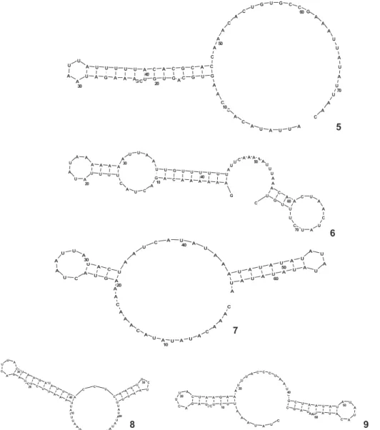
Potential secondary structures of the repeated sequences in the five pleurodiran turtles’ CR were constructed in this study, and the long repeated motif from each species formed a stable loop structure (Figs 5-9). In hornbills, these VNTRs’ stem-loop structures are believed to function as a termination se-quence in mitochondrial genome replication (DELPORT et al.
2002). In turtles, whether these VNTRs have some function or not needs further study, but it is noteworthy that the potential secondary structures of the repeated sequences were very simi-lar among P. unifilis, P. expansa, and P. subrufa (Figs 7-9).
Although the mechanism for generating the VNTRs in the mitochondrial genome is not well understood at present,
it is believed that the VNTRs can be used as molecular markers and provide sufficient phylogenetic information in molecular phylogenetics, population genetics, species identification, as well as genetic diversity and conservation, because they can be used to differentiate among genera, species, populations, and even individuals (ZARDOYA & MEYER 1998a, ZHANGet al. 2009).
Pelomedusids and podocnemidids were placed into Pelomedu-sidae (sensu lato) based on morphological characteristic (GAFFNEY
1975, GAFFNEY & MEYLAN 1988). Although DE BROIN (1988)
sug-gested that the extant Pelomedusinae and Podocneminae (GAFFNEY & MEYLAN 1988) should be recognized as two families,
i.e. Pelomedusidae (consisting of the extant African Pelusios
Figure 5-9.Secondary structure of repeat units constructed from VNTRs of the five pleurodiran turtles: (5) the 74 bp repeat unit of C. rugosa; (6) the 76 bp repeat unit of C. fimbriata; (7) the 65 bp repeat unit of P. subrufa; (8) the 69 bp repeat unit of P. expansa; (9) the 66 bp repeat unit of P. unifilis.
5
6
7
and Pelomedusa) and Podocnemidae (consisting of extant Madagascan Erymnochelys, and the extant South American Podocnemis and Peltocephalus), morphological studies still sup-port the monophyly of Pelomedusidae (sensu lato). Recently, molecular evidence has also suggested that the Pelomedusidae (sensu lato) may be monophyletic (GEOROGESet al. 1998, NOONAN
2000, FUJITAet al. 2004, NOONAN & CHIPPINDALE 2006, THOMSON
& SHAFFER 2010). In the present study, these secondary
struc-tures of VNTRs for P. subrufa (Pelomedusinae), P. expansa, and P. unifilis (Podocneminae) also supports the monophyly of Pelomedusidae (sensu lato). We suggest that comparative analy-ses of secondary structures of VNTRs may provide some poten-tial information for phylogenetic inferences.
ACKNOWLEDGMENTS
This research was supported by the National Natural Sci-ence Foundation of China (NSFC, #30970351), the Research Fund for the Doctoral Program of Higher Education of China (#20080370001), and the Key Laboratory of Biotic Environ-ment and Ecological Safety of Anhui province.
LITERATURE CITED
AMER, S.A. &Y. KUMAZAWA. 2009. Complete sequence of the
mitochondrial genome of the endangered Nile soft-shelled turtle Trionyx triunguis. Egyptian Journal of Experimental Biology (Zoology) 5: 43-50.
ANDERSON, S.; A.T. BANKIER; B.G. BARRELL; M.H.L. DE BRUIJN; A.R.
CNULSON; J. DROUIN; I.C.EPERON; D.P. NIERLICH; B.A. ROE; F. SANGER; P.H. SCHREIER; A.J.H. SMITH; R. STADEN & I.G. YOUNG. 1981. Sequence and organization of the human mitochondrial genome. Nature 290: 457-465.
BAKER, A.J. & H.D. MARSHALL. 1997. Mitochondrial control region
sequences as tools for understanding evolution, p. 51-82. In: D.P. MINDELL (Ed.). Avian Molecular Evolution and Systematics. San Diego, Academic Press.
BENSON, G. 1999. Tandem repeats finder: a program to analyze
DNA sequences. Nucleic Acids Research27 (2): 573-580. BREHM, A.; A.D. JAMES HARRIS; C.D. ALVES; J.D. JESUS; F.D. THOMARAT
& L.D. VICENTE. 2003. Structure and evolution of the mitochondrial DNA complete control region in the lizard Lacerta dugesii (Lacertidae, Sauria). Journal of Molecular Evolution 56 (1): 46-53.
BROUGHTON, R.E.& T.E. DOWLING. 1994. Length variation in mito-chondrial DNA of the minnow Cyprinella spiloptera. Genetics 138 (1): 179-190.
BROWN, G.G.; G. GADALETA; G. PEPE; C. SACCONE & E. SBISÀ. 1986.
Structural conservation and variation in the D-loop containing region of vertebrate mitochondrial DNA. Journal of Molecular Biology 192: 503-511.
BUROKER, N. E.; J.R. BROWN; T.A. GILBERT; P.J. O’HARA; A.T. BECKENBACH;
W.K. THOMASE & M.J. SMITH. 1990. Length Heteroplasmy of
Sturgeon Mitochondrial DNA: An Illegitimate Elongation Model. Genetics 144: 157-163.
CHANG, D. & D. CLAYTON. 1986. Identification of primary
trans-cription start sites of mouse mitochondrial DNA: Accurate in vitro initiation of both heavy- and light-strand transcriptions.
Molecular Cellular Biology 6: 1446-1453.
CLAYTON, D.A. 1982. Replication of animal mitochondrial DNA.
Cell 28:693-705.
DE BROIN, F. 1988. Les Tortues et le Gondwana. Examen des
rapports entre le fractionnement du Gondwana et la dispersion geographaique des tortues pleurodires a partir du Cretace. Stvdia Geologica Salmanticensia 2 (2): 103-142. DELPORT, W.; J.W. FERGUSON & P. BLOOMER. 2002. Characterization
and evolution of the mitochondrial DNA control region in hornbills (Bucerotiformes). Journal of Molecular Evolution 54: 794-806.
DODA, J.N.; C.T. WRIGHT & D.A. CLAYTON. 1981. Elongation of displacement-loop strands in human and mouse mitochondrial DNA is arrested near specific template sequences. Proceeding of the National Academy of Sciences of the United States of AME 78: 6116-6120.
DUNON-BLUTEAU, D.; M. VOLOVITCH & G. BRUN, 1985. Nucleotide sequence of a Xenopus lamis mitochondrial DNA fragment containing the D-loop, flanking tRNA genes and apocyto-chrome B gene. Gene 36: 65-78.
FORAN, D.R.; J.E. HIXSON & W.M. BROWN. 1988. Comparisons of
ape and human sequences that regulate mitochondrial DNA transcription and D-loop synthesis. Nucleic Acids Research 16: 5841-5861.
FU, Y.Y.; W.W. GU; Y.Z. LIU; J.Y. PENG & H.CHANG. 2006. Genetic
analysis of the polymorphism of mtDNA D loop and microsatellite loci in Tibet mini-pigs. Acta Laboratorium Animalis Scientia Sinica 14: 318-321.
FUJITA, M.K.; T.N. ENGSTROM; D.E. STARKEY & H.B. SHAFFER. 2004.
Turtle phylogeny: insights from a novel nuclear intron.
Molecular Phylogenetics Evolution 31: 1031-1040. FUMAGALLI, L.; P. TABERLET; L. FAVRE & J. HAUSSER. 1996. Origin and
evolution of homologous repeated sequences in the mitochondrial DNA control region of shrews. Molecular Biology Evolution 13: 31-46.
GAFFNEY, E.S. 1975. A phylogeny and classification of the higher
categories of turtles. Bulletin of the American Museum of Natural History 155: 391-436.
GAFFNEY, E.S. 1990. The comparative osteology of the Triassic
turtle Proganochelys. Bulletin of the American Museum of Natural History 194: 1-263.
GAFFNEY, E.S. & P.A. MEYLAN. 1988. A phylogeny of turtles, p. 157-219. In: M.J.BENTON (Ed.). The phylogeny and classification of Tetrapods. 1. Amphibians, Reptiles, Birds. New York, Systematics Association Special, vol. 35A.
GEMMELL, N.J.; P.S. WESTERN; J.M. WATSON & J.A. GRAVES. 1996.
mono-treme and therian mammals. Molecular Biology Evolution 13: 798-808.
GEOROGES, A.; J. BIRRELL; K.M. SAINT; W. MCCORD & S.C. DONNELLAN.
1998. A phylogeny for side-necked turtles (Chelonia: Pleurodira) based on mitochondrial and nuclear gene sequence variation.
Biological Journal of Linnnean Society 67: 213-246. GHIVIZZIANI, S.C.; C.S. MADSEN; M.R. NELEN; C.V. AMMINI & W.W.
HAUSWIRTH. 1994. In organelle foot print analysis of human
mitochondrial DNA: Human mitochondrial transcription factor A interactions at the origin of replication. Molecular Cellular Biology 14: 7717-7730.
GoricKI, S. & P. TRONTELJ. 2006. Structure and evolution of the mitochondrial control region and flanking sequences in the European cave salamander Proteus anguinus. Gene 378: 31-41.
HÄRLID, A.; A. JANKE & U. ARNASON. 1997.The mtDNA sequence
of the ostrich and the divergence between paleognathous and neognat-hous birds. Molecular Biology Evolution14: 754-761.
HOELZEL, A.R. 1993. Evolution by DNA turnover in the control
region of vertebrate mitochondrial DNA. Current Opinion in Genetics & Development 3: 891-895.
HOFACKER, I.L.; P.F. STADLER; S. BONHOEFFER & W. FONTANA. 1995.
The Vienna RNA package: A comprehensive package for RNA secondary structure calculation and analysis for Windows95/NT3.51. Available online at: http://www.tbi. univie.ac.at/RNA [Accessed: 04/VIII/2011].
JI, X.; X. WU; P. YAN & G. AMATO. 2008. Complete sequence and
gene organization of the mitochondrial genome of Siamensis Crocodile (Crocodylus siamensis) Molecular Biology Reports 35: 133-138.
JUNGT, S.O.; Y.M. LEE; Y. KARTAVTSEV; I.S. PARK; D.S. KIM & J.S. LEE. 2006. The complete mitochondrial genome of the Korean soft-shelled turtle Pelodiscus sinensis (Testudines, Trionychidae).
DNA Sequence 17 (6): 471-483.
KING, T.C.& R.L. LOW. 1987. Mapping of control elements in
the displacement loop region of bovine mitochondrial DNA.
Journal of Biological Chemistry 262: 6204-6213. KUMAR, S.; K. TAMURA & M. NEI. 1993. MEGA: Molecular
Evolu-tionary Genetics Analysis. Pennsylvania State University, University Park.
LARIZZA, A. 2002. Lineage Specificity of the Evolutionary Dynamics of the mtDNA D-Loop Region in Rodents. Journal of Molecular Evolution 54 (2): 145-155.
LIU, H.Z. 2002. The structure and evolution of the mtDNA
control region in fish: taking example for Acheilognathinae.
Progress in Natural Science 12 (3):266-270.
LOWE, T.M. & S.R. EDDY. 1997. tRNAscan-SE: a program for
improved detection of transfer RNA genes in genomic sequence. Available online at: http://lowelab.ucsc.edu/ tRNAscan-SE [Accessed: 04/VIII/2011].
MATHEWS, D.H.; M.E. BURKARD & D.H. TURNER. 1999. RNAstructure: A computer program for Windows95/NT. Available online
at http://rna.chem.rochester.edu [Accessed: 04/VIII/2011]. NEAR, T.J.; P.A. MEYLAN & H.B. SHAFFER. 2005. Assessing
concordance of fossil calibration points in molecular clock studies: an example using turtles. The American Naturalist 165: 137-146.
NILSSON, M.A. 2009.The structure of the Australian and South
American marsupial mitochondrial control region.
Mitochondrial DNA 20 (5-6): 126-38.
NOONAN, B.P. 2000. Does phylogeny of pelomedusid turtles
reflect vicariance due to continental drift? Journal of Biogeography 27: 1245-1249.
NOONAN, B.P.& P.T. CHIPPINDALE. 2006.Vicariant origin of Malagasy reptiles supports Late Cretaceous Antarctic landbridge. The American Naturalist 168: 731-741.
PENG, R.; B. ZENG; X. MENG; B. YUE; Z. ZHANG & F. ZOU. 2007. The
complete mitochondrial genome and phylogenetic analysis of the giant panda (Ailuropoda melanoleuca). Gene 397: 76-83.
QUINN, T.W. & A.C. WILSON. 1993 Sequence evolution in and
around the mitochondrial control region in birds. Journal of Molecular Evolution37: 417-425.
RAMIREZ,V.; P. SAVOIE & R. MORAIS. 1993.Molecular characterization and evolution of a duck mitochondrial genome. Journal of Molecular Evolution 37: 296-310.
RANDI, E. & V. LUCCHINI. 1998. Organization and Evolution of
the Mitochondrial DNA Control Region in the Avian Genus Alectoris. Journal of Molecular Evolution 47: 449-462. RAY, D.A. & L.D. DENSMORE. 2002. The Crocodilian Control
Region: General Structure, Conserved Sequences and Evolutionary Implications. Journal of Experimental Zoology (Molecular Development and Evolution) 294: 334-345.
RHODIN, A.G.J.; P.P. VAN DIJK; J.B. IVERSON & H.B. SHAFFER. 2010.
Turtles of the world, 2010 update: Annotated checklist of taxonomy and synonymy, Distribution, and Conservation Status, p. 000.85-000.164. In: A.G.J. RHODIN; P.C.H. PRITCHARD;
P.P. VAN DIJK; R.A. SAUMURE; K.A. BUHLMANN; J.B. IVERSON & R.A. MITTERMEIER (Eds). Conservation Biology of Freshwater Turtles and Tortoises: A Compilation Project of the IUCN/ SSC Tortoise and Freshwater Turtle Specialist Group Chelonian Research Monographs5. Checklist v3.2010, available online at: http://www.iucn-tftsg.org/cbftt, doi:10.3854/crm.5.000.
RITCHIE, P. & D. LAMBERT. 2000. A repeat complex in the
mito-chondrial control region of Adélie penguins from Antarctica.
Genome 43: 613-618.
RUOKONEN, M. & L. KVIST. 2002. Structure and evolution of the avian mitochondrial control region. Molecular Phylogenetics and Evolution 23: 422-432.
SACCONE, C.; G. PESOLE & E. SBISÀ. 1991. The main regulatory
SAITOU, N. & M. NEI. 1987. The neighbor-joining method: A
new method for reconstructing phylogenetic trees.
Molecular Biology and Evolution 4: 406-425.
SAMBROOK, J.& D. RUSSELL. 2001. Molecular Cloning: A
laboratory Manual. New York, Cold Spring Harbour Laboratory Press. 3rd ed.
SBISÀ, E.; F. TANZARIELLO; A. REYES; G. PESOLE & C. SACCONE. 1997. Mammalian mitochondrial D-loop region structural analysis: identification of new conserved sequences and their functional and evolutionary implications. Gene 205: 125-140.
SERB, J.M.; C.A. PHILLIPS & J.B. IVERSON. 2001.Molecular Phylogeny
and Biogeography of Kinosternon flavescens Based on Com-plete Mitochondrial Control Region Sequences. Molecular Phylogenetics and Evolution 1 (18): 149-162.
SUN, K.; J. FENG; L. JIN; Y. LIU; L. SHI & T. JIANG. 2009. Structure,
DNA sequence variation and phylogenetic implications of the mitochondrial control region in horse shoe bats.
Mammalian biology 74: 130-144.
THOMPSON, J.D.; T.J. GIBSON; F. PLEWNIAK; F. JEANMOUGIN & D.G.
HIGGINS. 1997. The ClustalX windows interface: flexible
strategies for multiple sequence alignment aided by quality analysis tools. Nucleic Acids Research 24: 4876 4882. THOMSON, R.C. & H.B. SHAFFER. 2010. Sparse Supermatrices for
Submitted: 05.IV.2011; Accepted: 24.VII.2011. Editorial responsibility: Marcio R. Pie
Phylogenetic Inference: Taxonomy, Alignment, Rogue Taxa, and the Phylogeny of Living Turtles. Systematic Biology 59
(1): 42-58.
WALBERG, M.W. & D.A. CLAYTON. 1981. Sequence and properties
of the human KB cell and mouse L cell D-loop regions of mitochondrial DNA. Nucleic Acids Research9: 5411-5421. XIONG, L.; L. NIE; X. LI & X. LIU. 2010. Comparison research and phylogenetic implications of mitochondrial control regions in four soft shelled turtles of Trionychia (Reptilia, Testudinata). Genes and Genomics 32:291-298.
ZARDOYA, R. & A. MEYER. 1998a. Cloning and characterization of
a microsatellite in the mitochondrial control region of the African side-necked turtle, Pelomedusa subrufa. Gene 216: 149-153.
ZARDOYA, R. & A. MEYER. 1998b. Complete mitochondrial genome
suggests diapsid affinities of turtles. Proceeding of the National Academy of Sciences of the United States of AME 95: 14226-14231.
ZHANG, Y.; L. NIE; Y. HUANG; Y. PU & L. ZHANG. 2009. The
Mito-chondrial DNA Control Region Comparison Studies of Four Hinged Turtles and its Phylogentic Significance of the Genus Cuora Sensu Lato (Testudinata: Geoemydidae). Genes & Genomics 31: 349-359.
Appendix. Species and associated GenBank accession numbers used in the present study.
Family name Species name GenBank accession number
Trionychidae Trionyx triunguis NC_012833
Apalone ferox FJ890514
Palea steindachneri FJ541030
Dogania subplana AF366350
Lissemys punctata NC_012414
Pelodiscus sinensis AY687385
Carettochelyidae Carettochelys insculpta FJ862792
Pelomedousidae Pelomedusa subrufa AF039066
Podocnemididae Podocnemis unifilis this study
Podocnemis expansa AY572983
Chelidae Chelus fimbriata this study
Chelodina rugosa this study
Geoemydidae Mauremys reevesi AY676201
Cuora aurocapitata AY874540
Cuora flavomarginata EU708434
Mauremys mutica DQ453753
Cuora mouhotii DQ659152
Sacalia quadriocellata EF088646
Cuora galbinifrons NC_014102
Appendix. Continued.
Family name Species name GenBank accession number
Chelydridae Chelydra serpentine NC_011198
Macrochelys temmincki EF071948
Emydidae Chrysemys picta AF069423
Trachemys scripta FJ392294
Cheloniidae Chelonia mydas AB012104
Eretmochelys imbricata NC_012398
Lepidochelys olivacea AM258984
Testudinidae Testudo kleinmanni DQ080048
Stigmochelys pardalis DQ080041
Malacochersus tornieri DQ080042
Manouria emys DQ080040
Indotestudo elongata DQ080043
Testudo horsfieldii DQ080045
Testudo graeca DQ080049
Indotestudo forstenii DQ080044
Testudo marginata DQ080047
Platysternidae Platysternon megacephalum DQ016387
Phasianidae Gallus gallus AY235571
Note: The taxononmical classification provided by Genbank entries was used for the species and the classification of RHODINet al. (2010) was used for the following four species: Macroclemys temminckii (Troost in Harlan, 1835) was replaced by Macrochelys temminckii (Troost, 1835); Chinemys reevesii (Gray, 1831) should be Mauremys reevesii (Gray, 1831); Geochelone pardalis (Bell, 1828) should be Stigmochelys