Interleukin 10
So-Hee Lim, Eunha Park, Boram You, Youngseob Jung, A-Reum Park, Sung Goo Park, Jae-Ran Lee
*Biomedical Proteomics Research Center, Korea Research Institute of Bioscience and Biotechnology, Daejeon, Korea
Abstract
Recently, it was found that microglia regulated synaptic remodeling of the developing brain, but their mechanisms have not been well understood. In this study, the action of microglia on neuronal synapse formation was investigated, and the primary target of microglial processes was discovered. When the developing microglia were applied to cultured hippocampal neurons without direct contact, the numbers of dendritic spines and excitatory and inhibitory synapses significantly increased. In order to find out the main factor for synaptic formation, the effects of cytokines released from microglia were examined. When recombinant proteins of cytokines were applied to neuronal culture media, interleukin 10 increased the numbers of dendritic spines in addition to excitatory and inhibitory synapses. Interestingly, without external stimuli, the amount of interleukin 10 released from the intact microglia appeared to be sufficient for the induction of synaptic formation. The neutralizing antibodies of interleukin 10 receptors attenuated the induction of the synaptic formation by microglia. The expression of interleukin 10 receptor was newly found in the hippocampal neurons of early developmental stage. When interleukin 10 receptors on the hippocampal neurons were knocked down with specific shRNA, the induction of synaptic formation by microglia and interleukin 10 disappeared. Pretreatment with lipopolysaccharide inhibited microglia from inducing synaptic formation, and interleukin 1 antagonized the induction of synaptic formation by interleukin 10. In conclusion, the developing microglia regulated synaptic functions and neuronal development through the interactions of the interleukin 10 released from the microglia with interleukin 10 receptors expressed on the hippocampal neurons.
Citation: Lim S-H, Park E, You B, Jung Y, Park A-R, et al. (β01γ) Neuronal Synapse Formation Induced by Microglia and Interleukin 10. PLoS ONE κ(11)μ eκ1β1κ. doiμ10.1γ71/journal.pone.00κ1β1κ
Editor: Laurent Groc, Institute for Interdisciplinary Neuroscience, France
Received March βκ, β01γ; Accepted October 10, β01γ; Published November ββ, β01γ
Copyright: © β01γ Lim et al. This is an open-access article distributed under the terms of the Creative Commons Attribution License, which permits unrestricted use, distribution, and reproduction in any medium, provided the original author and source are credited.
Funding: National Research Foundation of Korea (NRF) grant funded by the Korea government (β01βR1AβAβA0β0145β0 and β00λ-00κ7γ54 to J.R.L.) and a grant from KRIBB research initiative program (to J.R.L.). The funders had no role in study design, data collection and analysis, decision to publish, or preparation of the manuscript.
Competing interests: The authors have declared that no competing interests exist. * E-mailμ [email protected]
Introduction
Microglia are generally considered to be immunological sensors that survey neuronal diseases and viral attacks in the nerve system [1,β,γ]. The roles of microglia under normal physiological conditions, however, have been relatively neglected and have not been studied as much as the pathological roles [4,5,6,7,κ]. Recently, it has been reported that non-activated “resting” microglia dynamically extended and retracted their processes as if they were actively surveying the microenvironment in the brain [λ,10,11]. The interaction between the fractalkine receptors (CXγCR1) in microglia and the chemokine fractalkine (CXγCL1) in neurons was suggested as having significant roles in neuronal synaptic pruning and the regulation of synaptic transmissions during postnatal development in mice [1β,1γ]. Moreover, microglia were shown to engulf the presynaptic inputs in activity-dependent synaptic pruning processes through CRγ, the receptor of the
complement component Cγ on microglia in the postnatal retinogeniculate system [14].
shown to attenuate the long-term potentiation (LTP) in the hippocampus and its effects on synaptic plasticity were antagonized by interleukin 10 (IL-10) [β1]. Null mutant interleukin 1 receptor (IL-1 receptor) -/- mice showed impaired learning and synaptic plasticity, but displayed a memory rescue via the transplantation of wild type neural precursor cells [ββ]. IL-10 has been suggested to exert neuro-protective effects and to recover neurite outgrowth by decreasing glial activation and down-regulating microglial nitric oxide (NO) production [βγ,β4,β5]. IL-10-deficient (IL-10-/-) mice were shown to be less efficient than wild type mice in a test of hippocampal-dependent learning and memory, after the intraperitoneal injection of LPS [β6]. Recently, spinal cord neurons and cortical neurons were found to express interleukin 10 receptors (IL-10 receptors), and IL-10 appeared to have significant roles in neuronal development as well as neuronal protection [β7,βκ].
As previously mentioned, resting microglia have neuronal functions, including synaptic remodeling, during the development of central nervous systems [1β,1γ,14]. However, the primary target of these resting microglial processes has not yet been identified, and the mechanism of interaction between the microglia and the neuronal circuit is not well understood. In this paper, the induction of neuronal synapse formation by microglia was investigated using a co-culture system of hippocampal neurons and microglia without direct contact. The effects of cytokines which were known to be released from microglia were examined to find out the main factors for synaptic formation. Additionally, the expression of cytokine receptors on hippocampal neurons was discovered. To confirm the roles of cytokines, the expression of cytokine receptors was knocked down in hippocampal neurons, and then synaptic formation was analyzed. Our results suggest that microglia control neuronal synapse formation in developing neurons by releasing cytokines that interact with cytokine receptors expressed on hippocampal neurons.
Results
Neuronal synapse formation induced by microglia
Microglia were applied to hippocampal neurons to determine whether microglia could induce neuronal synapse formation without direct contact (Figure 1). Rat pups of postnatal Day 1 were used for the preparation of developing microglia. Floating microglia were harvested, plated on porous cell culture inserts, and then applied to cultured hippocampal neurons. Previously, several components released from astrocytes were suggested to induce neuronal synapse formation [β0]. Therefore, the preparation of microglia was performed very carefully to exclude astrocytes. The shape of the microglia was round and amoeboid (characteristics of “developing microglia”), when stained with anti-CD11b/c antibodies (specific marker of microglia) [βλ,γ0] (Figure 1A). The staining with anti-GFAP antibodies (specific astrocyte marker) showed that the prepared microglia were not contaminated with astrocytes. Moreover, the prepared microglia did not express mRNA of GFAP when RT-PCR was performed, and this again confirmed their purity (Figure 1B).
The effect of the developing microglia on synaptic formation was examined by visualizing the dendritic spine on hippocampal neurons that were transfected and overexpressed with green fluorescent protein (GFP). The microglia plated on the porous cell culture insert were applied to the cultured hippocampal neurons of days in vitro (DIV) κ, and the synaptic formation was analyzed at DIV 15. The density of the dendritic spines was significantly increased by addition of microglia compared with the control (no microglia) (Figure 1C). For the analysis of the excitatory and inhibitory synapses, the hippocampal neurons were stained with vGLUT or anti-vGAT antibodies (Figures 1D, 1E, respectively). The numbers of excitatory (vGLUT-stained) and inhibitory (vGAT-stained) synapses were also significantly increased by addition of microglia compared with the control.
Recently, it was found that ATP released from microglia acts on astrocytes through PβY1 purinergic receptors, which in turn regulate synaptic activity [1λ]. Reactive blue (RB), the antagonist of PβY receptors, was added to the co-culture system of microglia and hippocampal neurons to eliminate the induction of synaptic formation through PβY receptors (Figure S1). Synaptic formation was not reduced by RB at all, and this result shows that synaptic formation was not induced through PβY receptors on astrocytes. In conclusion, the developing microglia on porous cell culture inserts induced synaptic formation of hippocampal neurons without direct contact.
Neuronal synapse formation induced by IL-10 released from microglia
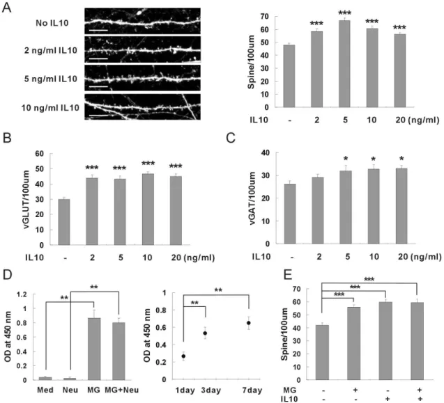
Microglia has been known to release many kinds of cytokines in response to external stimuli [5]. Because microglia induced neuronal synapse formation without direct contact, cytokines released from microglia could be main factors for synaptic formation. Therefore, the roles of cytokines were examined by applying recombinant proteins of cytokines to hippocampal neurons (Figure 2). When recombinant IL-10 was applied to hippocampal neurons at DIV κ and the synaptic formation was analyzed after one week, the density of the dendritic spines was significantly increased (Figure 2A). The numbers of excitatory synapses as well as the inhibitory synapses were also increased by application of IL-10 (Figure βB, βC). On the other hand, the recombinant IL-1 , TNF-α, IL-6, and IL-4 did not induce synaptic formation of hippocampal neurons at any concentrations (Figure S2A-2D). An ELISA assay demonstrated that an appreciable amount of IL-10 was released from the intact microglia without external stimuli in the neuronal culture media (Figure 2D). In order to confirm the roles of IL-10 released from microglia, recombinant IL-10 was applied to hippocampal neurons together with microglia (Figure 2E). Synaptic formation was not induced additively by application of exogenous IL-10 together with microglia. This result suggests that microglia and IL-10 use the same signal pathway for the induction of synaptic formation. In conclusion, microglia appear to induce neuronal synapse formation by releasing IL-10 into the culture media of hippocampal neurons.
New Functions of Microglia and Interleukin 10
Figure 1. Neuronal synapse formation induced by microglia. (A) Microglia were prepared from mixed glia without astrocytes. The prepared microglia were stained with antibodies of CD11b/c (red) and GFAP (green). CD11b/c and GFAP are specific markers of microglia and astrocytes respectively. The numbers of cells stained with antibodies of CD11b/c (microglia) or GFAP (astrocytes) were counted, and the purity of microglia was analyzed. Scale bar, 10 m.
(B) An RT-PCR assay was performed to determine the purity of microglia prepared from mixed glia. The expressions of Iba-1 (a specific marker of microglia) and GFAP (a specific marker of astrocytes) were analyzed.
(C) The density of dendritic spines was increased by application of microglia. Rat cultured hippocampal neurons were transfected at days in vitro (DIV) 7 with pSuper.neo-gfp, and then immunostained using anti-GFP antibodies at DIV 15 for analysis. Microglia plated on a porous cell culture insert were applied to hippocampal neurons of DIV κ. The density of dendritic spines increased when microglia was applied. Means±SEM. n=4κ dendrites for no microglia, 4κ for 0.β5 × 10⁵ microglia, 4κ for 0.5 × 10⁵ microglia, 4κ for
1.0 × 10⁵ microglia. **p<0.01 and ***p<0.001, by the Newman-Keuls multiple comparison test after application of one-way ANOVA,
F=λ.λ15, p<0.0001. Scale bar, 10 m.
(D) The number of excitatory synapses was increased by application of microglia. The cultured hippocampal neurons were immunostained using antibodies of GFP (green) and vGLUT, the excitatory synaptic marker (red). The addition of microglia increased the number of vGLUT. Means±SEM. n=40 dendrites for no microglia, 40 for 0.β5 × 10⁵ microglia, 40 for 0.5 × 10⁵
microglia, 40 for 1.0 × 10⁵ microglia. ***p<0.001, by the Newman-Keuls multiple comparison test after application of one-way
ANOVA, F=κ.β06, p<0.0001. Scale bar, 10 m.
(E) The number of inhibitory synapses was increased by application of microglia. Antibodies of GFP (green) and vGAT, the inhibitory synaptic marker (red), were used for the immunostaining of hippocampal neurons. The addition of microglia increased the number of vGAT. Means±SEM. n=40 dendrites for no microglia, 40 for 0.β5 × 10⁵ microglia, 40 for 0.5 × 10⁵ microglia, 40 for 1.0 ×
105 microglia. *p<0.05 and ***p<0.001, by the Newman-Keuls multiple comparison test after application of one-way ANOVA,
Figure 2. Neuronal synapse formation induced by IL-10 released from microglia. (A) The density of dendritic spines was increased by application of recombinant IL-10. Recombinant proteins of IL-10 were applied to hippocampal neurons of DIV κ and the density of dendritic spines was analyzed after one week. Means±SEM. n=γ1 dendrites for control (no IL-10), γ1 for β ng/ml IL-10, γ1 for 5 ng/ml IL-10, γ1 for 10 ng/ml IL-10, γ1 for β0 ng/ml IL-10. ***p<0.001 by the Newman-Keuls multiple comparison test after application of one-way ANOVA, F=15.66, p<0.0001. Scale bar, 10 m.
(B) The number of excitatory synapses was increased by application of recombinant IL-10. The application of recombinant IL-10 increased the number of vGLUT. Means±SEM. n=γ4 dendrites for control (no IL-10), γ5 for β ng/ml IL-10, γ1 for 5 ng/ml IL-10, γ1 for 10 ng/ml IL-10, γγ for β0 ng/ml IL-10. ***p<0.001 by the Newman-Keuls multiple comparison test after application of one-way ANOVA, F=16.γγ, p<0.0001.
(C) The number of inhibitory synapses was increased by application of recombinant IL-10. The application of recombinant IL-10 increased the number of vGAT. Means±SEM. n=γβ dendrites for control (no IL-10), βκ for β ng/ml IL-10, βλ for 5 ng/ml IL-10, β7 for 10 ng/ml IL-10, β7 for β0 ng/ml IL-10. *p<0.05 by the Newman-Keuls multiple comparison test after application of one-way ANOVA,
F=β.λ60, p=0.0ββ0.
(D) IL-10 was released from intact microglia without external stimuli. Microglia were plated on a porous cell culture insert, and incubated in a neuronal culture media. After γ~7 days, the amount of IL-10 released from the microglia was analyzed using an ELISA kit. An appreciable amount of IL-10 was released from microglia compared with controls (neuronal culture media only or hippocampal neuron only). **p<0.01 by the Newman-Keuls multiple comparison test after application of one-way ANOVA, F=4κ.β5,
p=0.001γ.
(E) Microglia and recombinant IL-10 use the same signal pathway for induction of synaptic formation. Addition of recombinant IL-10 did not additively enhance the induction of synaptic formation by microglia. Means±SEM. n=β7 dendrites for control (no microglia no IL-10), β7 for the application of 1.0 × 10⁵ microglia, βκ for 5 ng/ml of IL-10, β7 for microglia and IL-10. ***p<0.001 by the
Newman-Keuls multiple comparison test after application of one-way ANOVA, F=15.50, p<0.0001. doiμ 10.1γ71/journal.pone.00κ1β1κ.g00β
New Functions of Microglia and Interleukin 10
IL-10 receptors expressed on hippocampal neurons
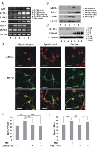
It is well known that IL-10 receptors are expressed on glial cells [1,κ]. Recently, it has been discovered that spinal neurons, cortical neurons, and retinal ganglion cells also have IL-10 receptors [β7,βκ,γ1]. Using the RT-PCR assay, the expressions of IL-10 receptors were examined on cultured hippocampal neurons (Figure 3A). Interestingly, the IL-10 receptor α was mainly expressed in early developing hippocampal neurons of DIV 7, and on the other hand, IL-10 receptor was expressed similarly in DIV 7 and DIV 15 neurons (Figure 3A, IL10Rα & IL10R , panels 1 and β). Because IL-10 receptors have been known to be expressed on glial cells, the question was raised whether the IL-10 receptor mRNA shown in panels 1 and β originated from hippocampal neurons [γβ]. The prepared microglia exhibited very strong expressions of IL-10 receptors, but the cultured neurons appeared not to be contaminated with microglia (Figure 3A, no Iba-1, a specific microglia marker, panels 1 and β). Although astrocytes appeared to grow with hippocampal neurons (Figure 3A, GFAP, panels 1 and β), the IL-10 receptor mRNA of the hippocampal neurons did not appear to originate from the astrocyte, as indicated by the IL-10 receptor α expression being stronger in DIV 7 neurons than in DIV 15 neurons, which is opposite to the GFAP expression (stronger in DIV 15 neurons than in DIV 7 neurons).
Then, the expressions of IL-10 receptor proteins were examined in cultured hippocampal neurons using anti-IL-10 receptor α antibody (Figure 3B). Similar to the mRNA expressions, the IL-10 receptor α proteins were expressed mainly in DIV 7 neurons (Figure 3B, panels 1 and β, Mw λ0-110 kDa) [γγ]. The developing rat brains of embryonic and early postnatal days also showed stronger expressions of IL-10 receptor α than did adult rat brains (Figure 3C). When an immunohistofluorescence was performed using anti-IL-10 receptor antibody, the hippocampal neurons expressed IL-10 receptor α not less than spinal cord neurons or cortical neurons did (Figure 3D).
In order to confirm the functions of IL-10 in synaptic formation, the neutralizing antibody of IL-10 receptor was applied to a co-culture system and the interaction between IL-10 and IL-10 receptor were inhibited (Figure 3E). The induction of synaptic formation by microglia disappeared when the neutralizing antibody of IL-10 receptor was applied. However, the anti-TNFα antibodies did not inhibit the induction of the synaptic formation by microglia (Figure 3F). As a result, synaptic formation was induced through the interaction of IL-10 released from the microglia with IL-10 receptors expressed on the hippocampal neurons of early postnatal development.
Neuronal synapse formation attenuated by the knockdown of IL-10 receptors
A specific shRNA was applied to attenuate the expression of IL-10 receptors on the hippocampal neurons, and the effects of knockdown on synaptic formation were examined (Figure 4). First, the efficiency of shRNA was evaluated by applying the lentivirus containing the shRNA of IL-10 receptor α to hippocampal neurons (Figure 4A). The IL-10 receptor α mRNA was decreased considerably with application of the shRNA.
Then two types of shRNA specific to rat IL-10 receptor α were applied to the hippocampal neurons of DIV 7, and the density of the dendrites was analyzed at DIV 15 (Figure 4B). The induction of synaptic formation via microglia and recombinant IL-10 disappeared entirely after application of shRNA. These results confirmed again the expression of IL-10 receptors on hippocampal neurons, and the induction of synaptic formation by the IL-10 released from the microglia. Thus the developing microglia appear to regulate neuronal development through the release of IL-10 bound to IL-10 receptors that are expressed in hippocampal neurons.
IL-1β antagonized IL-10 in neuronal synapse formation
It is known that the release of cytokines from microglia can be enhanced in response to treatment with bacterial endotoxin LPS [5]. The effect of LPS treatment on neuronal synapse formation was examined to determine whether microglia could regulate synaptic formation under pathological conditions (Figure 5A). When microglia were treated with LPS before application to hippocampal neurons, the induction of synaptic formation by microglia was attenuated. Treatment with 1 ng/ml LPS was sufficient to inhibit microglia from inducing synaptic formation. The RT-PCR assay showed that the expressions of IL-1 and TNFα on microglia were significantly increased by 1 ng/ml LPS, but on the other hand, IL-10 was not convincingly increased (Figure 5B). Although IL-1 and TNFα did not have any effect on neuronal synapse formation (Figure S2A, S2B), they could interfere with IL-10 and inhibit the induction of synaptic formation by microglia. Thus recombinant IL-1 or TNFα was applied to hippocampal neurons, together with recombinant IL-10, in order to investigate the crosstalk between cytokines. The induction of synaptic formation by IL-10 disappeared when IL-1 was applied to hippocampal neurons (Figure 5C). On the other hand, TNFα did not antagonize the effects of IL-10 on synaptic formation (Figure S3). These data suggest that the expression of IL-1 increased by LPS treatment attenuated the induction of synaptic formation by IL-10.
Discussion
From the present study, the developing microglia were newly found to induce synapse formation of hippocampal neurons by releasing the anti-inflammatory cytokine IL-10. IL-10 appears to interact with IL-10 receptors expressed on hippocampal neurons of early developmental stage. Without external stimuli, microglia released an appreciable amount of IL-10 for the induction of synaptic formation. Pro-inflammatory cytokines IL-1 antagonized the functions of IL-10 in the pathological conditions. When the IL-10 receptors were knocked down in the hippocampal neurons, the induction of synaptic formation was significantly attenuated. In conclusion, microglia could regulate the synaptic formation by releasing IL-10 that interacts with IL-10 receptor expressed on the developing hippocampal neurons.
Figure 3. IL-10 receptors expressed on hippocampal neurons. (A) Expression of IL-10 receptor mRNAs in hippocampal neurons. The expression of the IL-10 receptor was identified using RT-PCR. The mRNAs of IL-10 receptor α and were expressed in the hippocampal neurons. IL-10 receptor α was expressed mainly in hippocampal neurons of DIV 7. (1, cultured hippocampal neurons at DIV 7; β, cultured hippocampal neurons at DIV 15; γ, mixed glial culture at DIV 7; 4, mixed glial culture at DIV 15; 5, microglia) Quantification (DIV 15 neuron/ DIV 7 neuron)μ IL-10 receptor α, 0.61; IL-10 receptor , 1.06.
(B) Expression of IL-10 receptor proteins in cultured hippocampal neurons. Similar to the expression of mRNA, the IL-10 receptor α protein was expressed mainly in neurons of DIV 7. Anti-IL-10 receptor α antibodies (0.5 g/ml, Santa Cruz, sc-λκ5) were used for western blotting [β7]. Quantification (DIV 15 neuron/ DIV 7 neurons)μ IL-10 receptor α, 0.7γ.
(C) Expression of IL-10 receptor proteins in the developing rat brains. The IL-10 receptor α proteins were expressed mainly in the developing brains of embryonic and early postnatal days (E1κ~Pγ). Quantification of IL-10 receptor αμ E1κ, 0.γ0; P1, 0.β7; Pγ, 1.0; P7, 0.ββ; P14, 0.β0; PγW, 0.17; P6W, 0.15 (E, embryonic days; P, postnatal days).
(D) Images of IL-10 receptor expressions in cultured hippocampal neurons. Hippocampal neurons of DIV 6 were stained with antibodies of IL-10 receptor α (5 g/ml, Santa Cruz, sc-λκ5) (red) and MAPβ (the neuronal marker, green) after treatment with 4% formaldehyde and then -β0 °C methanol. Hippocampal neurons expressed IL-10 receptor proteins comparable to spinal neurons or cortical neurons.
(E) The induction of synaptic formation by microglia was antagonized by the neutralizing antibody of IL-10 receptor α. When anti-mouse IL-10 receptor α antibody was applied to the co-culture of anti-mouse microglia and anti-mouse hippocampal neurons, the density of dendritic spines was significantly decreased compared with the control (without anti-IL-10 receptor antibody). Means±SEM. n=γ0 dendrites for no microglia without IL-10 receptor antibody, βλ for the application of microglia only, βλ for no microglia with anti-IL-10 receptor antibody only, βλ for microglia with anti-anti-IL-10 receptor antibody. ***p<0.001 by the Newman-Keuls multiple comparison test after application of one-way ANOVA, F=17.γ5, p<0.0001.
(F) The induction of synaptic formation via microglia was not antagonized by the blocking antibody of TNFα. When anti-rat TNFα antibody was applied to the co-culture of rat microglia and rat hippocampal neurons, the density of dendritic spine was not decreased compared with control (without anti-TNFα antibody). Means±SEM. n=β7 dendrites for no microglia without anti-TNFα antibody, βκ for the application of microglia only, βλ for no microglia with anti-TNFα receptor antibody only, β7 for microglia with anti-TNFα antibody. **p<0.01 and ***p<0.001 by the Newman-Keuls multiple comparison test after application of one-way ANOVA,
F=λ.104, p<0.0001.
doiμ 10.1γ71/journal.pone.00κ1β1κ.g00γ
New Functions of Microglia and Interleukin 10
well understood. Our results show that IL-10 induces synaptic formation of hippocampal neurons, and IL-1 antagonizes the effects of IL-10 when endogenously released from microglia or
applied as recombinant proteins. It was reported that IL-1 receptors were also expressed on early postnatal neurons [γ1,γ4]. The developing microglia appear to regulate neuronal Figure 4. Neuronal synapse formation attenuated by the knockdown of IL-10 receptors. (A) The expressions of IL-10 receptors attenuated by shRNA in hippocampal neurons. The lentivirus expressing shRNA of IL-10 receptor α was applied to the cultured hippocampal neurons of DIV γ, and then RT-PCR was performed at DIV 7. The expression of IL-10 receptor α was decreased significantly by the application of shRNA. Quantification (shRNA/vector only)μ 15 A.U. shRNA, 0.7λ; β0 A.U. shRNA, 0.4λ; β5 A.U. shRNA, 0.β0 (A.U., arbitrary unit).
(B) Attenuated synaptic formation by application of shRNA of IL-10 receptor α. The induction of synaptic formation by microglia or recombinant IL-10 was dramatically attenuated when shRNA of IL-10 receptor α was applied to hippocampal neurons. Means±SEM.
n=βλ dendrites for vector only (shVec), βκ for shRNA of IL-10 receptor α #1 (shRNA#1), β6 for shRNA of IL-10 receptor α #β (shRNA#β), βλ for the application of microglia only, γ0 for microglia with shRNA#1, β6 for microglia with shRNA#β, βλ for the application of recombinant IL-10 (5 ng/ml) only, γ0 for IL-10 with shRNA#1, β7 for IL-10 with shRNA#β. ***p<0.001 by the Newman-Keuls multiple comparison test after application of one-way ANOVA, F=βγ.04, p<0.0001. Scale bar, 10 m.
Figure 5. IL-1β antagonized IL-10 in neuronal synapse formation. (A) The induction of synaptic formation by microglia was attenuated when microglia were pretreated with lipopolysaccharide (LPS). Means±SEM. n=βλ dendrites for no microglia, βλ for non-treated microglia, γ0 for microglia prenon-treated with 1 ng/ml LPS, γ0 for microglia prenon-treated with 10 ng/ml LPS, and βλ for microglia pretreated with 100 ng/ml LPS. ***p<0.001, by the Newman-Keuls multiple comparison test after application of one-way ANOVA, F=κ.7κ5, p<0.0001.
(B) Increased expressions of cytokine mRNAs after LPS treatment. An RT-PCR was performed after the microglia were treated with LPS at indicated concentrations. The mRNAs of IL-1 and TNFα were increased significantly by 1 ng/ml LPS, but the expression of IL-10 mRNA was not convincingly increased by 1 ng/ml LPS. Quantification (LPS treated/not treated)μ IL-10 by 1 ng/ml LPS, 1.57; IL-10 by 10 ng/ml LPS, 4.1γ; IL-10 by 100 ng/ml LPS, 4.06; IL-1 by 1 ng/ml LPS, 1λ.β; IL-1 by 10 ng/ml LPS, βκ.0; IL-1 by 100 ng/ml LPS, β5.κ; TNFα by 1 ng/ml LPS, 4.β6; TNFα by 10 ng/ml LPS, 5.07; TNFα by 100 ng/ml LPS, 4.4κ.
(C) The induction of synaptic formation by IL-10 was attenuated when IL-1 was applied together with IL-10. Recombinant proteins of IL-10 or IL-1 were applied to the cultured hippocampal neurons of DIV κ and the density of dendritic spines was analyzed after one week. IL-1 antagonized the induction of synaptic formation by IL-10. Means±SEM. n=γ0 dendrites for control (no IL-10 no IL-1 ), βκ for 5 ng/ml IL-10, β7 for 10 ng/ml IL-10, β6 for β ng/ml IL-1 , βκ for 5 ng/ml IL-1 , βλ for 10 ng/ml IL-1 , βλ for 5 ng/ml IL-10 plus 5 ng/ml IL-1 , γ0 for 10 ng/ml IL-10 plus 10 ng/ml IL-1 . **p<0.01 and ***p<0.001 by the Newman-Keuls multiple comparison test after application of one-way ANOVA, F=14.67, p<0.0001. Scale bar, 10 m.
doiμ 10.1γ71/journal.pone.00κ1β1κ.g005
New Functions of Microglia and Interleukin 10
functions including synaptic formation and synaptic plasticity by releasing cytokines such as IL-10 and IL-1 of which receptors are expressed on early postnatal neurons.
Microglia have been reported to engulf synaptic inputs and to play a role in activity-dependent synaptic pruning during postnatal brain development [1β,1γ,14]. These results which appear contradictory to ours, suggest that microglia function as a double-edged sword for synaptic formationμ microglia could remodel developing synapses both through direct contact and by indirect processes. Microglia appear to engulf synaptic materials for synaptic pruning and concurrently to release cytokines for induction of synaptic formation. When microglia were applied “directly” to developing hippocampal neurons (DIV κ), the density of dendritic spines was “not” increased after one week (Figure S4). This result appears to make sense because the induction of synaptic formation by IL-10 released from microglia could be countervailed by synaptic pruning derived from direct contact with microglia. In this context, it appears to be answerable for a recent report that synaptic formation was inhibited by “direct” application of microglia to the matured neurons [γ5]. Because IL-10 receptors are not expressed in the matured neurons, synaptic pruning could prevail and synaptic formation would be attenuated. When recombinant IL-10 was applied to the matured hippocampal neurons of DIV 14, synaptic formation was “not” induced after one week (DIV β1) (Figure S5). According to our results, the expression of IL-10 receptor α was significantly decreased in hippocampal neurons of DIV 15 (Figure 3A-3C). Therefore, IL-10 could regulate neuronal synapse formation in the early brain development.
Recently, IL-10 has attracted significant attention for potential therapeutic application because of its anti-inflammatory and immunosuppressive effects [β7,γ6,γ7,γκ]. IL-10 was suggested to have important roles for neuronal protection in glutamate-mediated neuronal cell death, focal brain ischemia, and spinal cord injuries. After central nervous system injuries, IL-10 was shown to improve neurological outcomes by reducing the number of apoptotic cells [γ6]. In transgenic mice expressing murine IL-10, the release of pro-inflammatory cytokines including TNFα, interferon- , and IL-1 were significantly reduced and the infarct size was reduced compared with wild type mice for middle cerebral artery occlusion [γ7]. The injection of herpes simplex virus-based vectors to express IL-10 in the spinal cord, increased the neuronal survival in the anterior quadrant of the spinal cord, and improved motor function after injury [γγ]. IL-10 appears to protect brains and to assist with neuronal functions under normal physiological conditions as well as in emergency situations.
Neurons were considered to be immune-privileged in a healthy brain; however, it has been known that the major factors of the immune system have significant roles in neuronal functions. Among them, MHC class I was suggested to regulate synaptic functions in the developing visual system, and to play major roles especially in postsynaptic activity [γλ,40,41]. In addition, C1q, the initiating protein of the classical complement pathway, was also shown to localize to immature synapses with Cγ and necessary for the developmental
pruning of retinogeniculate synapses [4β]. IL-6, a pro-inflammatory cytokine, was reported to inhibit the induction LTP in hippocampal slices [4γ]. These results suggest that many factors of immune systems have major roles in the maintenance of synaptic plasticity as well as in the regulation of synaptic formation [44,45,46]. Therefore, it would be very interesting to discover new roles of immune components including various cytokines and chemokines in neuronal functions and brain development.
Materials and Methods
Ethics statement
This study was performed in accordance with the regulations outlined by the Korean law. The animal experiment protocols were approved by the Animal Use and Care Committee of Korea Research Institute of Bioscience and Biotechnology (Permit Numberμ KRIBB-AEC-110ββ). Animals were sacrificed using COβ gas, and all efforts were made to minimize suffering.
Cytokines and antibodies
Recombinant cytokines were purchased from R&D systems (Minneapolis, MN, USA)μ rat IL-10 (547-RL), rat IL-1 (501-RL), rat TNFα (510-RT), rat IL-6 (506-RL), and rat IL-4 (504-RL). The following antibodies were usedμ anti-mouse IL-10 receptor α (AF-474-NA), anti-rat TNFα (MAB510) obtained from R&D systems (Minneapolis, MN, USA); CD11b/c (ab1β11), anti-GFAP (ab16λλ7) from Abcam (Cambridge, UK); anti-vGLUT (1γ5 γ0γ), anti-vGAT (1γ1 00β) from SYSY (Gottingen, Germany); anti-Iba-1 from Wako Pure Chemical Industries (Osaka, Japan); anti-MAPβ (M1406) from Sigma-Aldrich (Saint Louis, MO, USA); anti- -actin (4λ67) from Cell Signaling (Danvers, MA, USA); anti-IL-10 receptor α (sc-λκ5) from Santa Cruz Biotechnology (Dallas, TX, USA) [47,4κ]. The neutralizing antibody of IL-10 receptor α (R&D systems, AF-474-NA) or anti-rat TNFα antibody (R&D systems, MAB510), was applied to the culture media at a concentration of β g/ml.
Primer for RT-PCR
In these experiments, 1 g of total RNA was reverse transcribed in a β0 l reaction volume. PCRs were carried out using 1 l of the completed RT as template with the corresponding primersμ rat IL-10 mRNA (NM_01βκ54), TGCCTTCAGTCAAGTGAAGAC-γ’ (forward primer) and 5’-AAACTCATTCATGGCCTTGTA-γ’ (reverse primer); rat IL-1 (NM_0γ151β), 5’-CACCTTCTTTTCCTTCATCTTTG-γ’ (forward primer) and 5’-GTCGTTGCTTGTCTCTCCTTGTA-γ’ (reverse
primer); rat TNFα (NM_01β675),
CCCAGACCCTCACACTCAGAT-γ’ (forward primer) and 5’-TTGTCCCTTGAAGAGAACCTG-γ’ (reverse primer); rat IL-10 receptor α (NM_0571λγ), 5’-CTCGCTTCACAGTGGATGAA-γ’ (forward primer) and 5’-TAAATACGGTGGTGCGTGAA-γ’ (reverse primer); rat IL-10 receptor (NM_00110711), TCAGCATGGTGTGGTTCATT-γ’ (forward primer) and 5’-TCTTCCGTGATGATGCTCAG-γ’ (reverse primer); rat Iba-1
(NM_0171λ6), 5’-CCATGAAGCCTGAGGAAATTTCA-γ’
(reverse primer); rat GFAP (NM_01700λ), GAAACCAACCTGAGGCTGGAG-γ’ (forward primer) and 5’-GGCGATAGTCATTAGCCTCG-γ’ (reverse primer); rat GAPDH (NM_01700κ), 5’-CCCCCAATGTATCCGTTGTG-γ’ (forward primer) and 5’-TAGCCCAGGATGCCCTTTAGT-γ’ (reverse
primer); rat -actin (NM_0γ1144),
5’-AGAAGAGCTATGAGCTGCCTGACG-γ’ (forward primer) and 5’-TACTTGCGCTCAGGAGGAGCAATG-γ’ (reverse primer).
Production of shRNA of IL-10 receptor α
For knockdown of rat IL10 receptor α (NM_0571λγ), nt 7γκ -756 (CGTGGAATCCCGAATTAAC, shRNA of IL-10 receptor α #1) or nt 14βλ - 1447 (TACCAGAAGCAGACCAGAT, shRNA of IL-10 receptor α #β) was subcloned into pSuper.gfp/neo plasmid vector (OligoEngine, Seattle, WA, USA). pSuper.gfp/neo containing shRNA was transfected and expressed in rat cultured hippocampal neurons using the calcium phosphate method. The same sequence for shRNA was used for the production of lentivirus.
Preparation of mixed glial cell culture and harvest of floating microglia
Rats of postnatal 1 day were decapitated and the skulls were washed twice using pre-warmed L-15 media (Sigma, Saint Louis, USA) with penicillin/streptomycin. The brains were taken out of skulls, washed twice, and stripped of their meninges. Cortex were isolated without hippocampus, transferred to conical tube, and minced with Pasteur pipette in L-15 media. Minced cortex was centrifuged at 1β00 rpm for γ sec and the supernatant containing cells were transferred to new conical tube. Then the pellet was added with new L-15 media, re-suspended, and centrifuged again. The resulted supernatant was transferred to new conical tube, and this process was performed once more. The pooled supernatant containing cells was centrifuged at 1400 rpm 5 min and the resulted pellet was added with the glial media (DMEM media with 10% FBS, 1 mM L-glutamine, 1 mM sodium pyruvate, penicillin/streptomycin). The glial cells were re-suspended and transferred to cell culture dish pre-coated with poly-D-lysine. After 1 day, half of media was changed with new glial media and then twice in a week. The floating microglia cells were harvested at 7~14 days after plating and centrifuged at 1400 rpm 5 min. The pellet was re-suspended with the glial media and transferred to porous cell culture insert (Millicell-PCF, PIHP01β50, Millipore, USA). The prepared microglia were stabilized on cell culture insert overnight and then applied to cultured hippocampal neurons next day.
Preparation of cultured hippocampal neurons,
transfection of neurons, and immunohistofluorescence
Primary hippocampal neurons were prepared from embryonic day 1κ (E1κ) rats, grown on glass coverslip pre-coated with poly-D-lysine in serum-free neurobasal media (Invitrogen, USA) with glutamine and B-β7 serum-free supplement (Invitrogen, USA), and transfected using the calcium phosphate method at days in vitro (DIV) 7, as described previously [4λ]. Microglia plated on porous cell culture insert or IL-10 were applied to hippocampal neurons of
DIV κ. After one week, at DIV 15, hippocampal neurons were fixed in 4% (v/v) formaldehyde/4% (w/v) sucrose, permeabilized with 0.β% (v/v) Triton X-100 in phosphate-buffered saline, incubated with primary antibodies (anti-EGFP, anti-vGLUT, anti-vGAT, anti-IL-10 receptor α, anti-MAPβ, 1~5 g/ml) overnight at 4 °C, and finally incubated with Cyγ-, or FITC-conjugated secondary antibodies (1μ1000, or 1μβ50 dilution) (Jackson ImmunoResearch Laboratories, West Glove, PA, USA) for β h at room temperature. Microglia was fixed, permeabilized, incubated with primary antibodies (anti-CD11b/c, anti-GFAP) overnight at 4 °C, and finally incubated with Cyγ-, or FITC-conjugated secondary antibodies for β h at room temperature.
Image acquisition and quantification
Images captured by confocal microscopy (LSM 510 Meta, Zeiss, Gottingen, Germany) using a 6γx objective were analyzed blindly using MetaMorph software (Universal Imaging) [50]. The density of dendritic spines (0.4-β.5 m) and synaptic protein clusters were measured from γ0-40 dendrites of eight to ten neurons; the total dendritic length of ~50 m was measured from the first dendritic branching points. Means from multiple individual dendrites were averaged to obtain a population mean and SEM. All experiments were repeated more than three times with similar results.
Sample preparation for western blot analysis
Primary hippocampal neurons grown on cell culture dishes were lysed with ice-cold 1% (v/v) Triton X-100 in Dulbecco’s phosphate-buffered saline (pH 7.4) (Invitrogen, USA) containing inhibitors of proteases and phosphatases. After incubation on ice for γ0 min, the neuron lysates were centrifuged at 1β,000 rpm for γ0 min at 4 °C. The cleared extracts were mixed with SDS-PAGE sampling buffer, boiled for κ min, resolved on SDS-PAGE, and blotted to nitrocellulose membrane (Amersham, UK). After incubated using primary antibodies (antiIL10 receptor α, antiIba1, antiGFAP, anti -actin, anti-PSD-λ5, anti-α-tubulin, 0.β~0.5 g/ml) and then HRP-labeled secondary antibodies, the protein bands blotted on nitrocellulose membranes were detected with ECL (Pierce chemical co, USA).
Mixed glial cells grown on cell culture dishes were lysed with ice-cold 1% (v/v) Triton X-100 in Dulbecco’s phosphate-buffered saline containing inhibitors of proteases and phosphatases. The mixed glial lysates were cleared, and mixed with SDS-PAGE sampling buffer. Floating microglia were harvested, centrifuged, and lyzed with ice-cold 1% (v/v) Triton X-100 in Dulbecco’s phosphate-buffered saline containing inhibitors of proteases and phosphatases.
For the study of developing rat brain, brains were removed from embryonic day 1κ (E1κ), postnatal day 1, γ, 7, 14 (P1, Pγ, P7, P14 respectively), and postnatal γ, 6 weeks (PγW and P6W respectively) rats. Brains were lysed in ice-cold buffered sucrose (0.γβ M sucrose, 4 mM HEPES, 1 mM MgClβ, 0.5 mM
CaClβ, pH 7.γ) with protease inhibitors using a tissue
homogenizer, mixed with SDS-PAGE sampling buffer, resolved on SDS-PAGE, and blotted to nitrocellulose membrane.
New Functions of Microglia and Interleukin 10
Measurement of IL-10 released from microglia
The microglia prepared from the mixed glial culture were plated on a porous cell culture insert, and then incubated in a neuronal culture media. After γ~7 days, the amount of IL-10 released from the microglia was analyzed. The amount of IL-10 released from the mouse microglia was measured using an ELISA kit (BD OptEIA Set Mouse IL-10, BD Biosciences, San Diego, CA, USA). The ELISA assay was carried out according to manufacturer instructions. Briefly, standard or culture media containing IL-10 were added to each well of the ELISA plate, pre-bound with anti-IL-10 antibody, incubated for β hr at room temperature, and washed five times. Then detection antibody and HRP were incubated successively. Finally, TMB substrate solution was added, incubated for γ0 min in the dark, and the absorbance was measured at 450 nm with correction 570 nm.
Treatment of microglia with LPS
Microglia on cell culture inserts were treated with LPS for 4 h and then residual LPS was washed out before microglia was applied to the cultured hippocampal neurons of DIV κ. After one week, at DIV 15, the synaptic formation of hippocampal neurons was analyzed.
Statistics
Results are expressed as Means±SEM and one-way ANOVA followed by application of the Newman-Keuls multiple comparison test was used to assess statistical significance. A comparison was considered to be significant if P<0.05.
Supporting Information
Figure S1. Neuronal synapse formation not induced through purinergic receptor. Reactive blue (RB), the antagonist of PβY purinergic receptor, was added to the co-culture system of hippocampal neurons and microglia. Synaptic formation was not reduced by treatment with RB. Means±SEM.
n=40 dendrites for no microglia, 40 for 1.0 × 10⁵ microglia, 40
for 1.0 × 10⁵ microglia plus 5 ng/ml RB, 40 for 1.0 × 10⁵
microglia plus 50 ng/ml RB. ***p<0.001, by the Newman-Keuls multiple comparison test after application of one-way ANOVA,
F=βλ.16, p<0.0001. Scale bar, 10 m. (TIF)
Figure S2. Neuronal synapse formation not induced by various cytokines. (A) The density of dendritic spines was not increased by application of recombinant IL-1 . Recombinant proteins of IL-1 were applied to the neuronal culture media at DIV κ and after one week the density of dendritic spines was analyzed. Means±SEM. n=βλ dendrites for control (no IL-1 ), γ0 for β ng/ml IL-1 , γ0 for 5 ng/ml IL-1 , βλ for 10 ng/ml IL-1 , βλ for β0 ng/ml IL-1 . These differences are considered to be not statistically significant by the Newman-Keuls multiple comparison test after application of one-way ANOVA, F=1.100,
p=0.γ5λ1.
(B) The application of the recombinant TNFα did not induce synaptic formation significantly at any concentration. Means
±SEM. n=βκ dendrites for control (no TNFα, βκ for 0.0γ ng/ml TNFα, βκ for 0.γ ng/ml TNFα, βκ for γ ng/ml TNFα, γ0 for 10 ng/ml TNFα, γ0 for 100 ng/ml TNFα These differences are considered to be not statistically significant by the Newman-Keuls multiple comparison test after application of one-way ANOVA, F=1.7γ6, p=0.1βλ1.
(C) The application of the recombinant IL-6 did not induce synaptic formation significantly at any concentration. Means ±SEM. n=βλ dendrites for control (no IL-6), γ1 for 0.5 ng/ml IL-6, γ0 for 5 ng/ml IL-6, βλ for β0 ng/ml IL-6, βλ for 400 ng/ml IL-6. These differences are considered to be not statistically significant by the Newman-Keuls multiple comparison test after application of one-way ANOVA, F=β.ββ7, p=0.06λ0.
(D) The application of the recombinant IL-4 did not induce synaptic formation significantly at any concentration. Means ±SEM. n=β7 dendrites for control (no IL-4), βκ for β ng/ml IL-4, β7 for 5 ng/ml IL-4, βλ for 10 ng/ml IL-4. These differences are considered to be not statistically significant by the Newman-Keuls multiple comparison test after application of one-way ANOVA, F=β.γ4γ, p=0.077β.
(TIF)
Figure S3. TNFα did not antagonize the effects of IL-10. When TNFα was applied to hippocampal neurons together with IL-10, the induction of synaptic formation by IL-10 was not attenuated. TNFα could not antagonize the effects of IL-10 in synaptic formation. Means±SEM. n=βκ dendrites for control (no IL-10 no TNFα), βλ for 5 ng/ml IL-10, βλ for 5 ng/ml TNFα, γ0 for 5 ng/ml IL-10 plus 5 ng/ml TNFα. *p<0.05 and **p<0.01, by the Newman-Keuls multiple comparison test after application of one-way ANOVA, F=βγ.γβ, p<0.0001.
(TIF)
Figure S4. Direct application of microglia did not induce synaptic formation. When developing microglia were applied directly to the hippocampal neurons of DIV κ and synaptic formation was analyzed after one week, the density of dendritic spine was not increased. Means±SEM. n=βλ dendrites for control (no microglia), βκ for 1.0 × 10⁵ microglia plated on cell
culture insert, βκ for 0.1 × 10⁵ microglia applied directly, βλ for
0.5 × 10⁵ microglia applied directly, βλ for 1.0 × 10⁵ microglia
applied directly. ***p<0.001, by the Newman-Keuls multiple comparison test after application of one-way ANOVA, F=11.7γ,
p<0.0001. (TIF)
Figure S5. IL-10 did not induce synaptic formation in the matured neurons. When recombinant IL-10 (5 ng/ml) was applied to hippocampal neurons of DIV 14 and synaptic formation was analyzed after one week (DIV β1), the density of dendritic spine was not increased significantly. Means±SEM.
n=βκ dendrites for control (no IL-10), β7 for 5 ng/ml IL-10. These differences are considered to be not statistically significant by the Newman-Keuls multiple comparison test after application of one-way ANOVA, F=β.0ββ, p=0.ββκ1.
Acknowledgements
We would like to thank Dr. EH. Joe for introduction into preparing rat microglia, and Dr. K. Seok for preparing mouse microglia.
Author Contributions
Conceived and designed the experimentsμ SHL JRL. Performed the experimentsμ SHL EP BY YJ. Analyzed the dataμ EP ARP SGP. Contributed reagents/materials/analysis toolsμ JRL. Wrote the manuscriptμ BY JRL.
References
1. Barres BA (β00κ) The mystery and magic of gliaμ a perspective on their roles in health and disease. Neuron 60μ 4γ0-440. doiμ10.1016/j.neuron. β00κ.10.01γ. PubMedμ 1κλλ5κ17.
β. Kempermann G, Neumann H (β00γ) Micorgliaμ the enemy within? Science γ0βμ 16κλ-16λ0. doiμ10.11β6/science.10λβκ64. PubMedμ 1465747λ.
γ. David S, Kroner A (β011) Repertoire of microglial and macrophage responses after spinal cord injury. Nat Rev Neurosci 1βμ γκκ-γλλ. doiμ 10.10γκ/nrnγ05γ. PubMedμ β167γ7β0.
4. Dheen ST, Kaur C, Ling EA (β007) Microglial activation and its implications in the brain diseases. Curr Med Chem 14μ 11κλ-11λ7. doiμ 10.β174/0λβλκ67077κ05λ7λ61. PubMedμ 175041γλ.
5. Hanisch UK, Kettenmann H (β007) Microgliaμ active sensor and versatile effector cells in the normal and pathologic brain. Nat Neurosci 10μ 1γκ7-1γλ4. doiμ10.10γκ/nn1λλ7. PubMedμ 17λ6565λ.
6. Ji KA, Yang MS, Jeong HK, Min KJ, Kang SH, Jou I, Joe EH (β007) Resident microglia die and infiltrated neutrophils and monocytes become major inflammatory cells in lipopolysaccharide-injected brain. Glia 55μ 1577-15κκ. doiμ10.100β/glia.β0571. PubMedμ 17κβγλ75. 7. Ji KA, Eu MY, Kang SH, Gwag BJ, Jou I, Joe EH (β00κ) Differential
neutrophil infiltration contributes to regional differences in brain inflammation in the substantia nigra pars compacta and cortex. Glia 56μ 10γλ-1047. doiμ10.100β/glia.β0677. PubMedμ 1κγκ1656.
κ. Biber K, Neumann H, Inoue K, Boddeke HWGM (β007) Neuronal 'On' and 'Off' signals control microglia. Trends Neurosci γ0μ 5λ6-60β. doiμ 10.1016/j.tins.β007.0κ.007. PubMedμ 17λ50λβ6.
λ. Nimmerjahn A, Kirchhoff F, Helmchen F (β005) Resting microglial cells are highly dynamic surveillants of brain parenchyma in vivo. Science γ0κμ 1γ14-1γ1κ. doiμ10.11β6/science.1110647. PubMedμ 15κγ1717. 10. Stence N, Waite M, Dailey ME (β001) Dynamics of microglial activationμ
A confocal time-lapse analysis in hippocampal slices. Glia γγμ β56-β66. doiμ10.100β/10λκ-11γ6(β0010γ)γγμγ. PubMedμ 11β4174γ.
11. Wu LJ, Zhuo M (β00κ) Resting microglial motility is independent of synaptic plasticity in mammalian brain. J Neurophysiol λλμ β0β6-β0γβ. doiμ10.115β/jn.01β10.β007. PubMedμ 1κβ5616β.
1β. Paolicelli RC, Bolasco G, Pagani F, Maggi L, Scianni M et al. (β011) Synaptic pruning by microglia is necessary for normal brain development. Science γγγμ 1456-145κ. doiμ10.11β6/science.1β0β5βλ. PubMedμ β177κγ6β.
1γ. Hoshiko M, Arnoux I, Avignone E, Yamamoto N, Audinat E (β01β) Deficiency of the microglial receptor CXγCR1 impairs postnatal functional development of thalamocortical synapses in the barrel cortex. J Neurosci γβμ 15106-15111. doiμ10.15βγ/JNEUROSCI. 1167-1β.β01β. PubMedμ βγ1004γ1.
14. Schafer DP, Lehrman EK, Kautzman AG, Koyama R, Mardinly AR et al. (β01β) Microglia sculpt postnatal neural circuits in an activity and complement-dependent manner. Neuron 74μ 6λ1-705. doiμ10.1016/ j.neuron.β01β.0γ.0β6. PubMedμ ββ6γβ7β7.
15. Beattie EC, Stellwagen D, Morishita W, Bresnahan JC, Ha BK et al. (β00β) Control of synaptic strength by glial TNFα. Science βλ5μ ββκβ-ββκ5. doiμ10.11β6/science.1067κ5λ. PubMedμ 11λ10117. 16. Stellwagen D, Malenka RC (β006) Synaptic scaling mediated by glial
TNF-α. Nature 440μ 1054-105λ. doiμ10.10γκ/nature04671. PubMedμ 16547515.
17. Kaltschmidt B, Widera D, Kaltschmidt C (β005) Signaling via NF- B in the nervous system. Biochim Biophysic. Acta 1745μ βκ7-βλλ.
1κ. Santello M, Bezzi P, Volterra A (β011) TNFα controls glutamatergic gliotransmission in the hippocampal dentate gyrus. Neuron 6λμ λκκ-1001. doiμ10.1016/j.neuron.β011.0β.00γ. PubMedμ β1γκβ557. 1λ. Pascual O, Achoura SB, Rostaing P, Triller A, Bessis A (β01β)
Microglia activation triggers astrocyte-mediated modulation of excitatory neurotransmission. Proc Natl Acad Sci U S A 10λμ E1λ7-Eβ05. doiμ10.107γ/pnas.110476710λ. PubMedμ ββ167κ04.
β0. Christopherson KS, Ullian EM, Stokes CCA, Mullowney CE, Hell JW et al. (β005) Thrombospondins are astrocyte-secreted proteins that promote CNS synaptogenesis. Cell 1β0μ 4β1-4γγ. doiμ10.1016/j.cell. β004.1β.0β0. PubMedμ 15707κλλ.
β1. Kelly A, Lynch A, Vereker E, Nolan Y, Queenan P et al. (β001) The anti-inflammatory cytokine, interleukin (IL)-10, blocks the inhibitory effect of IL-1 on long term potentiation. J Biol Chem β76μ 45564-4557β. doiμ10.1074/jbc.M10κ757β00. PubMedμ 115κ1β75. ββ. Menachem-Zidon OB, Avital A, Ben-Menahem Y, Goshen I, Kreisel T
et al. (β011) Astrocytes support hippocampal-dependent memory and long-term potentiation via interleukin-1 signaling. Brain Behav Immun β5μ 100κ-1016. doiμ10.1016/j.bbi.β010.11.007. PubMedμ β10λγ5κ0. βγ. Silva SL, Vaz AR, Diógenes MJ, van Rooijen N, Sebastião AM et al.
(β01β) Neuritic growth impairment and cell death by unconjugated bilirubin is mediated by NO and glutamate, modulated by microglia, and prevented by glycoursodeoxycholic acid and interleukin-10. Neuropharmacology 6βμ βγλκ-β40κ. doiμ10.1016/j.neuropharm. β01β.0β.00β. PubMedμ ββγ61βγγ.
β4. Balasingam V, Yong VW (1λλ6) Attenuation of astroglial reactivity by interleukin-10. J Neurosci 16μ βλ45-βλ55. PubMedμ κ6ββ1β5. β5. Park KW, Lee HG, Jin BK, Lee YB (β007) Interleukin-10 endogenously
expressed in microglia prevents lipopolysaccharide-induced neurodegeneration in the rat cerebral cortex in vivo. Exp Mol Med γλμ κ1β-κ1λ. doiμ10.10γκ/emm.β007.κκ. PubMedμ 1κ160κ5β.
β6. Richwine AF, Sparkman NL, Dilger RN, Buchanan JB, Johnson RW (β00λ) Cognitive deficits in interleukin-10-deficient mice after peripheral injection of lipopolysaccharide. Brain Behav Immun βγμ 7λ4-κ0β. doiμ 10.1016/j.bbi.β00λ.0β.0β0. PubMedμ 1λβ7β4γλ.
β7. Zhou Z, Peng X, Insolera R, Fink DJ, Mata M (β00λ) Interleukin-10 provides direct trophic support to neurons. J Neurochem 110μ 1617-16β7. doiμ10.1111/j.1471-415λ.β00λ.06β6γ.x. PubMedμ 1λ575707.
βκ. Sharma S, Yang B, Xi XP, Grotta JC, Aronowski J et al. (β011) IL-10 directly protects cortical neurons by activating PI-γ kinase and STAT-γ pathways. Brain Res 1γ7γμ 1κλ-1λ4. doiμ10.1016/j.brainres. β010.11.0λ6. PubMedμ β11γκ740.
βλ. Zusso M, Methot L, Lo R, Greenhalgh AD, David S et al. (β01β) Regulation of postnatal forebrain amoeboid microglial cell proliferation and development by the transcription factor Runx1. J Neurosci γβμ 11βκ5-11βλκ. doiμ10.15βγ/JNEUROSCI.61κβ-11.β01β. PubMedμ ββκλ571β.
γ0. Kaur C, Ling EA, Wong WC (1λκ7) Localisation of thiamine pyrophosphatase in the amboeboid microglial cells in the brain of postnatal rats. J Anat 15βμ 1γ-ββ. PubMedμ βκβ0λ1β.
γ1. Diem R, Hobom M, Grötsch P, Kramer B, Bähr M (β00γ) Interleukin-1 protects neurons via the interleukin-1 (IL-1) receptor-mediated Akt pathway and by IL-1 receptor-independent decrease of transmembrane currents in vivo. Mol Cell Neurosci ββμ 4κ7-500. doiμ10.1016/ S1044-74γ1(0β)0004β-κ. PubMedμ 1β7β7445.
γβ. Ledeboer A, Brevé JJP, Wierinckx A, van der Jagt S, Bristow AF et al. (β00β) Expression and regulation of interleukin-10 and interleukin-10 receptor in rat astroglial and microglial cells. Eur J Neurosci 16μ 1175-11κ5. doiμ10.1046/j.1460-λ56κ.β00β.0ββ00.x. PubMedμ 1β405λ7κ.
γγ. Zhou Z, Peng X, Insolera R, Fink DJ, Mata M (β00λ) IL-10 promotes neuronal survival following spinal cord injury. Exp Neurol ββ0μ 1κγ-1λ0. doiμ10.1016/j.expneurol.β00λ.0κ.01κ. PubMedμ 1λ716γ66.
γ4. Huang Y, Smith DE, Ibáñez-Sandoval O, Sims JE, Friedman WJ (β011) Neuron-specific effects of Interleukin-1b are mediated by a novel isoform of the IL-1 receptor accessory protein. J Neurosci γ1μ 1κ04κ-1κ05λ. doiμ10.15βγ/JNEUROSCI.4067-11.β011. PubMedμ ββ15λ11κ.
γ5. Ji K, Akgul G, Wollmuth LP, Tsirka SE (β01γ) Microglia actively regulate the number of functional synapses. PLOS ONE κμ e56βλγ-e56βλγ. doiμ10.1γ71/journal.pone.0056βλγ. PubMedμ βγγλγ60λ. γ6. Bachis A, Colangelo AM, Vicini S, Doe PP, De Bernardi MA et al.
(β001) Interleukin-10 prevents glutamate-mediated cerebellar granule cell death by blocking caspase-γ-like activity. J Neurosci β1μ γ104-γ11β. PubMedμ 11γ1ββλ5.
γ7. De Bilbao F, Arsenijevic D, Moll T, Garcia-Gabay I, Vallet P et al. (β00λ) In vivo over-expression of interleukin-10 increases resistance to New Functions of Microglia and Interleukin 10
focal brain ischemia in mice. J Neurochem 110μ 1β-ββ. doiμ10.1111/j. 1471-415λ.β00λ.060λκ.x. PubMedμ 1λ457075.
γκ. Pestka S, Krause CD, Sarkar D, Walter MR, Shi Y et al. (β004) Interleukin-10 and related cytokines and receptors. Annu Rev Immunol ββμ λβλ-λ7λ. doiμ10.1146/annurev.immunol.ββ.01β70γ.1046ββ. PubMedμ 150γβ600.
γλ. Corriveau RA, Huh GS, Shatz CJ (1λλκ) Regulation of Class I MHC gene expression in the developing and mature CNS by neural activity. Neuron β1μ 505-5β0. doiμ10.1016/S0κλ6-6β7γ(00)κ056β-0. PubMedμ λ76κκγκ.
40. Huh GS, Boulanger LM, Du H, Riquelme PA, Brotz TM et al. (β000) Functional requirement for Class I MHC in CNS development and plasticity. Science βλ0μ β155-β15λ. doiμ10.11β6/science. βλ0.54λλ.β155. PubMedμ 1111κ151.
41. Goddard CA, Butts DA, Shatz CJ (β007) Regulation of CNS synapses by neuronal MHC class I. Proc Natl Acad Sci U S A 104μ 6κβκ-6κγγ. doiμ10.107γ/pnas.070β0βγ104. PubMedμ 174β0446.
4β. Stevens B, Allen NJ, Vazquez LE, Howell GR, Christopherson KS et al. (β007) The classical complement cascade mediates CNS synapse elimination. Cell 1γ1μ 1164-117κ. doiμ10.1016/j.cell.β007.10.0γ6. PubMedμ 1κ0κγ105.
4γ. Tancredi V, D’Antuono M, Cafè C, Giovedì S, Buè MC et al. (β000) The inhibitory effects of interleukin-6 on synaptic plasticity in the rat hippocampus are associated with an inhibition of mitogen-activated protein kinase ERK. J Neurochem 75μ 6γ4-64γ. PubMedμ 10κλλλγκ. 44. Gao X, Smith GM, Chen J (β00λ) Impaired dendritic development and
synaptic formation of postnatal-born dentate gyrus granular neurons in
the absence of brain-derived neurotrophic factor signaling. Exp Neurol β15μ 17κ-1λ0. doiμ10.1016/j.expneurol.β00κ.10.00λ. PubMedμ 1λ014λγ7.
45. Monteggia LM, Barrot M, Powell CM, Berton O, Galanis V et al. (β004) Essential role of brain-derived neurotrophic factor in adult hippocampal function. Proc Natl Acad Sci U S A 101μ 10κβ7-10κγβ. doiμ10.107γ/ pnas.040β141101. PubMedμ 15β4λ6κ4.
46. Todd KJ, Serrano A, Lacaille JC, Robitaille R (β005) Glial cells in synaptic plasticity. J Physiol λλμ 75-κγ. PubMedμ 1644607κ.
47. Hocke AC, Ermert M, Althoff A, Brell B, N’Guessan PD et al. (β006) Regulation of interleukin IL-4, IL-1γ, IL-10, and their downstream components in lipopolysaccharide-exposed rat lungs. Comparison of the constitutive expression between rats and humans. Cytokine γγμ 1λλ-β11. doiμ10.1016/j.cyto.β006.01.007. PubMedμ 165ββγ70. 4κ. Boyd ZS, Kriatchko A, Yang J, Agarwal N, Wax MB, et al. (β00γ)
Interleukin-10 receptor signaling through STAT-γ regulates the apoptosis of retinal ganglion cells in response to stress. Inv Oph Vis Sic 44μ 5β06-5β11
4λ. Park AR, Oh D, Lim SH, Choi J, Moon J et al. (β01β) Regulation of dendritic arborization by BCR Rac1 GTPase-activating protein, a new substrate of protein tyrosine phosphatase receptor T. J Cell Sci 1β5μ 451κ-45γ1. doiμ10.1β4β/jcs.10550β. PubMedμ ββ76750λ.