Clinicopathological Features and Prognosis of
Metaplastic Breast Carcinoma: Experience of
a Major Chinese Cancer Center
Yiqian Zhang1, Feng Lv1, Yiling Yang1, Xiaolong Qian1, Ronggang Lang1, Yu Fan1, Fangfang Liu1, Yaqing Li1, Shuai Li1, Beibei Shen1, Gordon A. Pringle2, Xinmin Zhang2, Li Fu1, Xiaojing Guo1*
1Department of Breast Pathology and Lab, Key Laboratory of Breast Cancer of Breast Cancer Prevention and Therapy, National Clinical Research Center of Cancer, Tianjin Medical University Cancer Institute and Hospital, Tianjin, China,2Department of Pathology and Laboratory Medicine, Temple University School of Medicine, Philadelphia, Pennsylvania, United States of America
*guoxiaojing0728@126.com
Abstract
Metaplastic breast carcinoma (MBC) is a rare heterogeneous group of primary breast malig-nancies, with low hormone receptor expression and poor outcomes. To date, no prognostic markers for this tumor have been validated. The current study was undertaken to evaluate the clinicopathologic characteristics, the response to various therapeutic regimens and the prognosis of MBCs in a large cohort of patients from Tianjin Medical University Cancer Hos-pital in China. Ninety cases of MBCs diagnosed in our hosHos-pital between January 2000 and September 2014 were retrieved from the archives. In general, MBCs presented with larger size, a lower rate of lymph node metastasis, and demonstrated more frequent local recur-rence/distant metastasis than 1,090 stage-matched cases of invasive carcinoma of no spe-cific type (IDC-NST), independent of the status of estrogen receptor, progesterone receptor and human epidermal growth factor receptor 2 expressions. The five-year disease-free sur-vival (DFS) of MBC was significantly worse than IDC-NST. Using univariate analysis, lymph node metastasis, advanced clinical stage at diagnosis, high tumor proliferation rate
assessed by Ki-67 labeling, and epidermal growth factor receptor (EGFR) overexpression/ gene amplification were associated significantly with reduced DFS, while decreased OS was associated significantly with lymph node metastasis and EGFR overexpression/gene amplification. With multivariate analysis, lymph node status was an independent predictor for DFS, and lymph node status and EGFR overexpression/gene amplification were inde-pendent predictors for OS. Histologic subtyping and molecular subgrouping of MBCs were not significant factors in prognosis. We also found that MBCs were insensitive to neoadju-vant chemotherapy, routine chemotherapy, and radiation therapy. This study indicates that MBC is an aggressive type of breast cancer with poor prognosis, and that identification and optimization of an effective comprehensive therapeutic regimen is needed.
a11111
OPEN ACCESS
Citation:Zhang Y, Lv F, Yang Y, Qian X, Lang R, Fan Y, et al. (2015) Clinicopathological Features and Prognosis of Metaplastic Breast Carcinoma: Experience of a Major Chinese Cancer Center. PLoS ONE 10(6): e0131409. doi:10.1371/journal. pone.0131409
Editor:Yves St-Pierre, INRS, CANADA
Received:April 16, 2015
Accepted:June 2, 2015
Published:June 26, 2015
Copyright:© 2015 Zhang et al. This is an open access article distributed under the terms of the Creative Commons Attribution License, which permits unrestricted use, distribution, and reproduction in any medium, provided the original author and source are credited.
Data Availability Statement:All relevant data are within the paper.
Funding:National Natural Science Foundation of China (Grant No. 81172531) to Xiaojing Guo, National Natural Science Foundation of China (Grant No. 81302294) to Yiling Yang, National Natural Science Foundation of China (Grant No.81302292) to Xiaolong Qian and National Natural Science Foundation of China (Grant No. 81202101) to Fangfang Liu.
Introduction
Metaplastic breast carcinoma (MBC) is a rare type of breast cancer accounting for 0.2–5% of
all invasive mammary carcinomas [1]. It encompasses a group of neoplasms characterized by differentiation of the neoplastic epithelium into squamous cells and/or mesenchymal-looking elements. The 2012 WHO Classification of Tumors of Breast divided MBC into subtypes based upon the morphologic components of the tumors. The majority of MBCs are triple negative for estrogen receptors (ER), progesterone receptors (PR) and human epidermal growth factor receptor 2 (HER2) expression (TNBC), and may express cytokeratin 5/6 (CK5/6) and/or epi-dermal growth factor receptor (EGFR). MBC tends to present as a large tumor mass with low
axillary lymph node metastasis (LNM) and poor prognosis [2,3]. The lack of ER, PR, and
HER2 expression [2,4,5] makes endocrine therapy and molecularly targeted therapy
ineffec-tive, and therefore adjuvant and/or neoadjuvant chemotherapy have become the mainstay of management, although no therapeutic regimen has proven to be effective. So far, validated prognostic markers have not been identified for the tumor. Different morphologic and biologic features of the tumor have been reported in patients from different ethnic groups, and the out-comes of MBC diagnosed in different regions vary significantly [6]. Because of this variability and the fact that little is known about the biologic characteristics of MBC in the Chinese popu-lation, this study was undertaken to evaluate MBC with regard to its clinicopathologic charac-teristics, its response to multi-disciplinary therapeutic regimens and its prognosis in a large cohort of patients from Tianjin Medical University Cancer Hospital, a major Chinese cancer center.
Materials and Methods
Ethics statement
All human breast tissues were collected with written informed consent from patients prior to participation in the study. The protocols for collection and analysis of the samples were approved by the Institutional Review Board of the Tianjin Medical University Cancer Institute and Hospital, in accordance with the current revision of the Helsinki Declaration. The study was approved by the Institutional Review Board of the Tianjin Medical University Cancer Institute and Hospital.
Case Selection
Ninety cases of MBC were identified from 30,216 cases of invasive breast carcinoma (0.3%) diagnosed between January 2000 and September 2014 at the Department of Breast Cancer Pathology and Research Laboratory, Tianjin Medical University Cancer Hospital, Tianjin,
China. All patients were female and the median age was 54.6 years (range 28–89 years). Family
history of breast cancer or BRCA gene mutation was noted in 4 of the patients. Prior to the can-cer diagnosis, a portion of the patients had annual mammography or ultrasonography screen-ing for breast cancer startscreen-ing from age 40. Seventy-seven (85.6%) patients received modified or radical mastectomy (MRM), 5 patients (5.6%) accepted breast-conserving surgery, and the other 8 (0.9%) patients received quadrectomy. Final clean surgical margins in excisional speci-mens were achieved in all the patients, either initially or in follow-up wider excision or mastectomy.
(IDC-NST) were randomly selected from the same time period as the control group, among which 193 cases were triple negative for ER, PR and HER2 expression (TN-IDC).
Immunohistochemistry
Immunohistochemistry (IHC) was performed on selected tumor sections using the avidin-biotin-immunoperoxidase technique for ER, PR, HER2, Ki-67, p53, CK5/6 and EGFR. All pri-mary antibodies were purchased from Abcam (Cambridge, MA, USA). Formalin-fixed paraffin embedded tissue sections were employed in each case using a standard protocol. The immune-reaction was evaluated independently by the 3 pathologists.
The ER, PR and HER2 status was determined using the criteria of the American Society of
Clinical Oncology/College of American Pathologists (ASCO/CAP) [7,8]. For ER and PR,
nuclear staining in1% of the tumor cells was considered positive. HER2 immunoreactivity
was evaluated on a standardized scale from 0–3 based on the intensity of membranous staining
and the proportion of invasive tumor cells stained, with strong complete membranous staining
in>10% of tumor cells (3+) considered positive. Ki-67 and p53 immunoreaction presented
with nuclear staining, and CK5/6 and EGFR with membranous and/or cytoplasmic stain. Ki-67 labelling index was calculated and high tumor proliferation was defined as a labelling index
14% [9]. Overexpression of p53 was defined as nuclear stain in10% tumor cells [10], and a
cut-off of>10% tumor cells stained was adopted for CK5/6 positivity [11]. EGFR
immunor-eaction was evaluated based on the staining intensity, with 0 indicating absence of staining, and 1+, 2+, and 3+ representing respectively weak, moderate, and strong staining intensity. EGFR overexpression was defined as 2+ or 3+ staining. Molecular classification of tumors was performed using the established criteria [12]. Tumors negative for ER, PR and HER2, and
posi-tive for CK5/6 and/or EGFR were classified as basal-like carcinoma [13–15].
Fluorescent in situ hybridization
Fluorescent in-situ hybridization (FISH) detection of HER2 gene amplification was performed in selected cases with equivocal IHC reaction for HER2 (2+), using FDA-approved PathVysion HER2 DNA Probe Kit (Abbott Laboratories). At least 20 invasive carcinoma cells in each case were evaluated to determine HER2 gene copies and the ratio of HER2 gene vs chromosome 17
centromere signals. HER2/CEP17 ratio>2.0 was considered positive HER2 gene amplification,
per the 2013 ASCO/CAP recommendations [8]. FISH for EGFR was performed using the LSI EGFR/CEP 7 Probe (Vysis) per manufacturer's instruction. EGFR gene amplification was
defined as a ratio of EGFR gene vs chromosome 7 centromere signals2.0. EGFR
FISH-amplified samples also included those with40% of tumor cells demonstrating4 copies of
EGFR gene.
Survival analysis
All patients were followed up for 1–173 months with a median of 59 months. They were
fol-lowed at 3-month intervals initially, then at 6-month intervals, and annually afterwards. Two patients were lost in follow-up. Patients were censored from the date of last follow-up visit or death from causes other than breast cancer, local or regional recurrences, or the development of a second primary carcinoma, including contralateral breast cancer. If a patient was con-firmed to have metastasis during follow-up without recurrence, the last follow-up visit date was used. Age, time to first recurrence, and survival time were calculated relative to the primary
diagnosis date. Kaplan–Meier survival curves were constructed, and between-group differences
was tested using Cox-proportional hazard analysis and expressed with a 95% confidence inter-val (CI).
Statistical analysis
The statistical analysis was performed with the use of software packages SPSS version 19.0. All
statistical tests were two-sided, and a P value of<0.05 was considered significant.
Results
Clinicopathological features
Characteristics of the 90 MBC patients and control patients are summarized inTable 1.
Com-pared to patients with IDC-NST or TN-IDC, patients with MBC displayed unique characteris-tics, such as larger tumor size (p = 0.000; p = 0.013) and less frequent lymph node metastasis (LNM; p = 0.000; p = 0.000). No significant differences in patient age and tumor stage were identified. As reported previously, a greater proportion of MBCs are TNBCs than tumors in the IDC-NST group (p = 0.000).
Table 1. Clinicopathological features of MBC, IDC-NST and TN-IDC.
Characteristics MBC IDC-NST P TN-IDC P
N(%) N(%) N(%)
Age 50 35(38.9) 502(46.1) 0.189 96(49.7) 0.088
>50 55(61.1) 588(53.9) 97(50.3)
LN metastasis* Yes 16(20.8) 541(50.8) 0.000 92(49.2) 0.000
No 61(79.2) 523(49.2) 95(50.8)
Primary tumor 1 19(21.1) 348(31.9) 0.000 47(24.4) 0.013
2 51(56.7) 674(61.8) 128(66.3)
3 19(21.1) 50(4.6) 14(7.3)
4 1(1.1) 18(1.7) 4(2.1)
Recurrence/metastasis Yes 22(24.4) 136(12.5) 0.001 29(15.0) 0.044
No 66(73.3) 954(87.5) 164(85.0)
Pathologic tumor stage I 12(13.3) 221(20.3) 0.061 32(16.6) 0.062
II 68(75.6) 688(63.1) 120(62.2)
III 10(11.1) 181(16.6) 41(21.2)
Type of surgery MRM 77(85.6) 1064(97.6) 0.000 187(96.9) 0.000
Other 13(14.4) 26(2.4) 6(3.1)
Chemotherapy Yes 74(82.2) 870(79.8) 0.583 172(89.1) 0.109
No 16(17.8) 220(20.2) 21(10.9)
Radiotherapy Yes 5(5.6) 222(20.4) 0.001 52(26.9) 0.000
No 85(94.4) 868(79.6) 141(73.1)
Endocrine therapy Yes 9(10.0) 512(47.0) 0.000 _ _
No 81(90.0) 578(53.0) _
Anti-HER2 therapy Yes 1(1.1) 127(11.7) 0.000 _ _
No 89(98.9) 963(88.3) _
TNBC Yes 64(71.1) 193(17.7) 0.000 _ _
No 26(28.9) 897(82.3) _
MBC, Metaplastic breast carcinoma; IDC-NST, Invasive carcinoma of no special type; TN-IDC, Triple negative invasive ductal carcinoma; MRM, Modified radical mastectomy; TNBC, Triple negative breast cancer;
*No lymph node information was available in 13 of the MBC cases infile.
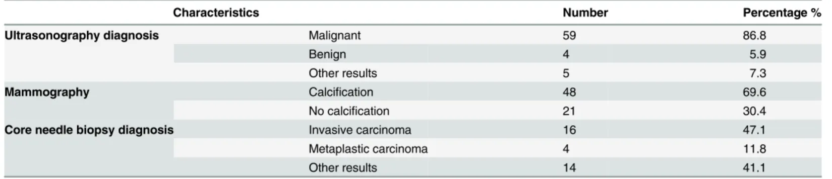
Malignancy was diagnosed in 59 of 68 (86.8%) patients with MBC in preoperative ultraso-nographic evaluation. Calcifications were found in 48 patients (48/69, 69.6%) in mammo-graphic evaluation. Histologic diagnosis of metaplastic carcinoma was only achieved in 4 of 34 patients (11.8%) in preoperative core needle biopsies (Table 2).
Seventy-seven (85.6%) patients received modified or radical mastectomy (MRM) and 5 patients (5.6%) accepted breast-conserving surgery. Three patients (1 spindle cell carcinoma, 1 squamous cell carcinoma, and 1 carcinoma with cartilaginous metaplasia) received neoadju-vant chemotherapy, but all failed with no reduction in tumor size observed. Postoperatively, paclitaxel plus anthracycline adjuvant chemotherapy was offered to 74 of the patients (82.2%), 3 patients (3.3%) received one of the neoadjuvant chemotherapeutic regimens (Paclitaxel+ epir-ubicin, or epirubicin + cyclophosphamide + docetaxel), and 5 patients (5.6%) had radiother-apy. Nine patients (10%) with ER and/or PR positive tumors accepted endocrine therapy, and 1 patient with tumor HER2 overexpression (1.1%) received adjuvant trastuzumab. Fewer patients in the MBC group received MRM (p = 0.000; p = 0.000), radiotherapy (p = 0.000; p = 0.001), endocrine therapy (p = 0.000) and anti-HER2 therapy (p = 0.000) than those in the TN-IDC and/or IDC-NST group (Table 1).
Histopathological features
The average tumor size of MBC was 4.01 cm (1.0–10.0 cm). LNM was identified in 16 of 77
cases (20.8%) with regional lymph node biopsy. The most common MBC subtype was spindle cell carcinoma (34.4%), followed by squamous cell carcinoma (31.1%) and MBC with mesen-chymal differentiation (24.5%). Fibromatosis-like subtype (4.4%) was the least common in this cohort of patients (Table 3). Among them, squamous cell carcinoma had the highest lymph node metastasis rate (25.9%, data not shown).
Table 2. Preoperative evaluation of MBC patients.
Characteristics Number Percentage %
Ultrasonography diagnosis Malignant 59 86.8
Benign 4 5.9
Other results 5 7.3
Mammography Calcification 48 69.6
No calcification 21 30.4
Core needle biopsy diagnosis Invasive carcinoma 16 47.1
Metaplastic carcinoma 4 11.8
Other results 14 41.1
doi:10.1371/journal.pone.0131409.t002
Table 3. Histologic subtypes of MBC.
Histologic subtypes Number Percentage %
Spindle cell carcinoma 31 34.4
Squamous cell carcinoma 28 31.1
Metaplastic carcinoma with mesenchymal differentiation 22 24.5
Mixed metaplastic carcinoma 5 5.6
Fibromatosis-like metaplastic carcinoma 4 4.4
Molecular Classification
Two cases were classified as luminal A type (2.2%), 17 cases were luminal B type (18.9%), 7 cases were HER2-overexpression type (7.8%), and 64 cases were triple-negative type (71.1%). Fifty eight cases (85%) of the triple negative tumors were basal-like type breast carcinomas (Table 4).
EGFR overexpression and copy number analysis
By immunohistochemistry, EGFR overexpression was identified in 52 of the 90 (57.8%) cases, and in 46 of the 64 triple-negative (71.9%) carcinomas. Squamous cell carcinomas had a signif-icantly higher proportion of EGFR overexpression (82.1%), compared to other subtypes
(p = 0.002;Table 5).
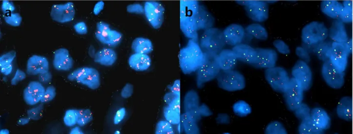
Forty-seven of 52 cases with EGFR overexpression were submitted for FISH analysis, and 14 of them (29.8%) demonstrated EGFR gene amplification (Fig 1a and 1b). Of these 14 cases, 5 (35.7%) showed increased EGFR copy number and 9 (64.3%) displayed high aneusomy.
Survival analysis
Recurrence and/or distant metastasis occurred in 22 patients (24.4%) during follow-up. The most common site of recurrence was the chest wall and the most common metastatic site was lung, followed by bone and brain. A 67.9% five-year DFS and a 78.7% of five-year OS were identified in this cohort of patients. In univariate analysis (Table 6), LNM (p = 0.000), advanced stage at diagnosis (p = 0.047), EGFR overexpression (p = 0.007), and high Ki-67 labeling (p = 0.026) of tumors were the significant predictive factors for reduced DFS, among which only LNM (p = 0.008) was significant in multivariate analysis. For OS, tumor lymph node metastasis (p = 0.000), EGFR overexpression (p = 0.049), and gene amplification (p = 0.002) were the significant predictive factors in univariate analysis, among which lymph node metastasis and EGFR gene amplification were significant predictive factors using multi-variate analysis (p = 0.001 and 0.022). Tumor size, morphologic subtype, biologic marker (ER, PR and HER2) expression, and therapeutic regimen were not significantly associated with patient survival.
Although not statistically significant, prognostic trends for certain morphologic subtypes of MBC were noted. Four patients with fibromatosis-like MBC had an average DFS of 64 months
Table 4. Molecular classification of MBC based upon the major component.
Molecular Classification Number Percentage %
Luminal A 2 2.2
Luminal B 17 18.9
HER2-overexpression 7 7.8
Triple-negative 64 71.1
(Basal-like) 58 64.4
doi:10.1371/journal.pone.0131409.t004
Table 5. EGFR overexpression of MBC.
Subtype of MBC EGFR P
-/1+ 2+/3+
Squamous cell carcinoma 5 23
others 33 29 0.002
and OS of 82 months, in contrast to a DFS of 51 months and OS of 55 months for patients with other histopathologic subtypes. Two patients with luminal A type MBC showing mesenchymal differentiation had an average DFS of 76 months and OS of 76 months, in contrast to a DFS of 51 months and OS of 56 months for patients with other molecular subtypes.
During the same period of follow-up, development of local recurrence or distant metastasis was identified in 136 patients (12.5%) with IDC-NST and in 29 patients (15.0%) in the
TN-IDC group (Table 1), significantly less than those with MBC (24.4%) (P = 0.001, P = 0.044, respectively).
Patients with MBC demonstrated a shorter five-year DFS (67.9% vs 88.9% vs 86%; p = 0.000, p = 0.001 respectively), and five-year OS (78.7% vs 93.0% vs 90.6%; p = 0.000, p = 0.021 respectively), when compared separately with survival in the IDC-NST and TN-IDC
groups of patients (Fig 2a–2d). Group comparison analysis among MBC, TN-IDC and
non-triple negative IDC (NTN-IDC) showed that patients with MBC had the worst five-year DFS and OS, followed by TN-IDC and NTN-IDC, which carried the most favorable five-year DFS (p = 0.000) and OS (p = 0.000) among the groups (Fig 2e and 2f).
Discussion
Previous studies have suggested that patients with MBC tend to present with larger tumors, less lymph node involvement, a higher proportion of triple-negative cases, and an increased
rate of distant metastasis, compared to IDC-NST [2,3]. MBC has been reported to have a
poorer prognosis than IDC [3,16] although comparable prognoses have been reported in cases
with matched stages. In this cohort of patients, with no significant difference in tumor stage among the groups of MBC, IDC-NST and TN-IDC. MBC had the largest average tumor size, the lowest rate of LNM, and the highest frequency of local recurrence and/or metastasis, and the worst prognosis measured by five-year DFS and OS. The results indicate that MBC has a poorer prognosis than that of IDC-NST and TNBC.
The 3-year DFS of MBC patients varies from 15% to 76% and the 3-year OS from 48% to
91% [5,17–19]. With longer follow-up, we identified a 67.9% year DFS and a 78.7%
five-year OS in our patients. Our findings are similar to those reported by Base et al in Korean
Fig 1. FISH analysis on EGFR gene amplification in MBC.Representative images of amplification of EGFR (a) and high aneusomy of EGFR (b). EGFR gene amplification was defined as a ratio of EGFR gene vs chromosome 7 centromere signals2.0. EGFR FISH-amplified samples also included those with
40% of tumor cells demonstrating4 copies of EGFR gene.
Table 6. Univariate analysis and multivariate analysis of MBC patient’s survivals.
Characteristics DFS OS
Univariate Multivariable Univariate Multivariable
5-yearDFS % P HR (95% CI) P 5-yearOS % P HR (95% CI) P
Age 50 72.9 0.280 86.1 0.272
>50 64.4 73.2
T stage 5cm 70.4 0.805 82.4 0.549
>5cm 62.6 71.7
N stage Negative 77.4 0.000 1.473–12.846 0.008 88.8. 0.000 2.175–24.631 0.001
Positive 29.2 44.2
Stage I 73.2 0.047 0.198–1.550 0.261 87.5 0.065
II 62.3 78.1
III 41.7 60.0
Feature of MBC Spindle 71.8 0.982 76.2 0.804
Squamous 63.4 75.5
Mesenchymal 69.2 80.8
Fibromatosis-like 66.7 100
Mixed 66.7 100
ER Negative 64.5 0.237 77.7 0.783
Positive 82.5 82.5
PR Negative 67.7 0.945 80.8 0.348
Positive 68.2 68.2
HER2 Negative 68.2 0.886 76.6 0.984
Positive 66.7 88.9
Ki67 0(0%-14%) 100 0.026 0.596–5.009 0.314 100 0.131
1(14%-50%) 83.4 87.1
2 (51%) 51.1 69.1
P53 <10% 80.8 0.179 85.4 0.324
10% 58.1 73.6
CK5/6 10% 63.8 0.673 78.7 0.859
>10% 70.1 78.5
EGFR - /1+ 87.1 0.007 0.920–9.084 0.069 95.0 0.049 0.596–12.866 0.194
2+/3+ 54.7 68.4
EGFR gene No amplification 73.5 0.103 84.5 0.002 1.209–11.904 0.022
Amplification 44.0 54.5
Chemotherapy Yes 64.5 0.445 76.1 0.237
No 83.6 90.0
Endocrine Yes 76.2 0.722 87.5 0.654
No 66.4 77.4
Operation MRM 66.8 0.711 77.7 0.656
Other 80.8 87.5
DFS, disease-free survival; OS, overall survival; ER, estrogen receptors; PR, progesterone receptors; HER2, human epidermal growth factor receptor 2; CK5/6, cytokeratin 5/6; MRM, modified radical mastectomy
Fig 2. Patient’s survival curves of MBC, IDC-NST and TN-IDC.Patients with MBC demonstrated shortened five-year DFS and five-year OS, when separately compared with those in the IDC-NST (a and b) and TN-IDC groups of patients (c and d). Group comparison analysis among MBC, TN-IDC and non-triple negative IDC (NTN-IDC) showed that patients with MBC had the worst five-year DFS and OS followed by TN-IDC, while NTN-IDC carried the most favorable five-year DFS and OS among the groups (e and f).
patients [4] and Gultekin et al in a Turkish population [17]. Multiple factors may contribute to the wide range of survival, including differences in patient population, classification of tumors, stage of tumors at presentation, patient management, and data collection.
The 90 cases of MBC culled from over 30,000 cases of invasive breast carcinomas (0.3%) diagnosed in the past 15 years in this cancer center of China showed that average tumor size
and percentage of TNBC were similar to those reported in the literature [2,3,5,17,20].
Note-worthy was the finding that the rates of lymph node metastasis (20.8%) and recurrence/ distant metastasis (24.4%) were lower in our patient population than the rates of lymph node
metastasis (24%-35%) [3,5,17] and recurrence/metastasis (26.9%-60%) reported in the
litera-ture [2,16,21].
MBC is a heterogeneous group of malignant breast tumors consisting of different morpho-logic subtypes with the frequency of subtypes showing considerable variation in different patient populations. Rakha et al [6] found that the most common subtype of MBC in Western countries was spindle cell carcinoma (34%), while squamous cell carcinoma (34%) was the most common in patients from Hong Kong and Singapore. Lai et al [3] reported that squamous cell carcinoma (35.6%) was the most common subtype in Taiwan, followed by carcinomas with osseous/chondroid differentiation (24.4%), sarcomatoid carcinoma (20%) and spindle cell car-cinoma (8.9%). Luini et al [20] showed that MBC with matrix-production was the most com-mon subtype (45.9%) in European patients, followed by carcinosarcoma (24.3%) and
squamous cell carcinomas (18.9%). Using the current WHO classification, we found that spin-dle cell carcinoma (34.4%) is the most common subtype of MBC in the Chinese population, followed by squamous cell carcinoma (31.1%) and carcinoma with mesenchymal differentia-tion (24.5%), similar to that of Western patients [6]. While the variadifferentia-tion in frequency of sub-types in different patient populations around the world is intriguing, variation in tumor classification and small case numbers in each study may be the principal factors contributing to the inconsistent frequency of tumor subtypes in the different studies. Further data collection is still needed to build up a valuable universal database.
Accurate diagnosis of MBC can be a challenge in preoperative core needle biopsies [21]. In our study, accurate diagnosis was made in only 11.8% of the cases. Tumor heterogeneity is pre-sumably the major contributing factor to the challenge. Therefore, surgical excision is the nec-essary procedure to achieve the final diagnosis, and the choices include mastectomy (modified or radical mastectomy MRM), lumpectomy, and breast-conserving surgery (BCS) [22].
Because MBC patients typically present with a large mass, MRM was often performed [3,23,
24]. The selection of surgical procedure may impact the 5 years-DFS of patients [16]. So far, there are no validated prognostic markers for MBC. Lee et al [16] reported that the subtype of MBC (non-squamous cell carcinoma vs squamous cell carcinoma) was associated with DFS, but its association with OS was not identified. Lester et al [25] found sarcomatoid MBCs were more aggressive than other triple-negative cancers. Sanguinetti et al [26] found tumor size had an important impact on patient outcome. Rakha et al [6], in their recent study of 405 MBC patients from a large international multicenter series, found that lymph node stage, lymphovascular invasion, histologic subtype, and chemotherapy were associated with lower breast cancer-specific survival and/or disease free interval. They found that spindle cell carcinoma had particularly aggressive biological behavior. In addition, significant differences in clinicopathologic features of MBC were described in patients from Western countries and from Asian countries, and they observed that MBC in Asian countries had a more favorable prognosis. Several of our findings differ from those of Rakha. In our cohort, the spindle cell car-cinoma was found to be the most common subtype of MBC and it did not lead to a worse DFS
or OS. Also, we couldn’t find a significant association between the histologic subtype of MBC
measured by Ki-67 staining, and EGFR overexpression were significantly associated with patient DFS in a univariate analysis. Among these factors, the status of lymph node metastasis was an independent prognostic factor for DFS in multivariate analysis. We also found that the rates of lymph node metastasis and EGFR gene amplification were significantly associated with patient OS both in univariate and multivariate analyses. However, tumor size, the status of tumor biomarkers (molecular subtypes), operative procedure, and postoperative therapy were not significant prognostic factors.
The inability of standard therapeutic protocols to effectively treat MBC has prompted a search for other therapeutic options, including those targeting EGFR. Aberrant signaling through EGFR overexpression is associated with neoplastic cell proliferation, migration, stro-mal invasion, resistance to apoptosis, and angiogenesis [27]. Bae et al [4] reported that MBC exhibited higher expression of EGFR compared to triple negative infiltrating ductal carcinoma. Reis-Filho et al [28] observed that 19 of 25 (76%) MBC cases exhibited EGFR expression. In our study, EGFR overexpression was identified in 57.8% of MBCs, and in 71.9% of the triple negative MBCs. Basal-like MBC lacking EGFR and KIT activating mutations may exhibit high EGFR copy numbers [29]. Reis-Filho al [28] reported EGFR gene amplification in 37% of the MBCs with EGFR overexpression. We found that 29.8% (14/47) of MBCs demonstrated EGFR gene amplification. These results beg the question of whether MBC patients with EGFR overex-pression and/or gene amplification might benefit from EGFR tyrosine kinase inhibitor and EGFR monoclonal antibody (cetuximab) therapies. We noted that squamous cell carcinoma had a significantly higher proportion of EGFR overexpression (82.1%) compared to other sub-types (p = 0.002). Whether squamous cell carcinoma will respond more favorably to cetuximab remains a valid clinical question to be answered. It seems reasonable to recommend a routine assessment of the EGFR status in MBC and to further explore this therapeutic option.
In summary, we report the clinicopathologic features and prognostic predictive markers in a large cohort of MBC patients from a major Chinese cancer center, and some unique features of MBC in the Chinese population have been noted. LNM is identified as an independent pre-dictive factor for unfavorable DFS, and LNM and EGFR overexpression/gene amplification are independent predictive factors for decreased OS. This study indicates that MBC is an aggressive type of breast cancer with poorer prognosis than IDC-NST and TN-IDC. New therapy target-ing EGFR in tumors with overexpression and/or gene amplification of EGFR is worthy of fur-ther exploration.
Acknowledgments
We thank the grant support from National Natural Science Foundation of China (Grant No. 81172531) to Xiaojing Guo, National Natural Science Foundation of China (Grant No. 81302294) to Yiling Yang, National Natural Science Foundation of China (Grant
No.81302292) to Xiaolong Qian and National Natural Science Foundation of China (Grant No. 81202101) to Fangfang Liu.
Author Contributions
Conceived and designed the experiments: XG LF XZ. Performed the experiments: YZ F. Liu YY XQ RL YF F. Lv YL SL BS. Analyzed the data: YZ F. Liu XG. Contributed reagents/materi-als/analysis tools: YY XQ RL YF BS. Wrote the paper: XG YZ XZ LF GP.
References
2. Song Y, Liu X, Zhang G, Song H, Ren Y, He X, et al. Unique clinicopathological features of metaplastic breast carcinoma compared with invasive ductal carcinoma and poor prognostic indicators. World J Surg Oncol. 2013; 11:129. doi:10.1186/1477-7819-11-129PMID:23738706
3. Lai HW, Tseng LM, Chang TW, Kuo YL, Hsieh CM, Chen ST, et al. The prognostic significance of meta-plastic carcinoma of the breast (MCB)—a case controlled comparison study with infiltrating ductal
carci-noma. Breast. 2013; 22(5): 968–973. doi:10.1016/j.breast.2013.05.010PMID:23787124
4. Bae SY, Lee SK, Koo MY, Hur SM, Choi MY, Cho DH, et al. The prognoses of metaplastic breast can-cer patients compared to those of triple-negative breast cancan-cer patients. Breast Cancan-cer Res Treat. 2011; 126(2): 471–478. doi:10.1007/s10549-011-1359-8PMID:21287362
5. Lim KH, Oh DY, Chie EK, Han W, Im SA, Kim TY, et al. Metaplastic breast carcinoma: clinicopathologic features and prognostic value of triple negativity. Jpn J Clin Oncol. 2010; 40(2): 112–118. doi:10.1093/
jjco/hyp139PMID:19887523
6. Rakha EA, Tan PH, Varga Z, Tse GM, Shaaban AM, Climent F, et al. Prognostic factors in metaplastic carcinoma of the breast: a multi-institutional study. Brit J Cancer. 2014; 112(2): 283–289. doi:10.1038/
bjc.2014.592PMID:25422911
7. Hammond ME, Hayes DF, Dowsett M, Allred DC, Hagerty KL, Badve S, et al. American Society of Clini-cal Oncology/College of American Pathologists Guideline Recommendations for ImmunohistochemiClini-cal Testing of Estrogen and Progesterone Receptors in Breast Cancer. Arch Pathol Lab Med. 2010; 134(7):e48–e72. doi:10.1043/1543-2165-134.7.e48PMID:20586616
8. Wolff AC, Hammond ME, Hicks DG, Dowsett M, McShane LM, Allison KH, et al. Recommendations for human epidermal growth factor receptor 2 testing in breast cancer: American Society of Clinical Oncol-ogy/College of American Pathologists clinical practice guideline update. J Clin Oncol. 2013; 31(31): 3997–4013. doi:10.1200/JCO.2013.50.9984PMID:24101045
9. Cheang MC, Chia SK, Voduc D, Gao D, Leung S, Snider J, et al. Ki67 index, HER2 status, and progno-sis of patients with luminal B breast cancer. J Natl Cancer Inst. 2009; 101(10):736–750. doi:10.1093/
jnci/djp082PMID:19436038
10. Altaf FJ, Mokhtar GA, Emam E, Bokhary RY, Mahfouz NB, Al AS, et al. Metaplastic carcinoma of the breast: an immunohistochemical study. Diagn Pathol. 2014; 9:139. doi:10.1186/1746-1596-9-139 PMID:25030022
11. Kim HM, Kim DH, Jung WH, Koo JS. Molecular classification of metaplastic carcinoma using surrogate immunohistochemical staining. Pathobiology. 2014; 81(2): 69–77. doi:10.1159/000354270PMID:
24356094
12. Steinman S, Wang J, Bourne P, Yang Q, Tang P. Expression of cytokeratin markers, ER-alpha, PR, HER-2/neu, and EGFR in pure ductal carcinoma in situ and DCIS with co-existing invasive ductal carci-noma of the breast. Ann Clin Lab Sci. 2007; 37(2):127–134. PMID:17522367
13. Blows FM, Driver KE, Schmidt MK, Broeks A, van Leeuwen FE, Wesseling J, et al. Subtyping of breast cancer by immunohistochemistry to investigate a relationship between subtype and short and long term survival: a collaborative analysis of data for 10,159 cases from 12 studies. PLoS Med. 2010; 7(5): e1000279. doi:10.1371/journal.pmed.1000279PMID:20520800
14. Nielsen TO, Hsu FD, Jensen K, Cheang M, Karaca G, Hu Z, et al. Immunohistochemical and clinical characterization of the basal-like subtype of invasive breast carcinoma. Clin Cancer Res. 2004; 10(16): 5367–5374. PMID:15328174
15. Kim MJ, Ro JY, Ahn SH, Kim HH, Kim SB, Gong G. Clinicopathologic significance of the basal-like sub-type of breast cancer: a comparison with hormone receptor and Her2/neu-overexpressing phenosub-types. Hum Pathol. 2006; 37(9):1217–1226. PMID:16938528
16. Lee H, Jung SY, Ro JY, Kwon Y, Sohn JH, Park IH, et al. Metaplastic breast cancer: clinicopathological features and its prognosis. J Clin Pathol. 2012; 65(5):441–446. doi:10.1136/jclinpath-2011-200586
PMID:22412048
17. Gultekin M, Eren G, Babacan T, Yildiz F, Altundag K, Guler N, et al. Metaplastic breast carcinoma: a heterogeneous disease. Asian Pac J Cancer Prev. 2014; 15(6):2851–2856. PMID:24761913 18. Al SA, El WA, Tulbah AM, Rahal MM, Ezzat AA. Metaplastic carcinoma of the breast clinical
presenta-tion, treatment results and prognostic factors. Acta Oncol. 2006; 45(2): 188–195. PMID:16546865 19. Esbah O, Turkoz FP, Turker I, Durnali A, Ekinci AS, Bal O, et al. Metaplastic breast carcinoma: case
series and review of the literature. Asian Pac J Cancer Prev. 2012; 13(9):4645–4649. PMID:23167395 20. Luini A, Aguilar M, Gatti G, Fasani R, Botteri E, Brito JA, et al. Metaplastic carcinoma of the breast, an
21. Park HS, Park S, Kim JH, Lee JH, Choi SY, Park BW, et al. Clinicopathologic features and outcomes of metaplastic breast carcinoma: comparison with invasive ductal carcinoma of the breast. Yonsei Med J. 2010; 51(6):864–869. doi:10.3349/ymj.2010.51.6.864PMID:20879052
22. Hu Q, Chen WX, Zhong SL, Li J, Luo Z, Tang JH, et al. Current progress in the treatment of metaplastic breast carcinoma. Asian Pac J Cancer Prev. 2013; 14(11):6221–6225. PMID:24377508
23. Tseng WH, Martinez SR. Metaplastic breast cancer: to radiate or not to radiate? Ann Surg Oncol. 2011; 18(1): 94–103. doi:10.1245/s10434-010-1198-6PMID:20585866
24. Nelson RA, Guye ML, Luu T, Lai LL. Survival outcomes of metaplastic breast cancer patients: results from a US population-based analysis. Ann Surg Oncol. 2015; 22(1):24–31. doi:
10.1245/s10434-014-3890-4PMID:25012264
25. Lester TR, Hunt KK, Nayeemuddin KM, Bassett RL, Gonzalez-Angulo AM, Feig BW, et al. Metaplastic sarcomatoid carcinoma of the breast appears more aggressive than other triple receptor-negative breast cancers. Breast Cancer Res Tr. 2012; 131(1):41–48.
26. Sanguinetti A, Lucchini R, Santoprete S, Farabi R, Fioriti L, Bistoni G, et al. Metaplastic carcinoma of the breast: Treatment, results and prognostic factors based on international literature. Ann Ital Chir. 2014; 85(2):109–113. PMID:24195912
27. Dancey JE, Freidlin B. Targeting epidermal growth factor receptor–are we missing the mark? Lancet.
2003; 362(9377):62–64. PMID:12853203
28. Reis-Filho JS, Milanezi F, Carvalho S, Simpson PT, Steele D, Savage K, et al. Metaplastic breast carci-nomas exhibit EGFR, but not HER2, gene amplification and overexpression: immunohistochemical and chromogenic in situ hybridization analysis. Breast Cancer Res. 2005; 7(6):R1028–1035. PMID:
16280056