Effects of Motor Tasks through Photos and Videos
in Patients After Stroke
Efeitos da Demonstração de Tarefas Motoras por Fotos e Vídeos
em Pacientes Pós-Acidente Vascular Encefálico
Fabíola Rodrigues de França Campos
a, Antônio Pereira Júnior
b& Tania Fernandes Campos
,baUniversidade Estácio de Sá, Rio de Janeiro, RJ, Brasil
& bUniversidade Federal do Rio Grande do Norte, Natal, RN, Brasil
Abstract
The aim of the present study was to examine the effects of motor tasks through photos and videos in post-stroke patients. Participants were 12 patients and 10 control people. Five functional tasks were presented in four different sequences and participants had to indicate the ones which reached the goal correctly. By ANOVA it was found that the response time of the patients was greater than of the control group (photos = patients: 4833 ± 310 ms, control: 1112 ± 76 ms, p = .0001 and videos = patients: 3655 ± 242 ms, control: 2451 ± 270 ms, p = .0001). Patients performed better with videos (p = .001). These results may infl uence therapeutic strategies and enable a discussion about a possible impairment of the mirror neuron system.
Keywords: Stroke, recognition, motor activities, activities of daily living, observational learning. Resumo
O objetivo deste estudo foi analisar os efeitos da demonstração de tarefas motoras através de fotos e vídeos em pacientes pós-AVE (acidente vascular encefálico). Participaram 12 pacientes e 10 contro-les. Cinco tarefas funcionais foram apresentadas em quatro sequências diferentes e os participantes indicavam aquela que alcançava o objetivo corretamente. Pela ANOVA verifi cou-se que o tempo de resposta dos pacientes foi maior (fotos = pacientes: 4833 ± 310 ms, controle: 1112 ± 76 ms, p
= 0,0001 e vídeos = pacientes: 3655 ± 242 ms; controle: 2451 ± 270 ms, p= 0,0001). Os pacientes apresentaram melhor desempenho com os vídeos (p= 0,001). Estes resultados podem infl uenciar as estratégias terapêuticas e permitir uma discussão a respeito de um possível impedimento do sistema de neurônios-espelho.
Palavras-chave: Acidente vascular encefálico, reconhecimento, atividades motoras, atividades da vida diária, aprendizagem observacional.
Mailing address: Departamento de Fisioterapia,
Uni-versidade Federal do Rio Grande do Norte, Av. Senador Salgado Filho, 3000, Natal, RN, Brazil 59066-800. E-mail: [email protected], fabiolafcampos@gmail. com and [email protected]
Acknowledgements: This research was supported by Support Program Graduate/Coordination of Improve-ment of Higher Education Personnel (PROAP/Capes) and National Council for Scientifi c and Technological Development (CNPq).
The motor rehabilitation of stroke patients is a long and tedious process that can rely on several therapeutic approaches currently available. The traditional correction/ facilitation method, for instance, uses therapeutic exercises and neuromuscular facilitation techniques to decrease
sensory-motor defi cits and promote motor improvement
(Knott & Kabat, 1954). A more recent approach, however, focuses on motor learning due to the practice or training of specifi c sequence of movements in functionally relevant contexts (Krakauer, 2006).
For instance, it had already been shown that motor learning is markedly improved when verbal instructions given to individuals are replaced by videotapes, fi lms, or photographs depicting the correct movement sequence that needs to be trained (Schmidt, 1991). A recent study with healthy subjects showed that learning by observation of videos, and the neural substrate of the observation-execution matching system, is organized to represent the goal of the learned motor task, rather than the specifi c effector used during learning itself (Williams & Gribble, 2012).
A modality of physical therapy based on observa-tion has been successfully tested for stroke rehabilitaobserva-tion (Celnik, Webster, Glasser, & Cohen, 2008; Ertelt et al., 2007) and has been called videotherapy indicating that the observation in combination with physical training may increase the effects of motor training after stroke and emphasizes the importance of action perception in neuro-rehabilitation (Ertelt, Hemmelmann, Dettmers, Ziegler, & Binkofski, 2012). The subsequent repetitive execution of the observed actions reinforces the cortical representation of action. The mirror neuron system provides a clearly de-fi ned neurophysiologic background for this new approach. Various studies were carry out regarding the clinical use of action observation for motor rehabilitation of sub-acute and chronic stroke patients, furthermore, because activa-tion of acactiva-tion representaactiva-tions by observaactiva-tion takes place also without the performance of active movements, the videotherapy is also potentially suitable for patients with acute strokes (Ertelt et al., 2012).
However, as far as we know, there is no study compar-ing the performance of stroke patients on the recognition of motor sequences displayed with either photographs or videos. In order to answer the question whether there is any difference in patient’s performance when trying to men-tally imitate movements displayed either as static (photos) or dynamic (videos) sequences, we used representations of activities of daily life (ADLs) and asked the patients which displayed the correct sequence of movements and which had more chance of reaching the implicit goal. Thus, the aim of this study was to examine the effects of the demonstration motor tasks through photos and videos in patients after stroke. We found the need to analyze what type of demonstration could improve the capability of ac-tion observaac-tion of patients in order to provide a basis for rehabilitation and to allow a discussion about a possible impediment of the mirror neuron system.
Material and Method
Subjects
The sample consisted of 12 stroke patients recruited from the physical therapy unit of a Public Hospital and 10 controls. We adopted the following inclusion criteria: age between 45 and 75 years, both gender, literate, fi rst clinically evident stroke (controlled via medical history, patient’s data in the respective clinic), unilateral cerebral
lesion and primarily motor symptoms (controlled by standard neurological examination), chronic stage of stroke (after 6 months), absence of serious cognitive disorders (controlled by medical evaluation), and non-use of sedatives, antidepressants, or neuroleptics. The inclusion criteria for the control group were: age between 45 and 75 years, both gender, literate, absence of pathology, and non-use of regular medication.
Procedures
The study was approved by the local Research Ethics Committee. All the participants were informed about the details of the procedures and signed a written informed consent. We kept a record of the patient’s medical his-tory, including lesion location, diagnosis, and duration of sequelae.
We used the NIHSS (National Institute of Health Stroke Scale) to determine the degree of neurological impairment of the patients. Awareness level, eye move-ments, visual fi eld, facial movements, motor function, and upper and lower limb ataxia were assessed, in addition to sensitivity, language, and the presence of dysarthria and spatial negligence (Caneda, Fernandes, Almeida, & Mugnol, 2006). To assess the overall health of control individuals we used the CIRS (Cumulative Illness Rating Scale; Parmelee, Thuras, Katz, & Lawton, 1995), which provides data on the status of six systems: cardiovascular/respiratory, gastrointestinal, genitourinary, musculoskeletal, integumentary, neuropsychological, and endocrine, with a severity scale of 0 to 4 points.
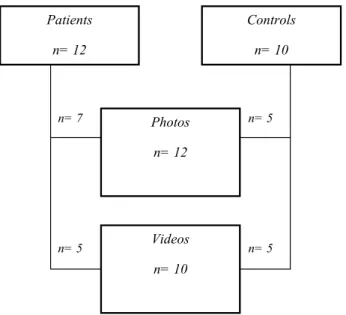
The patients and the controls were selected by simple randomization, according to the arrival at the physio-therapy service and eligibility criteria for inclusion either one of two demonstrations: with photos and using videos (Figure 1).
Figure 1. Schematic diagram of the study sample.
Patients
n= 12
Controls n= 10
Photos n= 12
n= 7 n= 5
n= 5
Videos n= 10
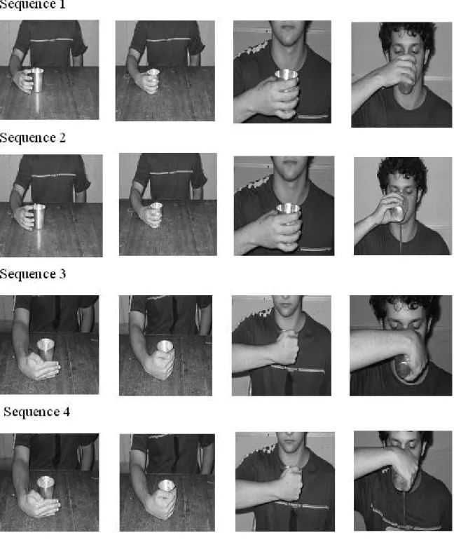
Five types of ADLs (drinking juice, combing hair, feeding, brushing teeth, and answering the telephone) were shown to each participant. Each activity was presented in 4 sequences, as shown in Figure 2, for 4 seconds. The sequences were presented individually on a computer screen, using either photos or videos. After, the 4 sequences were shown simultaneously on the screen and the subject
indicated which one displayed the correct sequence of movements and which one had more probability of reaching a goal. The screen was then turned off and the subject was informed whether he/she had been successful. The sequences previously randomized for each motor activity. In total, 24 trials were made for each activity; the subjects gave 120 responses for a total of 2640 data items.
Data Analysis
After performing a normality test (Shapiro-Wilk) to the data, we used the unpaired Student t test to compare the demographic and clinical characteristics of the sample. ANOVA (Tukey’s post-hoc test) was applied to to compare response times in the motor-sequence demonstration with the factors mode (photos or videos) and trials. We used the chi-square test to compare the frequency of correct and incorrect responses and frequency by gender and cerebral hemisphere affected. For statistical analysis we used the SPSS 14.0 (Statistical Package for the Social Science) software and the signifi cance level was set at 5%.
Results
The demographic and clinical characteristics of the sample are shown in Table 1. By analysis found no signifi -cant difference in the patients and controls between photos and videos regarding gender, age, schooling, the cognitive status (measured by MMSE) and general health (measured by CIRS). In patients no signifi cant difference was found regarding the affected hemisphere and neurological grade (measured by the NIHSS), but signifi cant difference was found regarding the sequelae time, in which patients with demonstration videos were longer sequel (Table 1).
Table 1
The Demographic and Clinical Characteristics of Stroke Patients and Controls According to the Groups of Photos and Videos
Variables
Patients Controls
Photos Videos p Photos Videos p
Gender(n/%) Women 1/12 1/20 .890 4/80 2/40 .068
Men 6/88 4/80 1/20 3/60
Age - years
(mean ±standard deviation) 62±23 62±26 .863 54±22 54±23 .658
Schooling – years
(mean ± standard deviation) 7±3 5±2 .186 11±4 10±5 .064
MEEM - scores
(mean ±standard deviation) 24±3 23±3 .715 28±1 27±1 .057
CIRS - scores
(mean ±standard deviation) 2.5±1.3 2.4±2.0 .844 1.6±1.0 1.6±1.2 .870
Affected hemisphere (n/%) Right 4/57 2/40 .867 - -
-Left 3/43 3/60 - -
-Sequelae time - months
(mean ±standard deviation) 11±7 25±14 .023 - -
-NIHSS - scores
(mean ±standard deviation) 1.6±1.3 4.2±3.2 .064 - -
-In the patients group we found a signifi cant difference in response times in the videos between the 1st and 2nd trials (p<.0001), and the 3rd and 4th trials (p<.0001). In the photos, there was a signifi cant difference between the 1st and 2nd trials (p<.0001; Figure 3A). In this group analysis showed a signifi cant difference between the recognition of videos and photos only the fi rst trial (p = .025).
Considering the average response time of the 24 trials in the recognition test, a difference was found between patients and controls on the photos (patients: 4833 ± 310 ms; control: 1112 ± 76 ms; p= .0001) as well as on the videos (patients: 3655 ± 242 ms; control: 2451 ± 270 ms;
p= .0001; Figure 4). In average, the patients had a longer response time in the photos sequences than in the videos (p= .001), whereas the inverse pattern was found for the control individuals (p=.001; Figure 4).
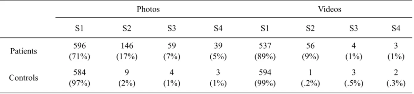
Analysis of the frequency of choosing one of the 4
movement sequences showed a signifi cant difference
between patients and controls (p= .0001). The patients chose the correct sequences (1 and 2) more frequently during videos (98%) than during photos (88%; Table 2). The controls, on the other hand, did not display any significant difference when comparing response
frequencies between the videos and photos (p=.166;
Table 2).
0 5000 10000 15000 20000 25000 30000
1 2 3 4 5 6 7 8 9 10 11 12 13 14 15 16 17 18 19 20 21 22 23 24
Ti
m
e
(m
s)
Trials
Videos
Photos A
0 5000 10000 15000 20000 25000 30000
1 2 3 4 5 6 7 8 9 10 11 12 13 14 15 16 17 18 19 20 21 22 23 24
Ti
m
e
(m
s)
Trials
Videos
Photos
B
Table 2
Absolute and Percentage Frequencies of Response Types in the Motor-Sequence Recognition Presented in Photos and Videos for the G roup of Patients and Controls
Photos Videos
S1 S2 S3 S4 S1 S2 S3 S4
Patients 596
(71%)
146 (17%)
59 (7%)
39 (5%)
537 (89%)
56 (9%)
4 (1%)
3 (1%)
Controls 584
(97%)
9 (2%)
4 (1%)
3 (1%)
594 (99%)
1 (.2%)
3 (.5%)
2 (.3%)
Discussion
Analysis of performance during the demonstration for photos and videos of motor tasks showed that the patients took longer to respond than the controls, suggesting that they had more diffi culty in both demonstrations. This re-sult may be related to a specifi c impairment of the mirror neuron system due to stroke (Buccino et al., 2004). Func-tional Magnetic Resonance Imaging (fMRI) has shown that action observation activates the premotor cortex in a somatotopic way (Avikainen, Forss, & Hari, 2002). Given the location of the stroke-impaired areas in our patients, it is possible that the lesions might have contributed to their poor performance in this study, which was based on movement sequences of activities using the upper limb.
Importantly, the analysis revealed a significant difference between the time of recognition of videos and photos on the fi rst trial and not the other trials, both for patients and for control subjects, possibly because the performance has stabilized quickly causing an effect “ceiling”. However, it was necessary to keep the data in the fi rst trial to total analysis to verify the best recognition pattern of motor sequences in tests with videos and photos in both groups (patients and controls). Thus, the results indicated that the patients took longer than control subjects to recognize the photos sequences than the videos sequences. The patients may have had diffi culty in grouping the individual motor activity sequences presented statically in the photos, and in using them to form a coherent motor imagery. An alternative explanation
0
1000
2000
3000
4000
5000
6000
Patients
Controls
Time (ms)
Photos
Videos
**
**
*
*
Figure 4. Mean and standard error of sequence recognition time of motor activities for the patients and the controls, presented in photos (separate parts) and videos (continuously).
is that the video-based presentation could have facilitated motor-sequence recognition by the patients. For instance, the sensory-motor system uses visual and proprioceptive signals differently when assessing a static or dynamic component of an activity (Chaminade, Ozto, Cheng, & Kawato, 2008). Visual information predominates when the static context of a motor action is being identifi ed and proprioceptive signals prevail with the dynamic components (Vogt & Thomaschke, 2007). According to a study by Stinear and coworkers (Stinear, Byblow, Steyvers, Levin, & Swinnen, 2006), a dynamic movement sequence is more effective for motor learning than a sequence of static visual images. These fi ndings suggest that the videos may have allowed a better understanding of the action intention, thereby facilitating the performance of the patients in the present study.
A comparison of the 4 response types showed that the patients chose more frequently the sequences in which the movement was being correctly executed instead of the sequences in which only the goal of the activity had been reached, irrespective of the correctness of the movement sequence. This result is in accordance with the proposal that the human mirror neuron system codes the intention associated with the observation of an everyday action being performed by others (Iacoboni & Mazziotta, 2007). The literature shows that when we watch human movements we are also covertly using our own movement representations, also we can use our own action system to understand the meaning of the behaviour of others. Action observation in this sense enables us to interact with other people and thus has a clear social function. This shared action system can use for learning new actions (de Vries & Mulder, 2007).
According to Carr and Shepherd (2006), if patients are given several explanations or demonstrations of the activity to be performed, before beginning this activity, this is suffi cient to provoke their memory of the movement and they may perform the entire activity without having to execute the components separately. The authors suggest that the environment needs to be organized so that the tasks are functionally relevant to the patient, with concrete goals such as: “reach and grab the glass on the table”, instead of “raise your arm” (Carr & Shepherd, 2006). In the present study, the presentation of continuous sequences using videos may have led to greater familiarity with daily activities; this also may explain the better performance of patients on the videos. Research suggests that videotherapy may have the advantage of to be easily administered at patient’s home who will perform it on her/his own In times of restricted resources in the health system, it is of paramount importance to develop cost-efficient rehabilitation programs (Ertelt et al., 2012).
During all the trials in the present study, the participants were informed of the results with respect to the move-ment presented in the sequences, such as the objective of the action. Many studies show that reduced frequency in knowledge of result has a greater benefi cial effect on learning, since this stimulates the activation of intrinsic feedback and error detection (Badets, Blandin, Wright, &
Shea, 2006; Page, Wilson, Shiel, Carter, & Norris, 2006; van Vliet & Wulf, 2006). Therefore, this is a fi nding that demonstrates the importance of feedback in the motor relearning process.
The results found in this study can not be extended to all patients with stroke, but they are representative for the studied sample. In conclusion, our results show that stroke patients are better able to recognize when motor move-ments sequences are presented for videos, so dynamically, and not for photos, so static. These results may infl uence therapeutic strategies during motor rehabilitation.
References
Avikainen, S., Forss, N., & Hari, R. (2002). Modulated activa-tion of the human SI and SII cortices during observaactiva-tion of hand actions. Neuroimage, 15, 640-646. doi:10.1006/ nimg.2001.1029
Badets, A., Blandin, Y., Wright, D. L., & Shea, C. H. (2006). Error detection processes during observational learning. Re-search Quarterly for Exercise and Sport, 77, 177-184. doi:1 0.1080/02701367.2006.10599352
Buccino, G., Binkofski, F., & Riggio, L. (2004). The mirror neuron system and action recognition. Brain and Language,
89, 370-376. doi:10.1016/S0093-934X(03)00356-0 Caneda, M. A., Fernandes, J. G., Almeida, A. G., & Mugnol, F.
E. (2006). Confi abilidade de escalas de comprometimento neurológico em pacientes com acidente vascular cerebral.
Arquivos de Neuro-Psiquiatria, 64, 690-697. doi:10.1590/ S0004-282X2006000400034
Carr, J. H., & Sherpherd, B. R. (2006). The changing face of neurological rehabilitation. Revista Brasileira Fisioterapia,
10, 147-156. doi:10.1590/S1413-35552006000200003 Celnik, P., Webster, B., Glasser, D. M., & Cohen, L.
G. (2008). Effects of action observation on physical training after stroke. Stroke, 39, 1814-1820. doi:10.1161/ STROKEAHA.107.508184
Chaminade, T., Oztop, E., Cheng, G., & Kawato, M. (2008). From self-observation to imitation: Visuomotor association on a robotic hand. Brain Research Bulletin, 75, 775-784. doi:10.1016/j.brainresbull.2008.01.016
De Vries, S., & Mulder, T. J. (2007). Motor imagery and stroke rehabilitation: A critical discussion. Journal of Rehabilitation Medicine, 39, 5-13. doi:10.2340/16501977-0020
Ertelt, D., Hemmelmann, C., Dettmers, C., Ziegler, A., & Binkof-ski, F. C. (2012). Observation and execution of upper-limb movements as a tool for rehabilitation of motor defi cits in paretic stroke patients: Protocol of a randomized clinical trial. BMC Neurology, 12, 42. doi:10.1186/1471-2377-12-42 Ertelt, D., Small, S., Solodkin, A., Dettmers, C., McNamara,
A., Binkofski, F., & Buccino, G. (2007). Action observation has a positive impact on rehabilitation of motor defi cits after stroke. Neuroimage, 36, T164-T173. doi:10.1016/j.neuroim-age.2007.03.043
Gallese, V., Fadiga, L., Fogassi, L., & Rizzolatti, G. (1996). Action recognition in the premotor cortex. Brain, 119, 593-609. doi:10.1093/brain/119.2.593
Iacoboni, M., & Mazziotta, J. C. (2007). Mirror neuron system: Basic fi ndings and clinical applications. Annals of Neurology,
62, 213-218. doi:10.1002/ana.21198
Krakauer, J. W. (2006). Motor learning: Its relevance to stroke recovery and neurorehabilitation. Current Opinion in Neurol-ogy, 19, 84-90.
Mukamel, R., Ekstrom, A. D., Kaplan, J., Iacoboni, M., & Fried, I. (2010). Single-neuron responses in humans during execution and observation of actions. Current Biology, 20, 750-756. doi:10.1016/j.cub.2010.02.045
Page, S. J., Szafl arski, J. P., Eliassen, J. C., Pan, H., & Cramer, S. C. (2009). Cortical plasticity following motor skill learning during mental practice in stroke. Neurorehabilitation and Neural Repair, 23, 382-388. doi:10.1177/1545968308326427 Page, M., Wilson, B. A., Shiel, A., Carter, G., & Norris, D.
(2006). What is the locus of the errorless-learning advantage?
Neuropsychologia, 44, 90-100. doi:10.1016/j.neuropsycholo-gia.2005.04.004
Parmelee, P. A., Thuras, P. D., Katz, I. R., & Lawton, M. P. (1995). Validation of the Cumulative Illness Rating Scale in a geriatric residential population. Journal of the American Geriatrics Society, 43, 130-137.
Sale, P., & Franceschini, M. (2012). Action observation and mirror neuron network: A tool for motor stroke rehabilitation.
European Journal of Physical and Rehabilitation Medicine,
48, 313-318.
Schmidt, R. A. (1991). Motor learning and performance: From principles to practice. Champaign, IL: Human Kinetics. Stefan, K., Cohen, L. G., & Duque, J. (2005). Formation of
a motor memory by action observation. The Journal of Neuroscience, 25, 9339-9346. doi:10.1523/JNEURO-SCI.2282-05.2005
Stinear, C. M., Byblow, W. D., Steyvers, M., Levin, O., & Swin-nen, S. P. (2006). Kinesthetic, but not visual, motor imagery modulates corticomotor excitability. Experimental Brain Research, 168, 157-164. doi:10.1007/s00221-005-0078-y Van Vliet, P. M., & Wulf, G. (2006). Extrinsic feedback for motor
learning after stroke: What is the evidence? Disability and Re-habilitation, 28, 831-840. doi:10.1080/09638280500534937 Vogt, S., & Thomaschke, R. (2007). From visuo-motor in-teractions to imitation learning: Behavioural and brain imaging studies. Journal of Sports Sciences, 25, 497-517. doi:10.1080/02640410600946779
Williams, A., & Gribble, P. L. (2012). Observed effector-independent motor learning by observing. Journal of Neu-rophysiology, 107, 1564-1570. doi:10.1152/jn.00748.2011