Cop
yright
© ABE&M t
odos os dir
eit
os r
eser
vados
.
The role of uncoupling protein 2
(UCP2) on the development of
type 2
diabetes mellitus
and
its chronic complications
Papel da proteína desacopladora 2 (UCP2) no desenvolvimento do diabetes melito tipo 2 e de suas complicações crônicas
Bianca Marmontel de Souza1,3, Taís Silveira Assmann1, Lúcia Maria Kliemann2,
Jorge Luiz Gross1,3, Luís Henrique Canani1,3, Daisy Crispim1,3
SUMMARY
It is well established that genetic factors play an important role in the development of type 2
diabetes mellitus (DM2) and its chronic complications, and that genetically susceptible subjects
can develop the disease after being exposed to environmental risk factors. Therefore, great efforts have been made to identify genes associated with DM2. Uncoupling protein 2 (UCP2) is expressed in several tissues, and acts in the protection against oxidative stress; in the negative regulation of insulin secretion by beta cells, and in fatty acid metabolism. All these mechanisms are associated with DM2 pathogenesis and its chronic complications. Therefore, UCP2 is a
can-didate gene for the development of these disorders. Indeed, several studies have reported that three common polymorphisms in UCP2 gene are possibly associated with DM2 and/or obesity.
Only a few studies investigated these polymorphisms in relation to chronic complications of diabetes, with inconclusive results. Arq Bras Endocrinol Metab. 2011;55(4):239-48
Keywords
UCP2; type 2 diabetes mellitus; diabetic retinopathy; diabetic nephropathy; DNA polymorphisms; haplotype
SUMÁRIO
Está bem estabelecido que fatores genéticos têm papel importante no desenvolvimento do diabetes melito tipo 2 (DM2) bem como de suas complicações crônicas e que indivíduos ge-neticamente suscetíveis podem desenvolver essa doença após exposição a fatores de risco ambientais. Assim, grandes esforços têm sido feitos para a identificação de genes associados ao DM2. A proteína desacopladora 2 (UCP2) é expressa em diversos tecidos e atua na proteção contra o estresse oxidativo, na regulação negativa da secreção de insulina pelas células-beta e no metabolismo dos ácidos graxos, mecanismos associados tanto à patogênese do DM2 como a suas complicações crônicas. Portanto, o gene UCP2 é um gene candidato para o
desenvolvi-mento dessas doenças. De fato, diversos estudos têm relatado que três polimorfismos comuns no gene UCP2 estão possivelmente associados ao DM2 e/ou à obesidade. Apenas poucos
es-tudos investigaram esses polimorfismos em relação às complicações crônicas do diabetes, obtendo resultados pouco conclusivos. Arq Bras Endocrinol Metab. 2011;55(4):239-48
Descritores
UCP2; diabetes mellitus tipo 2; retinopatia diabética; nefropatia diabética; polimorismos de DNA; haplótipo
1 Endocrinology Division,
Porto Alegre, RS, Brazil
2 Pathology Service, Hospital
de Clínicas de Porto Alegre, Porto Alegre, RS, Brazil
3 Graduate Studies Program in
Medical Sciences, Endocrinology, Universidade Federal do Rio Grande do Sul (UFRGS), Porto Alegre, RS, Brazil
Correspondence to: Daisy Crispim
Rua Ramiro Barcelos, 2350 prédio 12, 4° andar
90035-003 − Porto Alegre, RS, Brazil
Received on Feb/22/2011 Accepted on Apr/29/2011
INTRODUCTION
I
t is estimated that 7.6% of the Brazilian population have diabetes mellitus (DM) (1). This disease consti-tutes a serious public health problem because of its high prevalence, increased morbidity and mortality rates, andCop
yright
© ABE&M t
odos os dir
eit
os r
eser
vados
.
Type 2 DM (DM2) accounts for 90%-95% of DM cases worldwide and usually occurs in obese subjects over 40 years of age. It is characterized by hyperglyce-mia due to a combination of insulin resistance (IR) and inadequate compensatory insulin secretion (2). Chronic hyperglycemia is associated with long-term structural damage, dysfunction, and failure in several organs and tissues, which consequently lead to the de-velopment of chronic complications (2). These compli-cations are often categorized as microvascular (retino-pathy, nephropathy and neuropathy), or macrovascular (cardiovascular and cerebrovascular) (2). Among chro-nic microvascular complications of diabetes, diabetic retinopathy (DR) is the leading cause of blindness in adults (3); diabetic nephropathy (DN) is the most common cause of end-stage chronic renal disease and kidney transplants in many countries (4); and diabetic peripheral neuropathy (DPN) is responsible for 50%-75% of non-traumatic lower limb amputations (5). Ge-netic factors play an important role in the development of DM2 and its chronic complications, and genetically susceptible subjects can develop this disease after being exposed to environmental risk factors. Therefore, gre-at efforts have been made to identify genes associgre-ated with DM2 and its chronic complications (2).
Uncoupling protein 2 (UCP2) plays important ro-les in decreasing the production of reactive oxygen spe-cies (ROS) by mitochondria, regulating insulin secre-tion by pancreatic beta cells, and regulating free fatty acid (FFA) metabolism, which are mechanisms directly associated with the pathogenesis of DM or its chronic complications (6). Thus, the aim of this study was to review the role of UCP2 in relation to the development of DM2 or its chronic microvascular complications.
THE MITOCHONDRIAL RESPIRATORY CHAIN
(MRC)
Mitochondria are essential organelles in all eukaryotic cells, and regulate a number of key vital processes for cell survival and function, including energy production, redox control, calcium homeostasis, and certain meta-bolic and biosynthetic pathways. In addition, mitochon-dria are the main sources of ROS, and often play an es-sential role in physiological cell death mechanisms (7).
The main source of cell energy is the synthesis of adenosine triphosphate (ATP) from adenosine diphos-phate (ADP) and inorganic phosdiphos-phate (Pi), by means of oxidative phosphorylation carried out in the
mito-chondrial respiratory chain (MRC) (8). MRC is loca-ted in the mitochondrial inner membrane, and has ive multienzymatic complexes and two proteins responsi-ble for electron transport, cytochrome c and coenzy-me Q (Figure 1A). Oxidative phosphorylation involves coupling electron transport to the active pumping of protons across the mitochondrial inner membrane and generation of ATP in the MRC (7).
Oxidation of reduced nutrient molecules, such as carbohydrates, lipids, and proteins in cell metabolism generates electrons in the form of reduced hydrogen carriers (NADH and FADH2), which are referred as reduced cofactors. These reduced cofactors donate electrons to the MRC. The movement of electrons through the components of the MRC is driven by a redox potential along the chain. Complexes I, III and IV pump protons across the inner membrane, as elec-trons pass down the chain complexes. This produces an electrochemical potential difference across the inner membrane, known as proton-motive force, Δp, consis-ting mostly of an electrochemical gradient (membrane potential) and a small chemical gradient (difference in pH). The energy that is conserved in the proton gra-dient across the inner membrane is used by complex V (F1F0-ATP synthase) to synthesize ATP, as protons are transported back from the intermembrane space into the mitochondrial matrix. The inal destination for the electrons is the generation of molecular oxygen, which is reduced to water by complex IV, in the last step of the MRC. Therefore, the process of substrate oxidation and oxygen reduction is also called respiration (7,8).
ROS correspond to a variety of molecules and free radicals (chemical species with one unpaired electron) derived from the metabolism of molecular oxygen. Su-peroxide anion (O2− ), the product of an one-electron reduction of oxygen, is the precursor of most ROS, and a mediator in oxidative chain reactions (7). Dis-mutation of O2− (either spontaneously or by means of a reaction catalyzed by superoxide dismutases) produ-ces hydrogen peroxide (H2O2) which, in turn, may be fully reduced to water or, in the presence of ferrous or cuprous ions, may form the highly reactive hydroxyl radical ( OH) (7).
Cop
yright
© ABE&M t
odos os dir
eit
os r
eser
vados
.
ber of mitochondria and in the eficiency of oxidative phosphorylation, is closely associated with DM2 (10). Microarray analyses performed in biopsies of skele-tal muscle of DM2 patients and non-diabetic subjects from different populations have shown that PGC-1α (PPAR-g coactivator-1), the master control in the re-gulation of mitochondrial biogenesis, and NRF-1 (nu-clear respiratory factor-1), as well as their downstream target genes in oxidative metabolism, are decreased in DM2 patients (10,11). These indings were conirmed by enzymatic studies that reported that DM2 patients, or their insulin-resistant offspring, show a decline in mitochondrial oxidative activity (12), in insulin-sti-mulated ATP synthesis and fatty acid oxidation (13), and in the number of mitochondria found in skeletal muscle, compared with age-matched insulin-sensitive subjects (12).
Mitochondrial dysfunction and increased ROS pro-duction are involved in the repro-duction of lipid oxidation, and the increase in lipid content in muscle cells, exacer-bating IR (14). Decrease in fatty acid oxidation produ-ces increased intracellular levels of fatty acetyl-CoA and diacylglycerol. These molecules activate protein kinase C, p38/MAPK (mitogen-activated protein kinase) and JNK (jun N-terminal kinase) which, in turn, trigger a cascade of serines, leading to increased phosphorylation of serine residues of insulin receptor-1 (IRS-1). An incre-ase in serine phosphorylation blocks IRS-1 tyrosine resi-due phosphorylation, which inhibits the capacity of the receptor to phosphorylate the downstream target PI3K. Inactivation of PI3K impairs the translocation of glucose transporter type 4 (GLUT4) to the plasma membrane, and, consequently, leads to a decrease in glucose uptake in muscle cells upon insulin stimulation (14).
Furthermore, decreased ATP synthesis that results from mitochondrial dysfunction also plays an impor-tant role in decreasing insulin secretion in patients with DM2, since it may impair the regulation of K+ and Ca+ channels present in the cell membrane, and thereby inhibit exocytosis of insulin granules in beta cells (10).
UNCOUPLING PROTEINS (UCPS)
UCPs 1, 2, 3, 4 and 5 are members of an anion-car-rier protein family located in the mitochondrial inner membrane (6).These proteins have similarities in their structures, but different tissue expression in mammals. The original UCP, UCP1, is mainly expressed in brown adipose tissue, which is responsible for thermogenesis
Figure 1. Energy dissipation mediated by UCP2 and its roles in obesity and type 2 diabetes mellitus(DM2). 1A) UCP2 location and function in the mitochondrial respiratory chain (MRC). Numbers I-IV correspond to MRC complexes. F1Fo-ATP synthase is the fifth complex of MRC. During respiration, protons are pumped by MRC complexes and a proton electrochemical potential gradient is generated. The energy of the proton gradient drives the synthesis of ATP by the FoF1 complex. UCP2 catalyzes a regulated re-entry of protons into the matrix, uncoupling MRC and, consequently, reducing ATP synthesis. 1B) Main roles of UCP2 in obesity and DM2. ROS = reactive oxygen species. Q = coenzyme Q; C = cytochrome-c.
It is well-established that there is a strong positive correlation between mitochondrial inner membrane potential and production of ROS. At high membrane potentials, even a small increase in this potential greatly stimulates H2O2 production. Therefore, “mild uncou-pling”, i.e., a small decrease in membrane potential, has been suggested to have a natural antioxidant effect (9).
MITOCHONDRIAL DYSFUNCTION AND TYPE 2
DIABETES MELLITUS
In the past decade, several clinical and experimental studies have strengthened the hypothesis that mito-chondrial dysfunction, including reduction in the
num-Excess caloric load
↑ UCP2
↑ Energy dissipation
↓ Risk of obesity
↑ UCP2
↓ ROS production
↓ Risk of diabetic chronic complications
↑ UCP2
↓ Mitochondrial coupling
↓ ATP synthesis
↓ Insulin secretion
↑ Risk of DM2
1B 1A
Cop
yright
© ABE&M t
odos os dir
eit
os r
eser
vados
.
in newborns (15). It was recently shown that, under certain physiological conditions such as hyperglycemia, UCP1 is also expressed in skeletal muscle, white adipo-se tissue, retinal cells and pancreatic beta cells (16,17). UCP2 is widely distributed, whereas UCP3 is mainly restricted to the skeletal muscle, and UCP4 and UCP5/ BMCP1 are mainly expressed in the brain (6,15).
Over the last few years, several studies have shown that UCPs decrease metabolic eficiency by dissocia-ting substrate oxidation in the mitochondria from ATP synthesis, by means of the MRC. This is thought to be accomplished by promoting net translocation of pro-tons from the intermembrane space, across the inner mitochondrial membrane to the mitochondrial matrix, thereby dissipating the potential energy available for the conversion of ADP to ATP and, consequently, decrea-sing ATP production (6). This uncoupling effect then leads to homologue- and tissue-speciic functions, such as thermogenesis (UCP1), regulation of FFAs metabo-lism and transport (UCP2 and UCP3), reduction in ROS formation (UCP1-3 and UCP5/BMCP1), and re-gulation of ATP-dependent processes (UCP2) (15,18).
UNCOUPLING PROTEIN 2 (UCP2)
In 1997, Fleury and cols. (19) cloned and sequenced a gene homologous to UCP1 gene, later called UCP2.
UCP2 gene covers a 6.3 kb region on chromosome 11 (region 11q13), and has eight exons and seven introns (Figure 2). In humans, region 11q13 is linked to basal metabolic rate and body fat percentage (20). The trans-criptional gene unit is constituted by two non-coding exons followed by six exons that encode the 308 ami-no acids of the protein (19). Human UCP2 share 57% amino acid-sequence identity with human UCP1, and it is 71% identical to human UCP3 (8). In addition, the amino acid sequence of human UCP2 is 95% identical to mouse UCP2 (19).
UCP2 is expressed in a wide range of tissues and cell types, including brown and white adipose tissues, skele-tal muscle, heart, kidneys, liver, lungs, spleen, thymus, bone marrow, macrophages, brain, gastrointestinal tract, pancreatic islets and retinal cells (6,8,15,16,19). Although UCP2 is well expressed in many tissues at mRNA level, it would seem that UCP2 protein level is not simply proportional to mRNA concentration. For example, whereas UCP2 mRNA is found in heart, skeletal muscle, and brown adipose tissue, no UCP2 protein could be detected in these tissues (21). Thus,
Figure 2. Map of UCP2 gene locus on chromosome 11 (region 11q13). The eight exons (boxes) are numbered from left to right according to the transcriptional region. The black boxes represent the coding regions, and the light gray boxes represent the non-coding region, including 3’UTR region of exon 8. The vertical arrows show the main common polymorphisms associated with DM2 or its microvascular chronic complications. Figure adapted from http://www.nbci.nlm.nih.gov/gene/7351.
changes in the amount of UCP2 mRNA do not always relect the expression of the protein itself, which may be explained by different translational regulation of UCP2
gene among tissues, providing a mechanism by whi-ch its expression can be rapidly and strongly induced under stress conditions (21). PPAR (peroxisome pro-liferator-activated receptor)-α, PPAR-g, PGC-1α and SREBP-1c (sterol regulatory element binding protein--1c) transcription factors increase UCP2 gene expres-sion, while SIRT1 (sirtuin 1) and FOXA1 (forkhead box A1) factors inhibit its expression (22). UCP2 is also translationally regulated by an inhibitory upstre-am open reading frupstre-ame (ORF), which, when mutated, results in maximal UCP2 mRNA translation (23). Glu-tamine, an amino acid that has been implicated in the insulin secretion pathway, overcomes ORF inhibition and increases UCP2 eficiency (23).
It was proposed that both UCP2 and UCP3 only mildly uncouple respiration, slightly dissipating mem-brane potential energy, and thus slightly decreasing ATP production (8,18) (Figure 1A). Nonetheless, unlike UCP1, UCP2 and UCP3 only uncouple MRC after suitable induction by cold, ROS (particularly su-peroxide), ubiquinone, high levels of glucose and/or non-esteriied fatty acids, high impact exercise, sepsis, and hyperthyroidism. Their activities are inhibited by purine, such as ATP and GDP, and by interleukin-1b (24,25). As already mentioned, MRC uncoupling ge-nerated by UCP2 leads to protection from excess ROS production, while it also seems to be associated with inhibition of insulin secretion by beta cells and regu-lation of FFAs metabolism and transport (18,22,26).
po-Cop
yright
© ABE&M t
odos os dir
eit
os r
eser
vados
.
tential and decreasing ROS production (6). Since even “mild uncoupling” has a large effect on reducing ROS production, the hypothesis that UCP2 protects against oxidative stress is strongly supported and is now gene-rally accepted (6). Accordingly, UCP2 knockout mice have elevated ROS production in macrophages (27) and pancreatic beta cells (28). Moreover, rat clonal beta cell line (INS-1E) genetically modiied to overexpress
UCP2 gene presented increased survival after treat-ment with the free radical H2O2 (29). Likewise, beta cells exposed to oxidative stress attempted to overcome the toxicity caused by H2O2 by the induction of UCP2 mRNA (24). More recently, it was demonstrated that endothelial cells from bovine retinal cells incubated with high glucose levels increased UCP2 expression, which protected them from ROS damage derived from glucotoxicity, suggesting a protective role of this pro-tein in the pathogenesis of DR or other diabetic chro-nic complications (16).
An important coupling signal between glucose sen-sing and insulin secretion by pancreatic beta cells is the rise in ATP/ADP ratio. Increases in ATP/ADP ratio close the ATP-sensitive K+ channel in the mitochon-drial inner membrane. This causes membrane depola-rization, opening of voltage-gated calcium channels, and inlux of Ca2+ into the cytosol of beta cells, which subsequently triggers the exocytosis of granules contai-ning insulin. UCP2, by virtue of its proton leak activity, decreases the generation of ATP from glucose metabo-lism in beta cells, which consequently impairs glucose--stimulated insulin secretion (6,7,18,20). Accordin-gly, several studies have conirmed that UCP2 acts as a negative regulator of insulin secretion. For example,
UCP2 overexpression in rat islets totally suppresses glucose-stimulated insulin secretion (30). Additionally, pancreatic islets from UCP2 knockout mice (UCP2-/-) have increased insulin secretion in response to glucose. These mice have higher blood insulin and lower blood glucose than wild-type mice (26). Interestingly, double mutant Lepob/ob / UCP2-/- mice (i.e., obese and with
UCP2 gene deleted) have improved beta cell function independent of obesity (26).
FFAs are endogenous physiological regulators asso-ciated with increased UCP2 and UCP3 expression in a tissue-speciic manner (18,25). In preadipocyte cell lines, unsaturated fatty acids markedly induce UCP2 mRNA expression (31). Other cell lines derived from heart, li-ver and pancreatic islets also respond to the addition of different FFAs to the culture medium with an
increa-se in UCP2 mRNA levels (25). FFAs seem to regulate the UCP2 and UCP3 expression probably via PPAR-α, PPARg and SREBP-1c transcription factors (25).
Studies in proteoliposomes, isolated mitochondria and intact cells suggest that both UCP2 and UCP3 have a role in transporting FFAs or fatty acid hydro-peroxides (24,32). These proteins seem to export fatty acid anions outside of the mitochondrial matrix when a large excess of fatty acids is inside the mitochondria, protecting cells from the oxidative damage caused by excessive fatty acid peroxidation (lipotoxicity), main-ly of pomain-lyunsaturated fatty acids of the phospholipidic membrane. These fatty acids can bind to the superoxi-de produced by mitochondria, which converts them to 4-hydroxy-2-nonenal (HNE), an UCP2 activator. Af-ter activation by HNE, UCP2 is able to transport pro-tons across the mitochondrial inner membrane, thus increasing the uncoupling of MRC and, consequently, decreasing ROS formation (33).
Beta cell dysfunction can be produced by prolon-ged exposure of these cells to elevated glucose (glu-cotoxicity) and lipid (lipotoxicity) levels, conditions often associated with DM2. The exact mechanisms by which glucolipotoxicity triggers beta cell dysfunction are not well-known. However, evidence indicates that ROS production plays an important role in these me-chanisms (6). As already mentioned, it is known that beta cells exposed for a prolonged time to glucolipo-toxicity increase UCP2 expression in a way to protect them against damage caused by oxidative stress (22). Besides, it is clear that beta cells display low expression and activity of many of the enzymes involved in antio-xidant defense. Thus, an antioantio-xidant effect of UCP2 is of special importance in this cell type (29). However, increased UCP2 production leads to decreased insulin secretion, predisposing subjects to DM2 (6,22,32,33). Therefore, UCP2 is a candidate gene for the develop-ment of DM2, since altered expression of this gene can explain some key defects involved in this disease or its chronic complications (Figure 1B).
Polymorphisms in uncoupling protein 2 gene
associated with type 2 diabetes mellitus
polymor-Cop
yright
© ABE&M t
odos os dir
eit
os r
eser
vados
.
phism, which is an insertion/deletion of 45bp in the 3’ UTR region (3’ untranslated region) of exon 8 (36). Results of these studies have been variable: while some studies showed an association of one or more of these polymorphisms with obesity, reduced levels of insulin secretion by beta cells, IR and/or DM2 (34,37-45), other studies were unable to ind any association
be-tween these polymorphisms and these characteristics (35,36,46-48) (Table 1).
The A allele of the -866G/A polymorphism has been reported to increase UCP2 transcriptional acti-vity in transfected INS-1E cells derived from rat beta cells (36). However, data in human tissues have been conlicting, reporting either increased (36,50,51) or
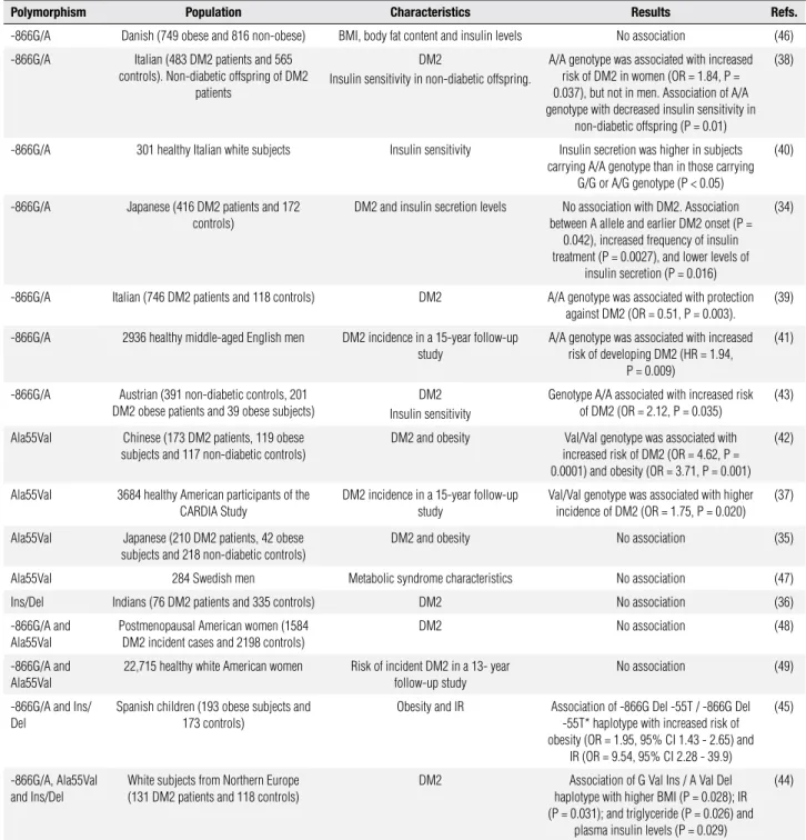
Table 1. Studies of the association between -866G/A, Ala55Val and Ins/Del polymorphisms in UCP2 gene and type 2 diabetes mellitus
Polymorphism Population Characteristics Results Refs.
-866G/A Danish (749 obese and 816 non-obese) BMI, body fat content and insulin levels No association (46)
-866G/A Italian (483 DM2 patients and 565 controls). Non-diabetic offspring of DM2
patients
DM2
Insulin sensitivity in non-diabetic offspring.
A/A genotype was associated with increased risk of DM2 in women (OR = 1.84, P = 0.037), but not in men. Association of A/A genotype with decreased insulin sensitivity in
non-diabetic offspring (P = 0.01)
(38)
-866G/A 301 healthy Italian white subjects Insulin sensitivity Insulin secretion was higher in subjects carrying A/A genotype than in those carrying
G/G or A/G genotype (P < 0.05)
(40)
-866G/A Japanese (416 DM2 patients and 172 controls)
DM2 and insulin secretion levels No association with DM2. Association between A allele and earlier DM2 onset (P =
0.042), increased frequency of insulin treatment (P = 0.0027), and lower levels of
insulin secretion (P = 0.016)
(34)
-866G/A Italian (746 DM2 patients and 118 controls) DM2 A/A genotype was associated with protection against DM2 (OR = 0.51, P = 0.003).
(39)
-866G/A 2936 healthy middle-aged English men DM2 incidence in a 15-year follow-up study
A/A genotype was associated with increased risk of developing DM2 (HR = 1.94,
P = 0.009)
(41)
-866G/A Austrian (391 non-diabetic controls, 201 DM2 obese patients and 39 obese subjects)
DM2 Insulin sensitivity
Genotype A/A associated with increased risk of DM2 (OR = 2.12, P = 0.035)
(43)
Ala55Val Chinese (173 DM2 patients, 119 obese subjects and 117 non-diabetic controls)
DM2 and obesity Val/Val genotype was associated with increased risk of DM2 (OR = 4.62, P = 0.0001) and obesity (OR = 3.71, P = 0.001)
(42)
Ala55Val 3684 healthy American participants of the CARDIA Study
DM2 incidence in a 15-year follow-up study
Val/Val genotype was associated with higher incidence of DM2 (OR = 1.75, P = 0.020)
(37)
Ala55Val Japanese (210 DM2 patients, 42 obese subjects and 218 non-diabetic controls)
DM2 and obesity No association (35)
Ala55Val 284 Swedish men Metabolic syndrome characteristics No association (47)
Ins/Del Indians (76 DM2 patients and 335 controls) DM2 No association (36)
-866G/A and Ala55Val
Postmenopausal American women (1584 DM2 incident cases and 2198 controls)
DM2 No association (48)
-866G/A and Ala55Val
22,715 healthy white American women Risk of incident DM2 in a 13- year follow-up study
No association (49)
-866G/A and Ins/ Del
Spanish children (193 obese subjects and 173 controls)
Obesity and IR Association of -866G Del -55T / -866G Del -55T* haplotype with increased risk of obesity (OR = 1.95, 95% CI 1.43 - 2.65) and
IR (OR = 9.54, 95% CI 2.28 - 39.9)
(45)
-866G/A, Ala55Val and Ins/Del
White subjects from Northern Europe (131 DM2 patients and 118 controls)
DM2 Association of G Val Ins / A Val Del haplotype with higher BMI (P = 0.028); IR (P = 0.031); and triglyceride (P = 0.026) and
plasma insulin levels (P = 0.029)
(44)
Cop
yright
© ABE&M t
odos os dir
eit
os r
eser
vados
.
decreased (44,52) UCP2 mRNA levels associated with -866A allele. Computational analyzes demonstrated that this polymorphism is involved in putative binding sites for speciic transcription factors, such as PAX6 (pai-red box gene 6) and HIF-1α (hypoxia-inducible factor-1α) (50). Esterbauer and cols. (50) hypothesized that preferential binding of some transcription factors to G or A allele in the promoter sequence may confer tissue--speciic advantages to either allele.
Non-diabetic subjects carrying the -866A allele have been shown to have decreased insulin secretion respon-se to intravenous and oral glucorespon-se and, therefore, have increased risk of developing DM2 (38,40,41,43). Simi-larly, DM2 patients carrying A allele have been shown to have signiicantly lower insulin secretion during in-travenous glucose tolerance test, and to require insulin therapy more frequently and earlier after diagnosis of the disease, than patients carrying the G/G genotype (34). In addition, some studies also suggest that A alle-le carriers also seem to have a higher waist-to-hip ratio, increased risk of metabolic syndrome, and higher levels of plasma markers of oxidative stress (51).
The Ins/Del polymorphism is located in the 3’ UTR region of the gene, only 158pb from the trans-cription stop codon. It also appears to be functional be-cause mRNA transcribed from the sequence containing the insertion allele displayed a shorter half-life in a fetal myoblast cell line than mRNA transcribed from the se-quence containing the deletion allele (53). In the other hand, Ala55Val (C/T) polymorphism causes a conser-vative amino acid change (alanine/valine) at position 55 of exon 4. Until now, there has been no evidence that this alteration generates a functional change in the protein. Thus, considering that the Ala55Val (C/T) polymorphism is tightly linked to the -866G/A poly-morphism (|D’| = 0.991), and partially linked to the Ins/Del polymorphism (|D’| = 0.879), this polymor-phism may not be a real disease-causing variant, but could simply be relecting the effects of -866G/A and Ins/Del polymorphism (54).
Studies on the association of the effects of Ala55Val and Ins/Del polymorphisms on DM2 or associated cha-racteristics have shown controversial results. Subjects carrying the Val/Val genotype of Ala55Val polymor-phism seem to have increased risk of developing DM2 and obesity, and higher BMI than subjects with Ala/Val or Ala/Ala genotypes (37,42,51,55). However, other studies indicated that this polymorphism is not indi-vidually associated with BMI, obesity, metabolic
syn-drome, DM2, and insulin secretion (35,44,47,48,51). Likewise, some studies did not ind any individual as-sociation between Ins/Del polymorphism and obesity, DM2 and/or insulin secretion (44-46,51), while other studies reported an association between the insertion allele and obesity (36,51).
Haplotype combinations constituted by UCP2 gene polymorphisms have also been found to be associated with BMI, obesity, insulin secretion and IR (44,45,48). Wang and cols. (44) reported that the G Val Ins / A Val Del haplotype (-866G/A, Ala55Val, and Ins/Del poly-morphisms) is associated with higher BMI (P = 0.028), IR (P = 0.031) and higher plasma insulin levels (P = 0.029) in northern European white subjects with and without DM2. Ochoa and cols. (45) investigated, in a group of Spanish obese and non-obese children, the association between -866G/A and Ins/Del polymor-phisms in the UCP2 gene and -55C/T (rs1800849) in
UCP3 gene with obesity and IR. Although they obser-ved no association between individual polymorphisms and these characteristics, the authors observed that the -866G Del -55T / -866G Del -55T haplotype was as-sociated with increased risk of obesity (OR = 1.9, 95% CI 1.4 - 2.6) and IR (OR = 9.5, 95% CI 2.3 - 39.9).
In brief, studies on these associations cited here indicated that the functional -866G/A polymorphism actually contributes to the biological variation of insu-lin secretion and, consequently, to the susceptibility to DM2. On the other hand, results reported by other studies on the effects of Ala55Val and Ins/Del poly-morphisms on DM2 or associated characteristics are still inconclusive.
Association between UCP2 gene polymorphisms and
chronic complications of diabetes
One of the main mechanisms linking hyperglycemia to diabetic microvascular and macrovascular complica-tions is mitochondrial overproduction of ROS (3,4). However, despite the recognized role of UCP2 in the protection against oxidative stress, only a few studies evaluated the association between UCP2 gene poly-morphisms and the occurrence of chronic complica-tions of diabetes.
Cop
yright
© ABE&M t
odos os dir
eit
os r
eser
vados
.
even more intense (OR = 0.28, 95% CI 0.1 - 0.7, P = 0.002) when A allele of the -866G/A polymorphism occurs in an haplotype containing T allele of -55C/T polymorphism in UCP3 gene. The same study reported no association between the -866G/A polymorphism and DN or DR.
In addition, Rudofsky and cols. (57) did not ob-serve any association between -866G/A polymor-phism and DPN, DN or DR in DM2 patients from Germany. Lindholm and cols. (53) reported that Ins/ Del polymorphism was not associated with micro- or macroalbuminuria in DM2 patients from Scandinavia. More recently, Crispim and cols. (54) showed that A Val Ins/A Val Ins haplotype (-866G/A, Ala55Val and Ins/Del polymorphisms) was associated with increased risk of proliferative DR, the most severe form of DR, in both DM2 patients (OR = 5.3, 95% CI 2.2 - 12.4, P < 0.00001) and type 1 DM patients (OR = 6.0, 95% CI 1.7 - 20.8, P = 0.005) from Rio Grande do Sul (Brazil). In addition to the inhibition of mitochondrial pro-duction of ROS, UCP2 may also regulate inlammation and apoptosis. These functions have important impli-cations for cardiovascular and cerebrovascular chronic complications of diabetes (58). Accordingly, Palmer and cols. (59) examined the impact of -866G/A poly-morphism on 5-year survival rate in a cohort of post--myocardial infarction patients, and observed no asso-ciation between this polymorphism and survival in the overall cohort. However, among DM2 patients, A/A and G/A genotype groups had signiicantly worse sur-vival than G/G diabetic patients (P < 0.05). Moreover, Cheurfa and cols. (60) reported an inverse association between A allele of -866G/A polymorphism and inci-dent cases of coronary artery disease in DM2 patients (hazard ratio = 0.88, CI 95% 0.8-0.9; P = 0.006). Stra-tiication by sex conirmed an allele association with coronary artery disease in men, whereas no association was observed in women (60).
In conclusion, DM2 and its chronic complications are multifactorial diseases associated with both genetic and environmental risk factors. Knowledge on factors associated with DM2 will allow us to better unders-tand the disease and its chronic complications, and may provide us with more effective approaches to treatment and prevention. UCP2 plays important roles in the de-crease in ROS formation by mitochondria, in negati-ve regulation of insulin secretion by beta cells, and in the regulation of FFA metabolism. These mechanisms are associated with the pathogenesis of DM2 or its
mi-crovascular complications and, in fact, several studies strongly suggest that -866G/A polymorphisms in the
UCP2 gene may contribute to the biological variation in insulin secretion and DM2 susceptibility. Therefore, further studies characterizing the molecular basis and regulatory mechanisms of UCP2 will enable better un-derstanding of the physiological role of this protein on the pathogenesis of obesity and DM2. Development of drugs that modulate the activity of UCP2 could, in the future, become new strategies for the treatment of DM2 or its chronic complications.
Acknowledgments: This study was partially supported by grants from the Conselho Nacional de Desenvolvimento Cientíico e Tec-nólogico (CNPq), and Fundo de Incentivo à Pesquisa e Eventos (FIFE) at Hospital de Clínicas de Porto Alegre. The authors would like to thank Haroldo Paraguassú de Souza for drawing igure 1.
Disclosure: no potential conlict of interest relevant to this article was reported.
REFERENCES
1. Malerbi D, Franco L. Multicenter study of the prevalence of diabe-tes mellitus and impaired glucose tolerance in the urban Brazilian population aged 30-69 yr. The Brazilian Cooperative Group on the Study of Diabetes Prevalence. Diabetes Care. 1992;15(11):1509-16. 2. American Diabetes Association. Diagnosis and classification of
diabetes mellitus. Diabetes Care. 2010;33 Suppl 1:S62-9. 3. Fong D, Aiello L, Gardner T, King G, Blankenship G, Cavallerano J, et
al. Retinopathy in diabetes. Diabetes Care. 2004;27 Suppl 1:S84-7. 4. Carpena M, Rados D, Sortica D, Souza B, Reis A, Canani L, et al.
Genetics of diabetic nephropathy. Arq Bras Endocrinol Metabol. 2010;54(3):253-61.
5. Bloomgarden Z. Diabetic neuropathy. Diabetes Care. 2008;31(3):616-21.
6. Fisler J, Warden CH. Uncoupling proteins, dietary fat and the me-tabolic syndrome. Nutr Metab (Lond). 2006;3:38.
7. Echtay K. Mitochondrial uncoupling proteins--what is their phy-siological role? Free Radic Biol Med. 2007;43(10):1351-71. 8. Dalgaard L, Pedersen O. Uncoupling proteins: functional
charac-teristics and role in the pathogenesis of obesity and Type II diabe-tes. Diabetologia. 2001;44(8):946-65.
9. Papa S, Skulachev V. Reactive oxygen species, mitochondria, apoptosis and aging. Mol Cell Biochem. 1997;174(1-2):305-19. 10. Wang C, Wang C, Wei Y. Mitochondrial dysfunction in insulin
in-sensitivity: implication of mitochondrial role in type 2 diabetes. Ann N Y Acad Sci. 2010;1201:157-65.
11. Patti M, Butte A, Crunkhorn S, Cusi K, Berria R, Kashyap S, et al. Coordinated reduction of genes of oxidative metabolism in hu-mans with insulin resistance and diabetes: Potential role of PGC1 and NRF1. Proc Natl Acad Sci U S A. 2003;100(14):8466-71. 12. Kelley D, He J, Menshikova E, Ritov V. Dysfunction of
mitochon-dria in human skeletal muscle in type 2 diabetes. Diabetes. 2002;51(10):2944-50.
Cop
yright
© ABE&M t
odos os dir
eit
os r
eser
vados
.
14. Lowell B, Shulman G. Mitochondrial dysfunction and type 2 dia-betes. Science. 2005;307(5708):384-7.
15. Erlanson-Albertsson C. Uncoupling proteins--a new family of proteins with unknown function. Nutr Neurosci. 2002;5(1):1-11. 16. Cui Y, Xu X, Bi H, Zhu Q, Wu J, Xia X, et al. Expression
modi-fication of uncoupling proteins and MnSOD in retinal endo-thelial cells and pericytes induced by high glucose: the role of reactive oxygen species in diabetic retinopathy. Exp Eye Res. 2006;83(4):807-16.
17. Sale M, Hsu F, Palmer N, Gordon C, Keene K, Borgerink H, et al. The uncoupling protein 1 gene, UCP1, is expressed in mamma-lian islet cells and associated with acute insulin response to glu-cose in African American families from the IRAS Family Study. BMC Endocr Disord. 2007;7:1.
18. Chan C, Saleh M, Koshkin V, Wheeler M. Uncoupling protein 2 and islet function. Diabetes. 2004;53 Suppl 1:S136-42.
19. Fleury C, Neverova M, Collins S, Raimbault S, Champigny O, Le-vi-Meyrueis C, et al. Uncoupling protein-2: a novel gene linked to obesity and hyperinsulinemia. Nat Genet. 1997;15(3):269-72. 20. Krauss S, Zhang C, Lowell B. The mitochondrial
uncoupling-pro-tein homologues. Nat Rev Mol Cell Biol. 2005;6(3):248-61. 21. Pecqueur C, Alves-Guerra M, Gelly C, Levi-Meyrueis C, Couplan
E, Collins S, et al. Uncoupling protein 2, in vivo distribution, in-duction upon oxidative stress, and evidence for translational re-gulation. J Biol Chem. 2001;276(12):8705-12.
22. Affourtit C, Brand M. On the role of uncoupling protein-2 in pan-creatic beta cells. Biochim Biophys Acta. 2008;1777(7-8):973-9. 23. Azzu V, Jastroch M, Divakaruni AS, Brand MD. The regulation and
turnover of mitochondrial uncoupling proteins. Biochim Biophys Acta. 2010;1797(6-7):785-91.
24. Esteves T, Brand M. The reactions catalysed by the mitochondrial uncoupling proteins UCP2 and UCP3. Biochim Biophys Acta. 2005;1709(1):35-44.
25. Thompson M, Kim D. Links between fatty acids and expression of UCP2 and UCP3 mRNAs. FEBS Lett. 2004;568(1-3):4-9.
26. Zhang C, Baffy G, Perret P, Krauss S, Peroni O, Grujic D, et al. Uncoupling protein-2 negatively regulates insulin secretion and is a major link between obesity, beta cell dysfunction, and type 2 diabetes. Cell. 2001;105(6):745-55.
27. Arsenijevic D, Onuma H, Pecqueur C, Raimbault S, Manning B, Miroux B, et al. Disruption of the uncoupling protein-2 gene in mice reveals a role in immunity and reactive oxygen species pro-duction. Nat Genet. 2000;26(4):435-9.
28. Krauss S, Zhang C, Scorrano L, Dalgaard L, St-Pierre J, Grey S, et al. Superoxide-mediated activation of uncoupling pro-tein 2 causes pancreatic beta cell dysfunction. J Clin Invest. 2003;112(12):1831-42.
29. Li L, Skorpen F, Egeberg K, Jørgensen I, Grill V. Uncoupling protein-2 participates in cellular defense against oxidative stress in clonal beta-cells. Biochem Biophys Res Commun. 2001;282(1):273-7.
30. Chan C, MacDonald P, Saleh M, Johns D, Marbàn E, Wheeler M. Overexpression of uncoupling protein 2 inhibits glucose-stimula-ted insulin secretion from rat islets. Diabetes. 1999;48(7):1482-6. 31. Reilly J, Thompson M. Dietary fatty acids Up-regulate the
ex-pression of UCP2 in 3T3-L1 preadipocytes. Biochem Biophys Res Commun. 2000;277(3):541-5.
32. Azzu V, Brand M. The on-off switches of the mitochondrial uncou-pling proteins. Trends Biochem Sci. 2010;35(5):298-307.
33. Brand M, Affourtit C, Esteves T, Green K, Lambert A, Miwa S, et al. Mitochondrial superoxide: production, biological effects, and activation of uncoupling proteins. Free Radic Biol Med. 2004;37(6):755-67.
34. Sasahara M, Nishi M, Kawashima H, Ueda K, Sakagashira S, Furu-ta H, et al. Uncoupling protein 2 promoter polymorphism -866G/A
affects its expression in beta-cells and modulates clinical profiles of Japanese type 2 diabetic patients. Diabetes. 2004;53(2):482-5. 35. Kubota T, Mori H, Tamori Y, Okazawa H, Fukuda T, Miki M, et al.
Mo-lecular screening of uncoupling protein 2 gene in patients with noninsulin-dependent diabetes mellitus or obesity. J Clin Endo-crinol Metab. 1998;83(8):2800-4.
36. Cassell P, Neverova M, Janmohamed S, Uwakwe N, Qureshi A, McCarthy M, et al. An uncoupling protein 2 gene variant is as-sociated with a raised body mass index but not Type II diabetes. Diabetologia. 1999;42(6):688-92.
37. Yu X, Jacobs DJ, Schreiner P, Gross M, Steffes M, Fornage M. The uncoupling protein 2 Ala55Val polymorphism is associated with diabetes mellitus: the CARDIA study. Clin Chem. 2005;51(8):1451-6. 38. D’Adamo M, Perego L, Cardellini M, Marini M, Frontoni S, Andreo-zzi F, et al. The -866A/A genotype in the promoter of the human un-coupling protein 2 gene is associated with insulin resistance and increased risk of type 2 diabetes. Diabetes. 2004;53(7):1905-10. 39. Bulotta A, Ludovico O, Coco A, Di Paola R, Quattrone A, Carella
M, et al. The common -866G/A polymorphism in the promoter re-gion of the UCP-2 gene is associated with reduced risk of type 2 diabetes in Caucasians from Italy. J Clin Endocrinol Metab. 2005;90(2):1176-80.
40. Sesti G, Cardellini M, Marini M, Frontoni S, D’Adamo M, Del Guer-ra S, et al. A common polymorphism in the promoter of UCP2 contributes to the variation in insulin secretion in glucose-tole-rant subjects. Diabetes. 2003;52(5):1280-3.
41. Gable D, Stephens J, Cooper J, Miller G, Humphries S. Varia-tion in the UCP2-UCP3 gene cluster predicts the development of type 2 diabetes in healthy middle-aged men. Diabetes. 2006 May;55(5):1504-11.
42. Xiu L, Weng J, Sui Y, Wang J, Yan J, Huang Z. [Common variants in beta 3-adrenergic-receptor and uncoupling protein-2 genes are associated with type 2 diabetes and obesity]. Zhonghua Yi Xue Za Zhi. 2004;84(5):375-9.
43. Krempler F, Esterbauer H, Weitgasser R, Ebenbichler C, Patsch J, Miller K, et al. A functional polymorphism in the promoter of UCP2 enhances obesity risk but reduces type 2 diabetes risk in obese middle-aged humans. Diabetes. 2002;51(11):3331-5. 44. Wang H, Chu W, Lu T, Hasstedt S, Kern P, Elbein S. Uncoupling
protein-2 polymorphisms in type 2 diabetes, obesity, and insulin secretion. Am J Physiol Endocrinol Metab. 2004;286(1):E1-7. 45. Ochoa MC, Santos JL, Azcona C, Moreno-Aliaga MJ,
Martínez--González MA, Martínez JA, et al. Association between obesity and insulin resistance with UCP2-UCP3 gene variants in Spanish children and adolescents. Mol Genet Metab. 2007;92(4):351-8. 46. Dalgaard L, Andersen G, Larsen L, Sørensen T, Andersen T,
Dri-vsholm T, et al. Mutational analysis of the UCP2 core promoter and relationships of variants with obesity. Obes Res. 2003;11(11):1420-7. 47. Rosmond R, Bouchard C, Björntorp P. Lack of association between the uncoupling protein-2 Ala55Val gene polymorphism and phe-notypic features of the Metabolic Syndrome. Biochim Biophys Acta. 2002;1588(2):103-5.
48. Hsu YH, Niu T, Song Y, Tinker L, Kuller LH, Liu S. Genetic variants in the UCP2-UCP3 gene cluster and risk of diabetes in the Women’s Health Initiative Observational Study. Diabetes. 2008;57(4):1101-7. 49. Zee RY, Ridker PM, Chasman DI. Mitochondrial uncoupling pro-tein gene cluster variation (UCP2-UCP3) and the risk of incident type 2 diabetes mellitus: the Women’s Genome Health Study. Atherosclerosis. 2011;214(1):107-9.
50. Esterbauer H, Schneitler C, Oberkofler H, Ebenbichler C, Paulwe-ber B, Sandhofer F, et al. A common polymorphism in the promo-ter of UCP2 is associated with decreased risk of obesity in middle--aged humans. Nat Genet. 2001;28(2):178-83.
Cop
yright
© ABE&M t
odos os dir
eit
os r
eser
vados
.
52. Oberkofler H, Iglseder B, Klein K, Unger J, Haltmayer M, Krem-pler F, et al. Associations of the UCP2 gene locus with asympto-matic carotid atherosclerosis in middle-aged women. Arterioscler Thromb Vasc Biol. 2005;25(3):604-10.
53. Lindholm E, Klannemark M, Agardh E, Groop L, Agardh CD. Puta-tive role of polymorphisms in UCP1-3 genes for diabetic nephro-pathy. J Diabetes Complications. 2004;18(2):103-7.
54. Crispim D, Fagundes N, dos Santos K, Rheinheimer J, Bouças A, de Souza B, et al. Polymorphisms of the UCP2 gene are as-sociated with proliferative diabetic retinopathy in patients with diabetes mellitus. Clin Endocrinol (Oxf). 2010;72(5):612-9. 55. Walder K, Norman RA, Hanson RL, Schrauwen P, Neverova M,
Jenkinson CP, et al. Association between uncoupling protein polymorphisms (UCP2-UCP3) and energy metabolism/obesity in Pima indians. Hum Mol Genet. 1998;7(9):1431-5.
56. Rudofsky G, Schroedter A, Schlotterer A, Voron’ko OE, Schlimme M, Tafel J, et al. Functional polymorphisms of UCP2 and UCP3 are
associated with a reduced prevalence of diabetic neuropathy in patients with type 1 diabetes. Diabetes Care. 2006;29(1):89-94. 57. Rudofsky G, Schrödter A, Voron’ko OE, Schlotterer A, Humpert
PM, Tafel J, et al. Promoter polymorphisms of UCP1, UCP2, and UCP3 are not associated with diabetic microvascular complica-tions in type 2 diabetes. Horm Metab Res. 2007;39(4):306-9. 58. Mattiasson G, Sullivan PG. The emerging functions of UCP2 in
he-alth, disease, and therapeutics. Antioxid Redox Signal. 2006;8(1-2):1-38.
59. Palmer BR, Devereaux CL, Dhamrait SS, Mocatta TJ, Pilbrow AP, Frampton CM, et al. The common G-866A polymorphism of the UCP2 gene and survival in diabetic patients following myocardial infarction. Cardiovasc Diabetol. 2009;8:31.