Morfometric study of arterial branching of the spleen compared
Morfometric study of arterial branching of the spleen compared
Morfometric study of arterial branching of the spleen compared
Morfometric study of arterial branching of the spleen compared
Morfometric study of arterial branching of the spleen compared
to radiological study
to radiological study
to radiological study
to radiological study
to radiological study
Estudo morfométrico da divisão arterial do baço comparado ao estudo
Estudo morfométrico da divisão arterial do baço comparado ao estudo
Estudo morfométrico da divisão arterial do baço comparado ao estudo
Estudo morfométrico da divisão arterial do baço comparado ao estudo
Estudo morfométrico da divisão arterial do baço comparado ao estudo
radiológico
radiológico
radiológico
radiológico
radiológico
LIANA FERREIRA ALENCAR SILVA1; LÍVIA MARA ALMEIDA SILVEIRA1; PAULA SÁTIRO TIMBÓ1; SANNA ROQUE PINHEIRO1; LARISSA VASCONCELOS
BARROS1; ANTONIO RIBEIRODA SILVA FILHO,TCBC-CE2
A B S T R A C T A B S T R A C T A B S T R A C T A B S T R A C T A B S T R A C T
Objective Objective Objective Objective
Objective: To study the distribution of the branches of the splenic artery and relate it to the radiological study of its intraparenchymal distribution, aiming to use this knowledge in partial splenectomy. MethodsMethodsMethodsMethodsMethods: In the macroscopic study, we used 60 human spleens which the splenic artery dissected from its origin to observe the division and the terminal branches directed to the spleen. We measured the distance between the visceral surface of the spleen and the terminal division of the splenic artery and the emergence of the polar branches. In the radiological study, we used 30 human spleens in which contrast was injected in the splenic artery to perform an arteriography and study the terminal division and polar branches. ResultsResultsResultsResultsResults: 93.34% of the spleens showed bifurcation and terminal pattern of division and 6.66% trifurcation. We identified secondary and tertiary side branches, having a relative frequency of 10% for type I, 17% for type II and 8.33% for both. The distance between the visceral surface of the spleen and terminal division was on average 2.89 cm and the emergence of type I polar artery was 4.85 cm and 2.39 cm for type II. In the 30 arteriographies we assessed the terminal division and bifurcation was observed in 90% of spleens and trifurcation in 10%, and the presence of polar arteries in 16% type I and type II in 20%. Conclusion
Conclusion Conclusion Conclusion
Conclusion: The splenic artery deisplays a bifurcation-type terminal division that can be viewed arteriographically. We highlight the existence of independent arterial segmentation in almost all cases (98%), similar in visceral and diaphragmatic surfaces of the spleen. Partial splenectomy is anatomical and the use of radiological methods becomes feasible in conservative treatment of splenic injuries.
Key words: Key words: Key words: Key words:
Key words: Splenectomy. Splenic artery. Spleen. Radiology, interventional.
Work performed in the Laboratory of Anatomy of the Christus Faculty Medical School Fortaleza, Ceará – CE, Brazil.
1. Medical School Graduate, Christus Faculty, Fortaleza, Ceará – CE, Brazil; 2. Professor, Anatomy, Christus Faculty Medical School, Fortaleza, Ceará – CE, Brazil.
INTRODUCTION
INTRODUCTION
INTRODUCTION
INTRODUCTION
INTRODUCTION
S
ince the dawn of humanity, the spleen has fascinated the ancient people due to the mysteries involved in its existence and, until today, it is an organ about which least is known. Until recently, many old ideas persisted, especially the one advocated by Aristotle (384-322 BC) in which the spleen had no major role in sustaining life. Today, it is known that the two most important activities of the spleen in humans, the phagocytic and immune, derive from its peculiar structure, when it comes to its cellular composition and richness of its irrigation. The spleen cells belong to the lymphoid tissue and to the mononuclear phagocytic system1.Advances in splenic surgery occurred. In 1952, King and Shumacker2 showed that splenectomized children had a higher susceptibility to infections. These publications prompted further studies on the functions of the spleen.
The proposition of conservative operations on the organ therefore gained greater relevance.
In Brazil, in 1959, Campos Christo3 has added to these developments by performing the first partial splenectomy based on the anatomical studies of Zappala4. For that surgeon, according to the orientation of the tissue architecture, any section of the spleen may be resected, preserving the rest of the organ. With capillary penetration studies, Silva Filho and Aragão5 confirmed the splenic arterial segmentation as independent zones.
182 Morfometric study of arterial branching of the spleen compared to radiological studyS i l v aS i l v aS i l v aS i l v aS i l v a
the anatomy and irrigation of this organ in order to make conservative operations, so as to preserve its protective function in the body.
This paper aims to study the distribution of the branches of the splenic artery directed to the spleen applied to the radiological study of its intraparenchymal distribution, aimed at employing this knowledge in partial splenectomy.
METHODS
METHODS
METHODS
METHODS
METHODS
For the macroscopic study, we used 60 spleens obtained from adult human cadavers of both sexes, without apparent disease, preserved in a 10% formaldehyde solution and belonging to the anatomy laboratory of the Christus Faculty Medical School.
The spleens were identified in the left subphrenic region and the splenic artery was dissected from its origin with the aim of studying its mode of division and its terminal branches directed to the spleen.
The arterial branching pattern was determined and reproduced schematically in a specific research protocol to which a photograph of each anatomical part was added.
Besides the classical anatomical dissection, measurements (caliper, precision of 0.01 cm) were taken of the distance between the visceral surface of the spleen (considered as the origin of the measurements) and the point of occurrence of the terminal division of the splenic artery and the emergence of polar branches. These values were also recorded in the research protocol for subsequent statistical analysis.
For the radiological study, in the performance of splenic arteriography, 30 apparently disease-free spleens were used, from adult human cadavers of both genders, without prior fixation, obtained from autopsies performed in the anatomy laboratory of the Christus Faculty Medical School.
The splenic artery was cannulated immediately after its origin in the celiac trunk, 20 ml of water being injected into the interior for cleaning of the vascular bed. Once completed the washing, we preceded to injection of 20 ml of 25% water-soluble radiographic contrast (Hypaque®), then performing the arteriography, carried out in the Radiology Service, Federal University of Ceará, in order to study the terminal division and polar branches of the splenic artery. The specimen was placed in Buck and the arteriographies were taken in the anteroposterior plane, keeping this incidence.
The statistical measures of the spleen consisted of calculating the mean, standard deviation, standard error of the mean, coefficient of variation, correlation coefficient and Student t test. The significance level of 5% (p <0.05) was accepted as a valid statistical limit.
RESULTS
RESULTS
RESULTS
RESULTS
RESULTS
The results of the macroscopic study are presented in table 1 and correspond to the first analysis of the type of terminal division of the splenic artery.
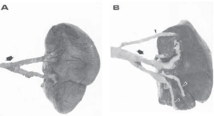
During dissection of the splenic artery, in order to study its extra-parenchymal branch, collateral secondary or tertiary branches directed to the ends of the spleen were identified. These vessels have received the designation of polar arteries and were subdivided into two types (Figure 1): 1) polar artery type I - collateral branch of the splenic artery originated prior to their terminal division, being long and relatively wide, directed to one of the spleen extermities, 2) type II polar artery - secondary or tertiary branch of the terminal division of the splenic artery, being short and thinner, also directed to one pole of the spleen. The absolute and relative frequencies of these branches are shown in table 2. The distance between the visceral surface of the spleen and the point of occurrence of these branches are exhibited on table 3.
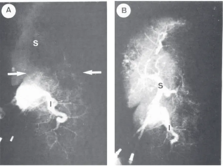
The results of arteriography are shown in figure 2 and tables 4 and 5. These results relate to the terminal division of the splenic artery and the polar branches type I and II.
DISCUSSION
DISCUSSION
DISCUSSION
DISCUSSION
DISCUSSION
Macroscopic study Macroscopic study Macroscopic study Macroscopic study Macroscopic study
The splenic artery has a terminal division, of bifurcation type, in most cases. The polar arteries are the branches that go to one extremity of the spleen and may be type I, a collateral, long, thick branch of the splenic artery, or type II, a secondary or tertiary branch of the spleen artery of lesser length and caliber. Both the division and the terminal polar arteries can be evidenced arteriographically.
According to the results of a study on the terminal division of the splenic artery in 60 fixed spleens, there was a bifurcation in 93.3% of the specimens, consistent with other studies in the literature6-9.
In relation to the trifurcation of the splenic artery, on its turn, it was found to occur in 6.7% of cases. One study cites an average terminal artery in 16.9% as a branch of the splenic artery7. In addition, there are studies that support its occurrence in 3.12%5 and in 10.6%10.
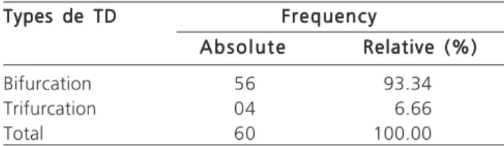
Table 1 Table 1 Table 1
Table 1 Table 1 – Types of Terminal Division (TD) of the splenic artery observed in the 60 dissected spleens.
Types de TD Types de TDTypes de TD
Types de TDTypes de TD FrequencyFrequencyFrequencyFrequencyFrequency A b s o l u t e
A b s o l u t e A b s o l u t e A b s o l u t e
A b s o l u t e Relative (%)Relative (%)Relative (%)Relative (%)Relative (%)
Bifurcation 56 93.34
Trifurcation 04 6.66
The study of polar branches of the splenic artery showed the polar type I present in 10% of cases, type II in 28.3% and spleens with type I and II in 8.3% of cases. These same values are consistent with those found in other publications7-9.
In this study, we evaluated the terminal division of the primary branches and polar branches of the splenic artery, verifying the average frequency of the length these branches between their origin, in the splenic artery trunk, and the splenic parenchyma, being 2.89 cm for the terminal division, 4.85 cm for the polar type I and 2.39 cm for the polar type II. This described average distance agrees with the range of values reported by other authors6,7.
Table 2 Table 2 Table 2 Table 2
Table 2 – Polar artery types (PA) identified in the 60 dissected spleens.
Typss de PA Typss de PA Typss de PA Typss de PA
Typss de PA FrequencyFrequencyFrequencyFrequencyFrequency A b s o l u t e
A b s o l u t eA b s o l u t e
A b s o l u t eA b s o l u t e Relative (%)Relative (%)Relative (%)Relative (%)Relative (%)
Type I 06 10.00
Type II 17 28.33
Type I + Type II 05 8.33
Total 28 46.66
Table 3 Table 3 Table 3 Table 3
Table 3 – Distance between the visceral surface of the spleen and: 1- the Terminal Division; and 2- the origin of polar arteries type I and type II.
Average Average Average Average
Average xxxxx SSSSS S -S -S -S -S - C V %C V %C V %C V %C V % M I NM I NM I NM I NM I N M A XM A XM A XM A XM A X
TD 2.89 0.91 0.13 31.51 1.04 5.05
PI 4.85 2.20 0.60 30.52 3.84 7.69
PII 2.39 1.54 0.29 50.89 0.93 5.25
TD: Terminal Division
PI: origin of polar arteries type I PII: origin of polar arteries type II x: average values
S: standard deviation
S-: standard error of the mean CV%: coefficient of variation MIN: minimum value MAX: maximum value
Radiological study Radiological study Radiological study Radiological study Radiological study
The results of arteriography in 30 spleens showed the terminal division of the splenic artery and its polar
Figure 1 Figure 1 Figure 1 Figure 1
184 Morfometric study of arterial branching of the spleen compared to radiological studyS i l v aS i l v aS i l v aS i l v aS i l v a
branches. The use of splenic arteriography is a means of comparative radiological study to the anatomical one, applied to radiological study to assess the splenic artery tree.
Studies show the importance of splenic arteriography in splenic trauma11, in expansive diseases12 and in diagnosis of splenic hematoma13 and delayed rupture of the spleen14. Furthermore, a study refers the use of ultrasound and computed tomography as a key means for diagnosing injuries of the spleen; it also states that ultrasound is the most reliable means of diagnosing these lesions15.
These observations render relevant the fact that it is feasible to angiographically identify (100%) the branches of the splenic artery terminal division and its polar branches. This is one more piece of data in guiding the surgeon when in need to identify those vessels.
Knowledge of the splenic artery intraparenchymal division is of great importance in surgical practice. It is for the branches of its terminal division or for its polar branches that the surgeon will turn when making a particular area of
Table 4 Table 4 Table 4
Table 4 Table 4 – Types of terminal Division (TD) observed in the 30 arteriographies performed.
Types de TD Types de TDTypes de TD
Types de TDTypes de TD FrequencyFrequencyFrequencyFrequencyFrequency A b s o l u t e
A b s o l u t e A b s o l u t e A b s o l u t e
A b s o l u t e Relative (%)Relative (%)Relative (%)Relative (%)Relative (%)
Bifurcation 27 90.00
Trifurcation 03 10.00
Total 30 100.00
Table 5 Table 5Table 5
Table 5Table 5 – Polar artery types observed in the 30 arteriographies performed.
Types of polar Types of polarTypes of polar
Types of polarTypes of polar FrequencyFrequencyFrequencyFrequencyFrequency a r t e r i e s
a r t e r i e sa r t e r i e s
a r t e r i e sa r t e r i e s A b s o l u t eA b s o l u t eA b s o l u t eA b s o l u t eA b s o l u t e Relative (%)Relative (%)Relative (%)Relative (%)Relative (%)
Polar arteries type I 05 16.00
Polar arteries type II 06 20.00
Total 11 36.00
Figure 2 Figure 2Figure 2 Figure 2
devascularization in order to perform a partial splenectomy. Therefore, it is necessary to know the vascular distribution, as well as its probable location. The measurement of the distance between these vessels and the visceral surface of the spleen is an additional parameter in guiding the surgeon to its faster identification.
According to the results of our study, we can infer that the splenic artery shows, in most cases, a
bifurcation-type terminal division that can be viewed arteriographically. We highlight the existence of independent arterial segmentation in almost all cases (98%), similar in the visceral and diaphragmatic surfaces of the spleen. Thus, one can conclude that partial splenectomy is anatomical and the use of radiological methods is feasible for the conservative treatment of splenic injuries.
R E S U M O R E S U M O R E S U M O R E S U M O R E S U M O
Objetivo: Objetivo: Objetivo: Objetivo:
Objetivo: Estudar a distribuição dos ramos da artéria esplênica dirigidos ao baço aplicado ao estudo radiológico da sua distribuição intraparenquimatosa, visando à utilização destes conhecimentos na esplenectomia parcial. Métodos:Métodos:Métodos:Métodos:Métodos: no estudo macroscópico, foram utilizados 60 baços humanos dos quais as artérias esplênicas foram dissecadas desde sua origem para visualizar a divisão terminal e os ramos dirigidos ao baço. Realizaram-se as medidas da distância entre a face visceral do baço e a divisão terminal da artéria esplênica e da emergência dos ramos polares. No estudo radiológico, utilizaram-se 30 baços humanos nos quais se injetou contraste nas artérias esplênicas para realizar as arteriografias e estudar a divisão terminal e ramos polares. Resultados:Resultados:Resultados:Resultados:Resultados: 93,34% dos baços apresentaram bifurcação como padrão de divisão terminal e 6,66% trifurcação. Identificaram-se ramos colaterais secundários e terciários tendo como freqüência relativa de 10% para o tipo I, 17% para o tipo II e 8,33% para ambas. A distância entre a face visceral do baço e a divisão terminal foi, em média, 2,89cm e para a emergência da artéria polar tipo I foi 4,85cm e 2,39cm para a tipo II. Nas 30 arteriografias realizadas, fez-se um estudo da divisão terminal no qual se observou bifurcação em 90% dos baços e trifurcação em 10%, além da presença de artéria polar tipo I em 16% e tipo II em 20%. Conclusão:Conclusão:Conclusão:Conclusão: a artéria esplênicaConclusão: apresenta divisão terminal do tipo bifurcação que pode ser visualizada arteriograficamente. Destaca-se a existência de segmentação arterial independente na quase totalidade dos casos (98%), semelhantes nas faces visceral e diafragmática do baço. A esplenectomia parcial é anatômica e torna-se factível o emprego de métodos radiológicos no tratamento conservador das lesões esplênicas.
Descritores Descritores Descritores Descritores
Descritores: Esplenectomia. Artéria esplênica. Baço. Radiologia intervencionista.
REFERENCES
REFERENCES
REFERENCES
REFERENCES
REFERENCES
1. Petroianu A. O Baço. 1a ed. São Paulo: CLR Balieiro; 2003.
2. King H, Shumacker HB Jr. Splenic studies. I. Susceptibility to infection after splenectomy performed in infancy. Ann Surg 1952; 136(2): 239-42.
3. Christo MC. Esplenectomias parciais regradas. Nota prévia sobre os três primeiros casos operados. Hospital 1959; 56:93-8. 4. Zappalá A. Estudo anatômico da divisão terminal da a. lienalis:
zonas arteriais do baço [tese de livre-docência]. Belo Horizonte: Universidade de Minas Gerais, Faculdade de Medicina; 1958. 5. Silva AR, Aragão AHM. Segmentação arterial do baço:
fundamento anatômico para a esplenectomia parcial. Rev bras cir 1988; 78(2):125-8.
6. Testut L, Latarjet A. Tratado de anatomia humana. Rio de Janeiro: Salvat; 1952.
7. Zappalá, A. Contribuição para o estudo da anatomia dos vasos e das “zonas vasculares lienais”. Dados anatômicos no homem e experimentais no cão, para aplicação na “lienectomia” parcial [tese de cátedra]. Recife: Universidade de Pernambuco, Faculdade de Medicina; 1959.
8. Esperança Pina JA. Territórios arteriais esplênicos. Lisboa: Universidade de Nova Lisboa; 1979.
9. Garcia-Porrero JA, Lemes A. Arterial segmentation and subsegmentation in the human spleen. Acta Anat 1988; 131(4):276-83.
10. Mandarim-Lacerda CA, Sampaio FJ, Passos MA. Vascular segmentation of the spleen in the newborn infant. Anatomical support for partial resection. J Chir 1983; 120(8-9):471-3.
11. Awe WC, Eidemiller L. Selective angiography in splenic trauma. Am J Surg 1973; 126(2):171-9.
12. Ekelund L, Göthlin J, Pettersson H. Angiography in expansile lesions of spleen. Am J Roentgenol Radium Ther Nucl Med 1975; 125(1):81-90.
13. Lande A, Bard R. Celiac arteriography following percutaneous splenoportography. Radiology 1975; 114(1):57-8.
14. Benjamin CI, Engrav LH, Perry FJ Jr. Delayed rupture or delayed diagnosis of rupture of the spleen. Surg Gynecol Obstet 1976; 142(2):171-2.
15. Mishalany HG, Mahour GH, Andrassy RJ, Harrison MR, Woolley MM. Modalities of preservation of the traumatized spleen. Am J Surg 1978; 136(6):697-700.
Received 25/03/2010
Accepted for publication 27/05/2010 Conflict of interest: none
Source of funding: none
How to cite this article: How to cite this article: How to cite this article: How to cite this article: How to cite this article:
Silva LFA, Silveira LMA, Timbó PS, Pinheiro SR, Barros LV, Silva Filho AR. Morfometric study of arterial branching of the spleen compared to radiological study. Rev Col Bras Cir. [periódico na Internet] 2011; 38(3). Disponível em URL: http://www.scielo.br/rcbc
Correspondence to: Correspondence to: Correspondence to: Correspondence to: Correspondence to: Antonio Ribeiro da Silva Filho