ABSTRACT
Sao Paulo Med J. 2008;126(3):190-3.
Case R
epor
t
Pedro PopoutchiCarlos Renato dos Reis Lemos
Julio César Rosa e Silva
Antônio Alberto Nogueira
Omar Feres
José Joaquim Ribeiro da Rocha
Postmenopausal intestinal
obstructive endometriosis: case
report and review of the literature
Discipline of Coloproctology, Department of Surgery and Anatomy,
Faculdade de Medicina de Ribeirão Preto (FMRP), Universidade de São
Paulo (USP), Ribeirão Preto, São Paulo, Brazil
CONTEXT: Endometriosis is characterized by the presence of endometrial tissue outside the uterine cavity, which is commonly detected in gynecological practice but rarely reported as a coloproctological disorder. The objective of the present report was to discuss a rare case of postmenopausal intestinal endometriosis simulating a malignant lesion, following a review of the literature.
CASE REPORT: A 74-year-old woman with com-plaints of hematochezia and tenesmus of two months’ duration accompanied by liquid feces and pelvic pain, but with no other gastrointestinal or gynecological complaints, was referred to our service. She had been menopausal for 22 years, with no hormone replacement treatment, and had undergone panhysterectomy three years before the referral to us, due to endometrial thickening and a right adnexal cyst. Five months before this referral, she had undergone laparotomy due to acute obstructive abdomen, which revealed a tumor mass involving the small bowel. Anato-mopathological examination of the enterectomy suggested a hypothesis of intestinal endometrio-sis. A proctological examination was normal. Computed tomography of the pelvis revealed thickening of the rectosigmoid transition and colonoscopy revealed friable tumor formation in the rectum. A biopsy of the lesion revealed muco-sal fragments of endometrial type, which led to a review of the previous anatomopathological examination. The patient underwent rectosig-moidectomy with protective transversotomy, with a good postoperative course, and anatomical examination confi rmed the intestinal endometrio-sis. The patient subsequently suffered a stenosing recurrence of the lesion and has undergone colostomy since then.
KEY WORDS: Intestines. Endometriosis. Surgery. Diagnosis. Postmenopause.
INTRODUCTION
Endometriosis is an estrogen-dependent disease that usually occurs in women during the menacme. Its etiopathogenesis is still a matter of controversy.1-3 However, occurrences
of endometriosis in patients with no menstrual fl ow,4,5 or the presence of lesions at sites where
there is no direct contact with menstrual fl ow, such as the lungs and intestine,6,7 raise the
hy-pothesis that metaplasia may develop in these areas and/or vascular transport may occur, especially if estrogen is not present.
The incidence of intestinal endometriosis ranges from 3 to 34%,8-10 and the sigmoid
and rectum are more commonly involved. This disorder is more frequent among women during the menacme6 and its importance lies
mainly in the need for a differential diagnosis with colon adenocarcinoma, which is the third most common type of cancer diagnosed in women.11
The objective of the present report was to describe and discuss a rare case of postmeno-pausal intestinal endometriosis simulating a malignant lesion, with emphasis on diagnostic and therapeutic methods, following a review of the literature.
CASE REPORT
A 74-year-old white woman from Itabuna, State of Bahia, Brazil, sought the Coloproctol-ogy Service of our institution with complaints of hematochezia and liquid feces of two months’ duration, as well as tenesmus and pelvic pain.
Her antecedents of interest included eight pregnancies, two abortions, six vaginal deliver-ies, menopause of 22 years’ duration, presence of diabetes and hypertension, and no hormone replacement treatment. She reached the men-arche at 13 years of age, and did not have any history of dysmenorrhea. She had undergone hysterectomy plus bilateral salpingo-oopho-rectomy three years before her referral to our
service, due to endometrial thickening, with a postoperative anatomopathological diagnosis of endometrial glandular hyperplasia and dis-crete cellular atypia. On the same occasion, a mass of chocolate-colored content that occlud-ed the upper third of the vagina and a paratubal cyst also containing chocolate-colored matter had been detected intraoperatively. Histologi-cal analysis led to a diagnosis of hematosalpinx with hemosiderin on the left side.
Five months before before her referral to our service, she was attended at the emergency service of Hospital das Clínicas, Faculdade de Medicina de Ribeirão Preto, Universi-dade de São Paulo (HC/FMRP/USP) with signs and symptoms of intestinal obstruction. She underwent exploratory laparotomy, which revealed a tumor mass surrounding some loops of the small intestine. Anatomopathological examination confi rmed the presence of intes-tinal endometriosis.
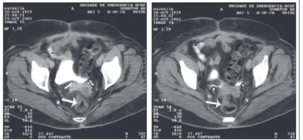
Physical and proctological examinations gave normal results, except for grade II obe-sity. Colonoscopy was requested (Figure 1), which revealed a friable and stenosing tumor formation in the upper rectum. A biopsy revealed mucosal fragments of endometrial type. Tomography of the pelvis (Figure 2) only showed parietal thickening in the rectosigmoid transition.
191
Sao Paulo Med J. 2008;126(3):190-3.
Figure 1. Computed tomography of the abdomen and pelvis of a 74-years-old woman, showing parietal thickening in the rectosigmoid transition.
Figure 2. Preoperative colonoscopy, showing friable and stenosing tumor formation in the upper rectum.
Figure 3. Surgical specimen of rectal endometriosis.
Figure 4. Extensive intestinal endome-triosis.
192
Sao Paulo Med J. 2008;126(3):190-3.
1. Abrao MS, Neme RM, Carvalho FM, Aldrighi JM, Pinotti JA. Histological classification of endometriosis as a predictor of re-sponse to treatment. Int J Gynaecol Obstet. 2003;82(1):31-40. 2. Abrão MS, Dias Jr JA, Podgaec S. Histórico e aspectos epidemi-ológicos da endometriose: uma doença prevalente e de conhe-cimento antigo. In: Abrão MS editor. Endometriose: uma visão contemporânea. Rio de Janeiro: Revinter; 2000. p. 149-68. 3. Seli E, Arici A. Endometriosis: interaction of immune and
endocrine systems. Semin Reprod Med. 2003;21(2):135-44. 4. Bellina JH, Schenck D. Large postmenopausal ovarian
endo-metrioma. Obstet Gynecol. 2000;96(5 Pt 2):846. 5. Goumenou AG, Chow C, Taylor A, Magos A. Endometriosis
arising during estrogen and testosterone treatment 17 years after abdominal hysterectomy: a case report. Maturitas. 2003;46(3):239-41.
6. Deval B, Rafii A, Felce Dachez M, Kermanash R, Levardon M. Sigmoid endometriosis in a postmenopausal woman. Am J Obstet Gynecol. 2002;187(6):1723-5.
7. Sakamoto S, Kishi K, Homma S, et al. [A case of catamenial pneumothorax due to diaphragmatic endometriosis confirmed by video-assisted thoracoscopic surgery]. Nihon Kokyuki Gakkai Zasshi. 2003;41(12):911-6.
8. Collin GR, Russel JC. Endometriosis of the colon. Its diagnosis and management. Am Surg. 1990;56(5):275-9.
9. Magtibay PM, Heppell J, Leslie KO. Endometriosis-as-sociated invasive adenocarcinoma involving the rectum in a postmenopausal female: report of a case. Dis Colon Rectum. 2001;44(10):1530-3.
10. Urbach DR, Reedijk M, Richard CS, Lie KI, Ross TM. Bowel resection for intestinal endometriosis. Dis Colon Rectum. 1998;41(9):1158-64.
11. Ferlay J, Bray F, Pisani P, Parkin DM. Globocan 2000. Cancer incidence, mortality and prevalence worldwide. Lyon: IARC Press; 2001.
12. Souza VCT, Baldin JA, Rocha AA, Moreira APT. Endometriose retal: relato de um caso. [Rectal endometriosis: report of a case]. Rev Bras Colo-Proctol. 1996;16(4):209-11.
13. Abrão MS, Machado MA, Campos FG, Habr-Gama A, Pinotti HW. Endometriose retal. [Rectal endometriosis]. Rev Hosp Clin Fac Med Sao Paulo. 1994;49(4):173-6.
14. Doniec JM, Kahlke V, Peetz F, et al. Rectal endometriosis: high sensitivity and specificity of endorectal ultrasound with an impact for the operative management. Dis Colon Rectum. 2003;46(12):1667-73.
15. Donnez J, Van Langendonckt A, Casanas-Roux F, et al. Current thinking on the pathogenesis of endometriosis. Gynecol Obstet Invest. 2002;54(Suppl 1):52-8; discussion 59-62.
Source of funding: None Conflicts of interest: None
Date of first submission: November 11, 2006 Last received: December 11, 2007 Accepted: May 5, 2008
REFERENCES
Anatomopathological examination confirmed recurrence of the disease, which caused ag-gressive stenosis of the anastomosis despite new transanal resections and dilatations. The patient has remained colostomized since then, and has not been in a suitable condition for intestinal transit reconstruction.
DISCUSSION
The rate of intestinal involvement in en-dometriosis cases is 3 to 34%.8-10 The rectum,
sigmoid colon, vermiform appendix, terminal ileus and cecum are the most affected seg-ments, in decreasing order of occurrence.10,12
The main symptoms are abdominal or pelvic pain, rectal pain, dysmenorrhea, dys-pareunia, constipation, tenesmus and rectal bleeding. More than 90% of these patients report some type of abdominal pain, while only 20% complain of rectal bleeding.10 The
symptoms are usually more marked during the menstrual period, but may also occur at any other time.13
Colorectal endometriosis is predominantly subserosal, rarely involving the muscularis or the mucosa. Colonoscopy is not always useful, but is of benefit by ruling out other lesions such as adenocarcinomas. Among the imaging examinations, endorectal ultrasound is particu-larly important. This has better sensitivity and specificity than computed tomography (CT) and nuclear magnetic resonance (NMR).14
The differential diagnoses of colonic lesions include adenocarcinomas, sarcomas, lymphomas, carcinomas and intestinal endo-metriosis.5 In cases of intestinal endometriosis,
the need for colectomy ranges from 0.1 to 0.7% of the cases8 since the rate of endometrial
carcinomas in ectopic endometrial tissue is very low. The presence of pelvic pain refractory to clinical treatment that originates in lesions of intestinal endometriosis and the obstructive nature of the disease are some of the few justi-fications for the procedure.10,12,13 In the present
case, the decision to proceed with surgery was due to the stenosing nature of the lesion.
Hormonal treatment should be consid-ered for childless young women, since this may lead to disappearance or reduction of the colorectal symptoms.10 Surgical treatment
should be instituted when the response to conservative treatment is inadequate or there are contraindications. In selected patients, in-testinal resection in combination with hyster-ectomy plus bilateral salpingo-oophorhyster-ectomy has yielded the best results.10
In advanced cases with extensive pelvic and rectal involvement, fibrosis and ureteral involvement usually impair the use of surgery, with frequent need for low resections and a protective stoma. Thus, in these cases, for bet-ter control over the disease, we chose hysbet-ter- hyster-ectomy plus bilateral salpingo-oophorhyster-ectomy in combination with rectal surgery.
The peculiar feature of the present case was the difficulty in explaining the appearance of the lesion in a patient who had been in a hypoestrogenic condition for 22 years and had undergone hysterectomy and oophorectomy three years before the present event. Moreover, she had no history suggesting the presence of endometriosis during the menacme (pelvic pain or infertility). In a case of this type, the hypothesis of intestinal tissue metaplasia should be considered.15
Advanced disease has been more strongly associated with pure or mixed undifferentiated disease. The results of Abrão et al. demon-strated that advanced disease with a worse outcome is related to higher prevalence of an undifferentiated pattern.1
CONCLUSION
193
Sao Paulo Med J. 2008;126(3):190-3. AUTHOR INFORMATION
Pedro Popoutchi, MD. Resident in the Discipline of Coloproctol-ogy, Department of Surgery and Anatomy, Faculdade de Medicina de Ribeirão Preto (FMRP), Universidade de São Paulo (USP), Ribeirão Preto, São Paulo, Brazil. Carlos Renato dos Reis Lemos, MD. Resident in the Discipline
of Coloproctology, Department of Surgery and Anatomy, Faculdade de Medicina de Ribeirão Preto (FMRP), Univer-sidade de São Paulo (USP), Ribeirão Preto, São Paulo, Brazil.
Julio César Rosa e Silva, MD. Attending physician, Department of Gynecology and Obstetrics, Faculdade de Medicina de Ribeirão Preto (FMRP), Universidade de São Paulo (USP), Ribeirão Preto, São Paulo, Brazil.
Antônio Alberto Nogueira, MD. Assistant professor, De-partment of Gynecology and Obstetrics, Faculdade de Medicina de Ribeirão Preto (FMRP), Universidade de São Paulo (USP), Ribeirão Preto, São Paulo, Brazil.
Omar Feres, MD. Assistant professor, Department of Surgery and Anatomy, Faculdade de Medicina de Ribeirão Preto (FMRP), Universidade de São Paulo (USP), Ribeirão Preto, São Paulo, Brazil.
José Joaquim Ribeiro da Rocha, MD. Assistant professor, De-partment of Surgery and Anatomy, Faculdade de Medicina de Ribeirão Preto (FMRP), Universidade de São Paulo (USP), Ribeirão Preto, São Paulo, Brazil.
Address for correspondence:
José Joaquim Ribeiro da Rocha
Disciplina de Coloproctologia, Departamento de Cirurgia e Anatomia
Faculdade de Medicina de Ribeirão Preto da Univer-sidade de São Paulo (FMRP-USP)
Av. Bandeirantes, 3.900 — Cidade Universitária Ribeirão Preto (SP) — Brasil — CEP 14048-900 Tel. (+ 55 16) 3602-2593
Fax. (+ 55 16) 3633-0836 E-mail: [email protected] E-mail: [email protected]
Copyright © 2008, Associação Paulista de Medicina
RESUMO
Endometriose intestinal obstrutiva na pós-menopausa: relato de caso e revisão da literatura CONTEXTO: A endometriose caracteriza-se pela presença de tecido endometrial fora da cavidade uterina, e a etiopatogenia ainda apresenta controvérsias. O objetivo desta publicação é apresentar e discutir, após revisão da literatura, um raro caso de endometriose intestinal na pós-menopausa que simulava uma lesão maligna.
RELATO DE CASO: Mulher de 74 anos apresentou-se com queixas de hematoquezia e tenesmo há dois meses. Relatou também aparecimento de fezes líquidas e dor pélvica no mesmo período, negando outras queixas gastrointestinais ou ginecológicas. Como antecedentes de interesse, revelou que era menopausada há 22 anos, sem terapia de reposição hormonal e realizou uma pan-histerectomia há três anos por espessamento endometrial e cisto anexial direito. Há cinco meses foi submetida a laparotomia exploradora por abdome agudo obstrutivo, com o achado de uma massa tumoral envolvendo alças de delgado. O exame anatomopatológico da enterectomia sugeriu a hipótese de endometriose intestinal. O exame proctológico era normal. A tomografia computadorizada da pelve mostrou um espessamento da transição retossigmóide e a colonoscopia, uma tumoração friável e estenosante no reto alto. A biópsia da lesão revelou fragmentos de mucosa tipo endometrial, que motivou a revisão do anatomopatológico anterior. A paciente foi submetida a retossigmoidectomia abdominal com transversostomia protetora, tendo boa evolução no pós-operatório. O anatomopatológico confirmou endometriose intestinal. Evoluiu com recidiva estenosante da lesão e pemanece colostomizada desde então.