Rev Odontol UNESP. 2013 Jan-Feb; 42(1): 13-19 © 2013 - ISSN 1807-2577
ARTIGO ORIGINAL
Prevalence of
Candida
spp. during radiographic
examination in Diabetes mellitus patients
Prevalência de Candida spp. durante o exame radiográico em pacientes diabéticos
Flávia Cristina VOLPATO
a, Juliana Rico PIRES
b, Isis do Rosário da Costa MARTINEZ
a,
Silvana Regina Perez ORRICO
c, Marciano Pires da COSTA
d,
Denise Madalena Palomari SPOLIDÓRIO
e, Andréa GONÇALVES
caFaculdade de Odontologia, UNESP – Univ Estadual Paulista, 14801-903 Araraquara - SP, Brasil bDepartamento de Periodontia e Implantodontia, UNIFEB – Centro Universitário da Fundação Educacional
de Barretos, 14783-226 Barretos - SP, Brasil
cDepartamento de Diagnóstico e Cirurgia, Faculdade de Odontologia, UNESP – Univ Estadual Paulista, 14801-903 Araraquara - SP, Brasil
dDepartamento de Bioquímica, Faculdade de Ciências Farmacêuticas, UNESP – Univ Estadual Paulista, 14801-902 Araraquara - SP, Brasil
eDepartamento de Fisiologia e Patologia, Faculdade de Odontologia, UNESP – Univ Estadual Paulista, 14801-903 Araraquara - SP, Brasil
Resumo
Introdução: Relata-se que indivíduos diabéticos são mais susceptíveis a infecções por Candida que indivíduos saudáveis, especialmente se doença periodontal estiver associada. Objetivo: Este estudo propôs avaliar a prevalência de colonização por Candida spp. durante o exame radiográico em pacientes diabéticos e não diabéticos. Material e método: Vinte e seis pacientes com Diabetes mellitus do tipo 2 e 20 pacientes sem Diabetes mellitus, apresentando periodontite crônica e Candida spp. na saliva, foram avaliados. Durante o exame radiográico, amostras de saliva foram coletas: da mucosa oral, do ilme radiográico periapical convencional, sensor radiográico digital (CDR) e bloco de mordida do posicionador de ilmes. Unidades formadoras de colônia (cfu/mL) e identiicação das leveduras do gênero Candida foram avaliadas.
Resultado: A mucosa oral de ambos os grupos mostrou maior colonização por Candida spp. quando comparada com outras superfícies coletadas (p < 0.05). Nos pacientes diabéticos, a mucosa da região esquerda superior mostrou níveis mais altos de colonização. Nos pacientes não diabéticos, a região de molar superior direito mostrou o nível mais alto de colonização durante o exame no posicionador, no sensor e no lado do ilme periapical que não ica voltado para a radiação X. Os níveis de Candida spp. na saliva foram similares entre diabéticos (média = 3.0 × 10⁶) e não diabéticos
(média = 3.8 × 10⁶). Conclusão: Nenhuma diferença na colonização por Candida spp. (cfu/mL) em pacientes diabéticos
e não diabéticos foi observada nas cinco superfícies coletadas e nas regiões radiográicas simuladas. Candida albicans foi a espécie prevalente de Candida spp. encontrada em todas as amostras.
Descritores: Doenças periodontais; diabetes mellitus; radiografia dentária digital.
Abstract
Introduction: It is suggested that individuals with diabetes are more susceptible to Candida infections than healthy people, especially if periodontal infection is associated. Objective: This study evaluated the prevalence of colonization by Candida spp. during radiographic examination in diabetic and non-diabetic patients. Material and methods: Twenty-six patients with type 2 diabetes mellitus and 20 patients without diabetes mellitus, presenting chronic periodontitis and presence of Candida spp. in saliva were evaluated. During radiographic examination, samples of saliva were collected from: oral mucosa, conventional radiographic periapical film, digital x-ray sensor (CDR), and bite block of the receptor-positioning device. Colony forming units (cfu/mL) and identification of Candida yeasts were assessed. Result: Oral mucosa from both groups showed the highest colonization with Candida spp. if compared with others surfaces collected (p < 0.05). In diabetic patients, the mucosa of the upper left regions showed higher levels of colonization. In non-diabetic patients, the upper right molar region showed the highest level of colonization during the examination of the receptor-positioning device, the sensor and the non-sensitive film. Candida spp. levels in saliva were similar between diabetics (mean = 3.0 × 10⁶) and non-diabetics
(mean = 3.8 × 10⁶). Conclusion: No difference in Candida spp. colonization (cfu/mL) in diabetics and non-diabetic
patients was observed for the five collected surfaces and the simulated radiographic region. Candida albicans was the prevalent species of Candida spp. found on all the samples.
INTRODUCTION
lnfection-control practices are designed to create and maintain a safe clinical environment to eliminate or minimize disease transmission during patient treatment1. here is high potential
for cross-contamination of equipment and environmental surfaces with blood or saliva when taking dental radiographs2.
Saliva has always been considered a potentially infectious material in dental infection control2. White, Glaze3 found that
dental healthcare workers can transfer oral microorganisms from the patient’s oral cavity to radiographic equipment during routine intraoral radiography. hese microorganisms remain viable on radiographic equipment for at least 48 hours.
Traditionally, intraoral radiographs are acquired using ilms. Since the introduction of digital radiography to dentistry, many dental schools and private practices have adopted digital imaging methods for acquiring radiographs. Digital radiology promises many advantages over traditional ilm-based techniques4,5.
Digital radiography sensors come into contact with mucous membranes and are considered semicritical devices. hey should be cleaned and ideally heat-sterilized or high-level disinfected between patients. However, there is a variety in the ability of digital radiographic sensors to be sterilized or high-level disinfected2. Semicritical items that cannot be reprocessed
by heat sterilization or high-level disinfection should, at a minimum, be barrier protected by a plastic barrier sheaths to reduce gross contamination during use that is not guaranteed from contamination2,6.
Previous reports mentioned that individuals with diabetes present oral complications more frequently than healthy people7. Commonly, these oral complications are associated
with fungi, for instance, Candida spp., which presents as white or white-yellow color, creamy, convex colonies, with a smooth and bright appearance, that are moist with a typical smell8. These oral complications are also associated
with Candida species that have frequently been isolated from the oral cavities of patients with diabetes mellitus9-15.
Besides, evidence indicates a strong correlation between the severity of periodontitis and diabetes, based on the fact that periodontal infection is associated with poor glycemic control in this particular group of patients7,16,17. Furthermore,
it has been established that the higher occurrence rate of yeasts is associated with immunocompromised patients with periodontal diseases, such as patients with diabetes12,13,18.
Moreover, patients with long-standing, poorly controlled diabetes are at risk of developing oral candidiasis19. Although
authors had related that 40% of the patients colonized with candidal species had no clinical signs of oral candidosis20.
A number of candidal species were recovered from the oral cavity of insulin-treated diabetic patients. C. albicans was the most commonly recovered species, recovered from 85% of the diabetic patients. C. dubliniensis was the second most commonly occurring species20.
Considering the lack of dada about the prevalence of colonization by Candida spp. in diabetic and non-diabetic patients
with periodontal disease during radiographic examination, this study was carried out in order to determine what surfaces would be contaminated. herefore, the aim of this study was to determine the prevalence of colonization by Candida spp. in diabetic and non-diabetic patients during radiographic examination, on diferent surfaces.
MATERIAL AND METHODS
1.
Sample
his study was approved by the Ethics in Human Research Committee of the Dental School (protocol number 79/03). All volunteers were informed about the aims and methods of this study, and gave their written consent to participate. he sample size calculation was based on previous studies21-23. A post-hoc
statistical power calculation test was performed based on cfu/mL for both groups and all evaluated surfaces, and the sample size was estimated in 20 patients per group, considering a power of 80%.
As inclusion criteria, all subjects that participated in this study must have been presenting chronic periodontitis and presenting Candida spp. in their saliva, regardless of gender, race, social group, age, oral hygiene, and nutrition habits. he following exclusion criteria were considered: history of antibiotic therapy within the previous 6 months and anti-inlammatory drugs within the previous 3 months; current history of immunosuppression; local or systemic use of antifungal drugs and use of mouthrinses.
2.
Periodontal Analyses
Chronic periodontitis was established as probing pocket depth (PPD) and clinic attachment loss (CAL) ≥ 4 mm and bleeding on probing in more than three sites in non-adjacent teeth24.
3.
Diabetes Analyses
he glycated hemoglobin exam (HbA1c; A1c) was requested for diabetic patients. he patient was classiied as having adequate metabolic control when presenting results from the glycated hemoglobin test A1c < 7%25.
4.
Saliva Analyses
he saliva collection was performed without stimulation, by asking the patients to retain their saliva and then deposit it in a funnel and bottles previously identiied and sterilized, until obtaining 3 mL of saliva for microbiological analysis. he tubes containing saliva were dispersed by vortexing for 60 seconds (Vortex, Marconi Equipamentos para Laboratório Ltda., Piracicaba, SP, Brazil) and the saliva was diluted in a decimal series of 10–1 to 10–4 in phosphate-bufered saline. he saliva was
Out of a total 153 periodontitis patients evaluated, 26 patients with diabetes mellitus and 20 patients without diabetes mellitus that presenting Candida spp. in their saliva were included in this study.
5.
Radiographic Examination
For all participants who required full-mouth radiographic examination, a set of 14 radiographs was used. When the patient already had a radiographic examination, a simulation of the examination was performed with written consent from the participant. hen, 26 diabetic and 20 non-diabetic patients were submitted to radiographic simulation/examination.
Before using the conventional periapical radiographic ilm (Kodak Insight, Eastman Kodak Company, Rochester, NY, USA) this material was placed in a plastic barrier that was sealed (Odontobrás, Ribeirão Preto, SP, Brazil) for 4s. he CDR sensor (Schick Technologies, Long Island City, NY, USA) was also protected with a plastic barrier. he conventional ilm and the CDR sensor were then disinfected by rubbing with sterile gauze dipped in 70% alcohol.
he intraoral radiographic examination (or the simulation) using conventional periapical radiographic ilm was taken with a Rinn ilm holder (XCP Instruments, Elgin, IL, USA). Initially radiographs were taken with conventional ilm from the regions solicited, ater that, the CDR sensor was maintained in the mouth for 30 seconds, with the patient’s ingers, simulating the technique on ive randomized regions in all patients. he regions were: upper right molar (Urm), upper right bicuspid (Urb), upper right cuspid and lateral incisor (Urc), upper central incisor (Uci), upper let cuspid and lateral incisor (Ulc), upper let bicuspid (Ulb), upper let molar (Ulm), lower let molar (Llm), lower let bicuspid (Llb), lower let cuspid and lateral incisor (Llc), lower central incisor (Lci), lower right cuspid and lateral incisor (Lrc), lower right bicuspid (Lrb), and lower right molar (Lrm).
6.
Microbiological Examination
Before the radiographic examination, saliva was collected from the oral mucosa of each patient with a sterile swab that was humidiied in sterile phosphate bufered saline (PBS) solution. Each swab was then rubbed onto a culture plate of Sabouraud Dextrose Agar (SDA, Acumedia, Neogen, Lansing, MI, USA)26
for posteriori microbiological analyses.
Ater the radiographic examination or simulation in each region, the conventional radiographic periapical ilm and the randomized regions of the sensor, still with the plastic barrier, were pressed into the SDA plate using sterile clinic tweezers; this procedure was carried out on both sides of the radiographic ilm and only on one side of the sensor. Only one side of the sensor is submitted to X-ray which was used because the other side presents a wire connection to the computer and this side would damage the procedure of pressure of the sensor into the DAS plate. Material was also collected from the surfaces of the RINN device, which remained in contact with the radiographic ilm, using a sterile swab, which was then maintained in sterilized
saline solution that was then rubbed onto the culture medium on the plate. Samples were incubated at 37 °C for 48 hours, for posteriori microbiological analyses of quantity (cfu/mL) and identiication of Candida species.
7.
Identiication of Candida spp.
Ater colony growth, the counting of the inoculum was performed in colony forming units per mL (cfu/mL). he characteristic colony morphology of the Candida yeasts was demonstrated using a stereoscopic microscope (Carl Zeiss do Brasil Ltda, Brazil) and diferent colonies were identiied by Gram staining. A test in CHROMagar
Candida chromogenic medium (Difco, BD) was performed to identify the Candida species27,28
and other identifying tests were also carried out according to Sandven29 and Sullivan, Coleman30, i.e., germ tube formation,
carbohydrate fermentation, carbohydrate assimilation, and thermotolerance assay31.
8.
Statistical Analyses
he values of colony-forming units per mL (cfu/mL) for diabetic and non-diabetic patients were compared, considering independent variables of the study the surfaces collected (mucosa, both sides of the conventional radiographic periapical ilm, CDR sensor and bite block of the receptor-positioning device) and the simulated radiographic region. he non-parametric Kruskal-Wallis test was used for comparison of the surfaces of collection in diabetic and non-diabetic patients while the Wilcoxon test was used for comparison of the radiographic regions for each type of surface collected. ANOVA and t-test were used for comparison the diabetic and non-diabetic patients with CAL, using BioStat 4.0 sotware (Belém, PA, Brazil).
RESULTS
he age range of subjects was 18- to 67-years old (male mean = 49.9 ± 0.1 years; female mean = 47.5 ± 1.8 years). Out of 46 participants, 11 (55%) patients without diabetes were female and nine (45%) were male, while 14 (53.9%) diabetic patients were female and 12 (46.1%) were male.
he total distribution of the concentration of glycated hemoglobin was similar in diabetic patients, considering that 25% male and 25% female presented less than 7% (good metabolic control) and 20% male and 30% female presented more than 8% (inadequate metabolic control).
Considering the percentage of patients with CAL ≥ 4 mm, it is possible to note that there was no statistical diference (ANOVA, t-test) between diabetic (male = 63.8%; female = 48.3%) and non-diabetic (male = 76.5%; female = 50.7%) patients.
surfaces on simulating regions, it was observed that the upper right molar (Urm) region showed the highest level of colonization during the examination of the receptor-positioning device, the sensor and the non-sensitive ilm on non-diabetic patients. In diabetic patients, the mucosa of the upper let regions (Ulb and Ulm) showed higher levels of colonization (Table 1).
Table 1 shows statistical diferences for the cfu/mL mean, when the collection surface was the conventional radiographic ilm, on the non-sensitive side, when compared to the oral mucosa in the upper right molar region of diabetic patients. he lower let molars, lower right bicuspid and molar region also demonstrated statistical diferences between the oral mucosa
and the receptor-positioning device. here was a statistically signiicant diference between the colonization of the surface of the device and the sensor for the lower right molar (Krukal-Wallis, p < 0.005).
he mean cfu/mL of non-diabetic patients was statistically diferent in the oral mucosa and receptor-positioning device for the lower let molar region (Kruskal-Wallis, p < 0.05; Table 1). When a Wilcoxon test (p < 0.05) was applied to the data of Table 1, a statistical diference was observed in the cfu/mL mean of the upper right molar for the sensitive ilm surface.
Candida spp. in saliva did not difer signiicantly between diabetic (mean = 3.0 × 10⁶) and non-diabetic patients
Table 1. Means and standard deviations (in parentheses) of cfu/mL of diabetic patients according to collected surfaces and simulated radiographic region
Simulated region
Surfaces – diabetic Surfaces – non diabetic
Device Sensor Sensitive ilm
Non-sensitive
ilm
Mucosa Device Sensor Sensitive ilm
Non-sensitive
ilm
Mucosa
Urm 3.7 (11.4) 4.3 (6.6) 2.4 (4.9) 2.7a (7.1) 22.5a (69.8) 4.8 (10.5) 19.1 (23.8) 5.4 (26.0) 15.5 (10.6) 18.2 (39.5)
Urb 7.9 (26.5) 15.6 (41.4) 5.2 (23.2) 8.7 (11.4) 26.8 (75.0) 4.2 (10.3) 10.9 (27.2) 5.4 (28.8) 12.8 (9.4) 73.1 (164.8) Urc 6.4 (15.6) 1.8 (2.2) 2.6 (12.5) 5.8 (6.9) 43.2 (84.8) 1.5 (4.9) 9.3 (28.0) 3.0 (17.1) 9.2 (7.5) 30.3 (51.9) Uci 4.2 (13.3) 8.9 (16.7) 2.8 (9.4) 4.0 (6.0) 45.6 (93.5) 0.6 (1.3) 1.7 (2.4) 0.8 (5.5) 3.6 (1.5) 60.4 (125.4) Ulc 4.3 (14.0) 7.1 (10.9) 3.9 (13.4) 7.1 (9.9) 34.3 (56.8) 1.7 (3.9) 11.3 (30.1) 3.6 (13.4) 6.8 (8.7) 46.1 (75.4) Ulb 3.6 (5.8) 6.5 (8.3) 8.0 (41.0) 12.1 (18.5) 61.9 (112.8) 0.7 (1.5) 15.3 (42.2) 5.2 (17.8) 7.6 (16.1) 16.0 (41.0) Ulm 1.9 (4.8) 10.8 (22.0) 4.9 (99.0) 5.1 (11.4) 63.9 (112.4) 0.9 (1.7) 5.9 (8.6) 1.8 (11.1) 6.0 (2.5) 17.2 (32.8) Llm 2.9b (6.4) 5.9 (8.0) 11.8 (22.8) 9.1 (29.4) 45.6b (75.4) 3.9a (11.1) 14.2 (32.9) 7.5 (23.7) 9.1 (25.0) 56.0a (151.1)
Llb 0.6 (1.2) 5.7 (7.0) 7.7 (23.1) 10.5 (15.9) 21.2 (47.3) 1.4 (4.7) 13.1 (28.8) 3.7 (10.8) 5.4 (7.0) 16.0 (15.9) Llc 1.5 (5.0) 10.7 (17.8) 8.4 (14.4) 6.2 (21.3) 17.6 (18.8) 1.9 (7.8) 7.2 (20.9) 3.1 (37.1) 12.2 (7.0) 15.3 (16.9) Lci 1.0 (3.6) 4.3 (8.9) 4.6 (21.8) 5.7 (10.3) 10.6 (27.5) 0.7 (1.7) 10.0 (21.0) 5.6 (19.1) 7.4 (14.8) 15.6 (21.0) Lrc 6.6 (27.8) 9.7 (26.1) 4.5 (15.6) 6.3 (10.2) 13.0 (43.5) 0.6 (1.7) 8.3 (18.8) 3.7 (9.8) 3.9 (8.9) 14.1 (60.0) Lrb 0.4c (1.0) 10.0 (22.8) 5.8 (14.2) 5.6 (15.9) 20.7c (48.3) 2.0 (6.7) 3.2 (7.5) 2.9 (2.3) 3.8 (5.2) 9.3 (21.9)
Lrm 1.5de (3.5) 22.0d (22.0) 7.8 (23.4) 9.0 (13.8) 39.8e (75.7) 1.7 (3.7) 9.4 (15.6) 4.1 (9.7) 4.6 (5.8) 38.4 (102.2) Same letters difer statistically (Kruskal-Wallis, p < 0.05). Abbreviations: Urm = upper right molar, Urb = upper right bicuspid, Urc = upper right cuspid and lateral incisor, Uci = upper central incisor, Ulc = upper let cuspid and lateral incisor, Ulb = upper let bicuspid, Ulm = upper let molar, Llm = lower let molar, Llb = lower let bicuspid, Llc = lower let cuspid and lateral incisor, Lci = lower central incisor, Lrc = lower right cuspid and lateral incisor, Lrb = lower right bicuspid, Lrm = lower right molar.
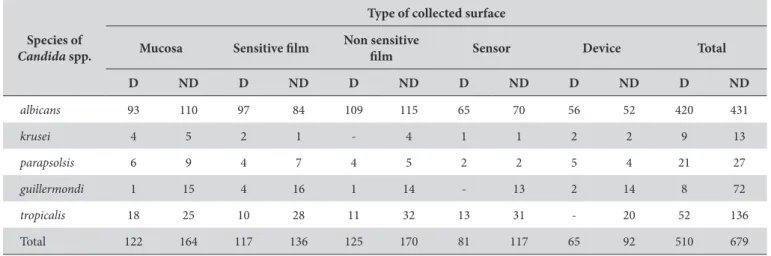
Table 2. Frequency of identiied species of Candida spp., according to the collected surface for diabetic and non-diabetic patients
Species of
Candida spp.
Type of collected surface
Mucosa Sensitive ilm Non sensitive
ilm Sensor Device Total
D ND D ND D ND D ND D ND D ND
albicans 93 110 97 84 109 115 65 70 56 52 420 431
krusei 4 5 2 1 - 4 1 1 2 2 9 13
parapsolsis 6 9 4 7 4 5 2 2 5 4 21 27
guillermondi 1 15 4 16 1 14 - 13 2 14 8 72
tropicalis 18 25 10 28 11 32 13 31 - 20 52 136
Total 122 164 117 136 125 170 81 117 65 92 510 679
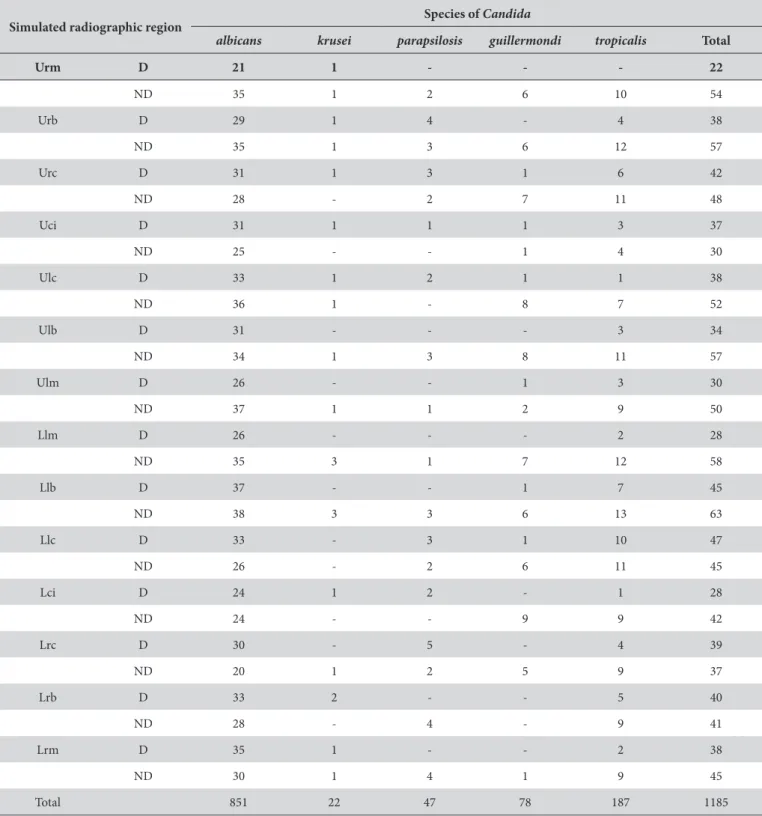
Table 3. Frequency of identiied species of Candida spp., according to simulated-radiographic region for diabetics and non-diabetics
Simulated radiographic region Species of Candida
albicans krusei parapsilosis guillermondi tropicalis Total
Urm D 21 1 - - - 22
ND 35 1 2 6 10 54 Urb D 29 1 4 - 4 38 ND 35 1 3 6 12 57 Urc D 31 1 3 1 6 42 ND 28 - 2 7 11 48 Uci D 31 1 1 1 3 37
ND 25 - - 1 4 30
Ulc D 33 1 2 1 1 38
ND 36 1 - 8 7 52
Ulb D 31 - - - 3 34 ND 34 1 3 8 11 57 Ulm D 26 - - 1 3 30
ND 37 1 1 2 9 50
Llm D 26 - - - 2 28 ND 35 3 1 7 12 58 Llb D 37 - - 1 7 45 ND 38 3 3 6 13 63 Llc D 33 - 3 1 10 47 ND 26 - 2 6 11 45 Lci D 24 1 2 - 1 28
ND 24 - - 9 9 42
Lrc D 30 - 5 - 4 39
ND 20 1 2 5 9 37
Lrb D 33 2 - - 5 40
ND 28 - 4 - 9 41
Lrm D 35 1 - - 2 38
ND 30 1 4 1 9 45
Total 851 22 47 78 187 1185
Abbreviations: Urm = upper right molar, Urb = upper right bicuspid, Urc = upper right cuspid and lateral incisor, Uci = upper central incisor, Ulc = upper let cuspid and lateral incisor, Ulb = upper let bicuspid, Ulm = upper let molar, Llm = lower let molar, Llb = lower let bicuspid, Llc = lower let cuspid and lateral incisor, Lci = lower central incisor, Lrc = lower right cuspid and lateral incisor, Lrb = lower right bicuspid, Lrm = lower right molar. D = diabetic, ND = non-diabetic. (mean = 3.8 × 10⁶). Table 2 shows the high prevalence of
Candida albicans for all collected surfaces from diabetic and non-diabetic patients, followed by Candida tropicalis. Tests for the identiication of Candida dubliniensis were performed, but this specie was not found in any patients. he device surface was the surface that carried the least number of Candida colonies and the non-sensitive ilm for non-diabetics was the surface that presented the highest Candidaalbicans colonization.
DISCUSSION
his study did not conirm the hypothesis that the presence of Candida spp. is more frequent in the oral cavity of diabetic than non-diabetic patients, as has been observed in other studies9,11,14,15.
his result may be due to the fact that half of the sample had good metabolic control. According to Hill et al.32, diabetes by itself does
not put a patient at risk of developing fungal infections, unless his/her metabolic control is poor. It is speculated that there is a tendency towards a greater presence of Candida spp. in the oral mucosa of non-diabetic patients in this study. However, due to data analyses of statistical values and considering the extreme values shown by this data set, statistical diferences were not observed, with the exception of some regions shown in Tables 2 and 3. his lack of diference suggests that studies about intraoral techniques should consider that the radiographic receptor or the receptor-positioning device may be infected by Candida spp. in a similar manner to the patient’s oral mucosa, independently of whether the patient is diabetic or not. Oten, students that are initiating their studies consider the oral cavity to be potentially infected, but handle the radiographic receptor as if it were not infected.
As regards the distribution of the participants in this study, measurements of glycated hemoglobin suggested that patients who presented glycated hemoglobin of higher than 8% did not have an increased risk of fungal infection. In contrast, a higher occurrence of fungal infections was shown by Hill et al.32 in patients with
concentrations of glycated hemoglobin of higher than 12%. he limitation of studies of small sample sizes of patients with diabetes should also be taken into consideration, since such sample sizes may prevent the analysis of risk factors for higher prevalence of fungal infections, such as metabolic control33.
C. albicans was the most commonly identiied candidal species in this and other studies12,20,34,35.
According to Willis et al.20, forty per cent of patients colonized
with candidal species had no signs of oral candidosis and a number of candidal species were recovered from the oral cavity of insulin-treated patients. C. albicans was the most commonly recovered species, being recovered from 85% of the diabetic patients. C. dubliniensis was the second most commonly occurring candidal species. he oral cavity relects the state of systemic health more frequently than any other part of the body and increased susceptibility to general and oral supericial infections with yeasts has long been associated with diabetes mellitus.
Manfredi et al.13 observed that diabetic patients with dentures
had more species of Candida, with the exception of Candida albicans, isolated from their mouths than dentate diabetics. his study showed a higher prevalence of Candida albicans, followed by C. tropicalis and C. guillermondi. Similarly, a previous study reported a non-signiicant trend towards a prevalence of species other than C. albicans in non-diabetic patients compared to diabetic patients13.
In conclusion, there was no diference in colonization (cfu/mL) for the Candida spp. between diabetic and non-diabetic patients with periodontal disease, when considering the ive collected surfaces and the simulated-radiographic regions studied. Candida albicans was the prevalent species of Candida spp. found on the collected surfaces and simulated radiographic regions, followed by Candida tropicalis.
ACKNOWLEDGEMENTS
his study was supported by FAPESP (process #2004/10589-2, 2005/00987-3, 2003/00890-4 and 1999/03026-1). We would like to acknowledge the subjects for their valuable participation.
REFERENCES
1. Bartolini JA, Chariton DG, Flint DJ. Infection control practices in dental radiology. Gen Dent. 2003;51:264-71.
2. Centers for Disease Control and Prevention. Guidelines for Infection Control in Dental Health-Care Settings — 2003. MMWR 2003;52(No. RR-17):[2;31].
3. White SC, Glaze S. Interpatient microbiological cross-contamination ater dental radiographic examination. J Am Dent Assoc. 1978;96:801-4. 4. Kalathingal SM, Moore S, Know S, Schuster G, Shrout MK, Plummer K. An evaluation of microbiologic contamination on phosphor plates in a dental school. Oral Surg Oral Med Oral Pathol Oral Radiol Endod. 2009;107:279-82. http://dx.doi.org/10.1016/j.tripleo.2008.05.025 5. van der Stelt PF. Filmless imaging: the uses of digital radiography in dental practice. J Am Dent Assoc. 2005;136:1379-87.
6. Hokett SD, Honey JR, Ruiz F, Baisedn MK, Hoen MM. Assessing the efectiveness of direct digital radiography barrier sheaths and inger cots. J Am Dent Assoc. 2000;131:463-7.
7. Taylor GW, Borgnakke WS. Periodontal disease: associations with diabetes, glycemic control and complications. Oral Dis. 2008;14:191-203. http://dx.doi.org/10.1111/j.1601-0825.2008.01442.x
8. Spolidorio DMP, Boriollo MFG, Estrela C, Spolidorio LC. Diferentes métodos fenotípicos para isolamento e identiicação de espécies de Candida. ROBRAC: Rev Odontol Brasil Central. 2009;18:18-26.
9. Lamey PJ, Darwaza A, Fisher BM, Samaranayake LP, Macfarlane TW, Frier BM. Secretor status, candidal carriage and candidal infection in patients with diabetes mellitus. J Oral Pathol. 1988;17:354-7. http://dx.doi.org/10.1111/j.1600-0714.1988.tb01549.x
10. Farah CS, Ashman RB, Challacombe SJ. Oral candidosis. Clin Dermatol. 2000;18:553-62. http://dx.doi.org/10.1016/S0738-081X(00)00145-0
11. Guggenheimer J, Moore PA, Rossie K, Myers D, Mongelluzzo MB, Block HM et al. Insulin-dependent diabetes mellitus and oral sot tissue pathologies. II. Prevalence and characteristics of Candida and candidal lesions. Oral Surg Oral Med Oral Pathol Oral Radiol Endod. 2000;89:570-6. http://dx.doi.org/10.1067/moe.2000.104477
13. Manfredi M, McCullough MJ, Al-Karaawi ZM, Hurel SJ, Porter SR. he isolation, identiication and molecular analysis of Candida spp. isolated from the oral cavities of patients with diabetes mellitus. Oral Microbiol Immunol. 2002;17:181-5. http://dx.doi.org/10.1034/ j.1399-302X.2002.170308.x
14. Belazi M, Velegraki A, Fleva A, Gidarakou I, Papanaum L, Baka D, et al. Candidal overgrowth in diabetic patients: potential predisposing factors. Mycoses. 2005;48:192-6. http://dx.doi.org/10.1111/j.1439-0507.2005.01124.x
15. Kumar BV, Padshetty, NS, Bai KY, Rao MS. Prevalence of Candida in the oral cavity of diabetic subjects. J Assoc Phys Indian. 2005;53:599-602. 16. Diabetes and periodontal diseases. Committee on Research, Science and herapy. American Academy of Periodontology.
J Periodontol. 2000;71:664-78. http://dx.doi.org/10.1902/jop.2000.71.4.664
17. Christgau M, Palitzsch KD, Schmalz G, Kreiner U, Frenzel S. Healing response to non-surgical periodontal therapy in patients with diabetes mellitus: clinical, microbiological, and immunological results. J Clin Periodontol. 1998;25:112-24. http://dx.doi.org/10.1111/ j.1600-051X.1998.tb02417.x
18. Moore LV, Moore WE, Riley C, Brooks CN, Burmeister JA, Smibert RM. Periodontal microlora of HIV positive subjects with gingivitis or adult periodontitis. J Periodontol. 1993;64:48-56. http://dx.doi.org/10.1902/jop.1993.64.1.48
19. Lamster IB, Lalla E, Borgnakake WS, Taylor GW. he relationship between oral health and diabetes mellitus. J Am Dent Assoc. 2008;139 (Suppl):19S-24S.
20. Willis AM, Coulter WA, Fulton CR, Hayes JR, Bell PM, Lamey P-J. Oral candidal carriage and infection in insulin-treated diabetic patients. Diabet Med. 1999;16:675-9. http://dx.doi.org/10.1046/j.1464-5491.1999.00134.x
21. Colombo APV, Teles RP, Torres MC, Souto R, Rosalém Jr. W, Mendes MCS et al. Subgingival microbiota of Brazilian subjects with untreated chronic periodontitis. J Periodontol. 2002;73:360-9. http://dx.doi.org/10.1902/jop.2002.73.4.360
22. Sardi JCO, Duque C, Camargo GACG, Holing JF, Gonçalves RB. Periodontal conditions and prevalence of putative periodontopathogens and Candida spp. in insulin-dependent type 2 diabetic and non-diabetic patients with chronic periodontitis - a pilot study. Arch Oral Biol. 2011;56:1098-105. http://dx.doi.org/10.1016/j.archoralbio.2011.03.017
23. Melton JJ, Redding SW, Kirkpatrick WR, Reasner CA, Ocampo GL, Venkatesh A, et al. Recovery of Candida dubliniensis and other Candida species from the oral cavity of subjects with periodontitis who had well-controlled and poorly controlled type 2 diabetes: a pilot study. Spec Care Dentist. 2010;30:230-4. http://dx.doi.org/10.1111/j.1754-4505.2010.00159.x
24. Armitage G. Development of a classiication system for periodontal diseases and conditions. Ann Periodontol. 1999;4:1-6. http://dx.doi. org/10.1902/annals.1999.4.1.1
25. American Diabetes Association. Standards of medical care in diabetes -2010. Diabetes Care. 2010;33( Suppl 1):S11-61. http://dx.doi. org/10.2337/dc10-S011
26. Williams DW, Lewis MAO. Isolation and identiication of candida from the oral cavity. Oral Dis. 2000;6:3-11. http://dx.doi. org/10.1111/j.1601-0825.2000.tb00314.x
27. Beighton D, Ludford R, Clark DT, Brailsford SR, Pankhurst CL, Tinsley GF, et al. Use of CHROMagar Candida Medium for Isolation of Yeasts from Dental Samples. J Clin Microbiol. 1995;33:3025-7.
28. Pfaller MA, Houston A, Cofmann S. Application of CHROMagar Candida for rapid screening of clinical specimens for Candida albicans,
Candida tropicalis, Candida krusei, and Candida (Torulopsis) glabrata. J Clin Microbiol. 1996;34:58-61.
29. Sandven P. Laboratory identiication and sensitivity testing yeast isolates. Acta Odontol Scand. 1990;48:27-36. http://dx.doi. org/10.3109/00016359009012731
30. Sullivan D, Coleman D. Candida dubliniensis: Characteristics and identiication. J Clin Microbiol. 1998;36:329-34.
31. Pinjon E, Sullivan D, Salkin I, Shanley D, Coleman D. Simple, inexpensive, reliable method for diferentiation of Candida dubliniensis
from Candida albicans. J Clin Microbiol. 1998;36:2093-5.
32. Hill LVH, Tan MH, Pereira LH, Embil JA. Association of oral candidiasis with diabetic control. J Clin Pathol. 1989;42:502-5. http://dx.doi. org/10.1136/jcp.42.5.502
33. Soell M, Hassan M, Miliauskaite A, Haïkel Y, Selimovic D. he oral cavity of elderly patients in diabetes. Diabetes Metab. 2007;33:S10-8. http://dx.doi.org/10.1016/S1262-3636(07)80053-X
34. Negrato CA, Tarzia O. Buccal alterations in diabetes mellitus. Diabetol Metab Syndr. 2010;2:1-11. http://dx.doi.org/10.1186/1758-5996-2-3 35. Manfredi M, McCullough MJ, Vescovi P, Al-Kaarawi ZM, Porter SR. Update on diabetes mellitus and related oral diseases. Oral
Dis. 2004;10:187-200. http://dx.doi.org/10.1111/j.1601-0825.2004.01019.x
CONFLICTS OF INTERESTS
he authors declare no conlicts of interest.
CORRESPONDING AUTHOR
Andréa Gonçalves
Departamento de Diagnóstico e Cirurgia, Faculdade de Odontologia, UNESP – Univ Estadual Paulista, Rua Humaitá, 1680, 14801-903 Araraquara - SP, Brasil
e-mail: [email protected]