Rev Odontol UNESP. 2015 May-June; 44(3): 157-162 © 2015 - ISSN 1807-2577
ORIGINAL ARTICLE
Doi: http://dx.doi.org/10.1590/1807-2577.0021
Evaluation of postoperative pain ater endodontic treatment with
foraminal enlargement and obturation using two
auxiliary chemical protocols
Avaliação da dor pós-operatória de tratamentos endodônticos realizados com ampliação e
obturação foraminal utilizando dois protocolos de substâncias químicas auxiliares
Marcelle Louise Sposito BOURREAU
a*, Adriana de Jesus SOARES
a,
Francisco José de SOUZA-FILHO
a†aFaculdade de Odontologia São Leopoldo Mandic, Campinas, SP, Brasil †in memorian
Resumo
Objetivo: Este estudo clínico prospectivo randomizado analisou a influência de duas substâncias químicas auxiliares, com diferentes potenciais de toxicidade, na dor pós-operatória observada em 301 tratamentos endodônticos concluídos em uma única sessão, com ampliação do forame apical e sobre-extensão de cimento para o periápice. Material e método: Foram usados gel de clorexidina a 2% (CHX 2% gel; n = 145) e hipoclorito de sódio a 5,25% (NaOCl 5,25%; n = 156). A incidência de dor pós-operatória e desconforto foi avaliada em 24 horas, e foi expressa em porcentagem. O teste exato de Fischer e o teste de Qui Quadrado foram utilizados para comparar a variação da dor pós-operatória. Os fatores analisados foram dor prévia, estado pulpar, idade e número de canais radiculares. Resultado: Nos dentes com dor prévia e instrumentados com CHX 2% gel, a incidência de dor pós-operatória foi 22.22% (6/27), contra 11.11% (3/22) nos dentes instrumentados com NaOCl 5,25%. Nos dentes sem dor prévia e instrumentados com CHX 2% gel, a incidência de dor pós-operatória foi 5.08% (6/118), contra 2.33% (3/129) nos dentes instrumentados com NaOCl 5,25%, sem diferenças estatisticamente significativas entre os grupos. Os resultados mostraram que a dor prévia exerceu uma influência significativa no estado pós-operatório (p < 0,001). Após 24 horas, 93,7% (282/301) dos dentes não apresentaram dor, ao passo que 6,3% (19/301) tiveram algum nível de dor pós-operatória e fizeram uso de uma ou duas doses da medicação. Conclusão: Diante dos resultados, podemos concluir que a substância química auxiliar não está associada à dor pós-operatória.
Descritores: Tratamento do canal radicular; clorexidina; hipoclorito de sódio; dor pós-operatória.
Abstract
Aim: This prospective randomized clinical study examined the influence of two different auxiliary chemical substances on postoperative pain in 301 single-visit endodontic treatments, with enlargement of the apical foramen and extrusion of cement into the periapical region. Material and method: The two auxiliary chemicals used were 2% chlorhexidine (2% CHX gel; n = 145) and 5.25% sodium hypochlorite (5.25% NaOCl; n = 156). The incidence of postoperative pain and discomfort was assessed at 24 hours and expressed as percentages. The Fisher exact test and the Chi-square test were used to compare variation in postoperative pain. The variables analyzed were previous pain, pulp status, age, and number of root canals. Result: In teeth with previous pain instrumented with 2% CHX gel, the incidence of postoperative pain was 22.22% (6/27) versus 11.11% (3/22) in teeth instrumented with 5.25% NaOCl. In teeth without previous pain instrumented with 2% CHX gel, the incidence of postoperative pain was 8.5% (6/118) versus 2.33% (3/129) in teeth instrumented with 5.25% NaOCl, with no statistically significant difference between the groups. Results showed that previous pain had a significant influence on postoperative status (p < 0.001). After 24 hours postoperatively, 93.7% (282/301) of the teeth had no pain and 6.3% (19/301) had some level of pain, and used one or two doses of medication. Conclusion: Based on the results, it can be concluded that the auxiliary chemical substances had no influence on postoperative pain.
INTRODUCTION
Pain and discomfort immediately ater endodontic treatment are signiicant problems for dentists and patients1-3, and their occurrence and management are of fundamental importance in endodontics.
A number of factors reported in the related literature have been associated with the process of postoperative pain, including the presence of preoperative pain1-3, pulp and periapical changes1,2 as well as location and tooth type3,4. Other factors described in the literature may also be associated with postoperative symptoms, such as number of visits4-6, original treatment or retreatment2, iatrogenic technical procedures associated with chemical7 or mechanical8 injuries, as well as injury caused by microorganisms and their products9,10.
Among mechanical factors, some authors found no correlation of overinstrumentation and overilling with postoperative pain11-15. Nevertheless, overinstrumentation with enlargement of the apical foramen can predispose to extrusion of auxiliary chemicals. herefore, the objective of the present prospective randomized study was to assess the inluence of diferent auxiliary chemical substances on postoperative pain in 301 single-visit endodontic treatments with the enlargement of the apical foramen and extrusion of endodontic cement into the periapical region.
MATERIAL AND METHOD
he present study was approved by the institutional ethics committee for research involving human subjects, under process number 2011/0115. hree hundred and one single-visit endodontic treatments were performed in 240 patients aged 13-79 years by the same endodontist. Oral and written consent was obtained from all participants. Data related to personal information, general health status, probable diagnosis, treatment indicated, and pre-, intra- and post-operative procedures were recorded and iled. All patients seeking treatment at the research venue during the study period who did not meet the exclusion criteria were selected by order of arrival for inclusion in the study. Given that pulp and periapical diagnoses and the tooth to be treated were not known, this selection was carried out randomly. Primary endodontic treatments and retreatments of all dental groups were included in the study. Teeth with open apexes, root resorptions, dental trauma, treatments not inished within a single session, root canals in which patency of the apical foramen was not achieved and teeth without overilling, were all excluded from the study.
Pulp and periapical diagnoses of the teeth were determined using periapical radiographs and the cold pulp vitality test. he initial clinical exam included checking for the presence of pain, istula, swelling, sensitivity on palpation and cold pulp tests. All examinations were carried out on both afected and control (contralateral unafected) teeth. Intraoral periapical radiographs were used to determine the presence of any periapical lesions.
he teeth were divided into 2 groups according to the auxiliary chemical used. In Group 1 (n = 145), the auxiliary chemical used for root canal preparation was 2% chlorhexidine gel (2% CHX gel; Essencial Pharma, Itapetininga, SP, Brazil), whereas in Group
2 (n = 156) 5.25% sodium hypochlorite (5.25% NaOCl; Fórmula & Ação, São Paulo, SP, Brazil) was used. he auxiliary chemical substances were introduced into the root canals using a 3 mL hypodermic syringe with a 20 × 5.5 needle, only to act during the action of the instruments. For irrigation of the root canals in both groups, physiological saline solution was employed, introduced into the root canals under pressure using a 5 mL hypodermic syringe with a 20 × 5.5 needle upon each change of instrument. he auxiliary chemical substance was reintroduced ater irrigation with physiological saline solution.
All teeth, irrespective of pulp and periapical diagnoses, were treated using the procedures outlined below. All patients were previously anesthetized (2% lidocaine with 1:100.000 adrenalin; DFL, Taquara, RJ, Brazil). Canals were instrumented using the crown-down technique which entailed removal of caries and restorations; standard access opening; rubber dam isolation; decontamination and enlargement of the cervical and middle thirds using a Hero 20/.06 rotary instrument (HERO 642; MicroMega, Besançon, Franche-Comté, France) with concomitant use of the auxiliary chemical. When required, enlargement of the canal body was complemented using Gates-Glidden #4 to #2 drills (Dentsply-Maillefer; Ballaigues, Jura-Nords Vaudois, Switzerland) in the crown-apex direction to promote adequate tapering. he apical third was explored using a #10 K-type hand ile (Hi-5; Miltex, York, PA, USA) for progressive decontamination until achieving patency. Actual root canal length was deined using an electronic apex locator (Novapex; Forum Engineering Technologies, Richon LeZion, Israel) by step-back withdrawal of the patency instrument to point zero. he working length (WL) was established as 1 mm beyond the actual root canal length in order to overinstrument the apical foramen area, keeping this area clean and debris-free. Subsequently, canal instrumentation and shaping was performed using rotary instruments (Mtwo system; VDW, Bayerwaldstraße Munich, Germany ) numbers 10/.04, 15/.05, 20/.06 and 25/.06, according to the manufacturer’s recommendations. Root canals in both groups were copiously irrigated with 5 mL of physiological saline solution under pressure upon each change of instrument. Ater root canal preparation and shaping, the inal diameter of the foramen was determined (anatomical inishing ile) by establishing the size of the K-type hand ile (CC+; VDW, Bayerwaldstraße, Munich, Germany) which provided the best it within the prepared apical foramen. his diameter was taken as the reference for calibrating the master gutta-percha cone (Konne, Belo Horizonte, MG, Brazil) for illing the root canal. In the absence of an apical stop for anchoring, the diameter for calibration of the cone was deined as two sizes larger than the inal diameter of the apical foramen. he root canal was illed with chlorhexidine gel and the cone shaped (by applying apical pressure) to give a snug it against the root canal walls until attaining an ideal lodgment point approximately 2 mm short of the actual root canal length, as veriied radiographically.
Jordan, UT, USA) and paper points calibrated to the actual root canal length (Endopoints, Paraíba do Sul, RJ, Brazil).
Root canals were illed with endodontic sealer (Pulp Canal Sealer EWT; (SybronEndo, Orange, CA, USA) using the De Deus obturation technique16. Sealer was placed in the root canal with the aid of the gutta-percha cone to ill the whole length of the canal. he gutta-percha cone was then placed at the lodgment point for thermoplasticization and vertical compaction.
he cervical portion of the root canal was sealed with Coltosol (Vigodent, Bonsucesso, RJ, Brazil) and the coronal access cavity was restored using composite resin or glass iber post cementation, as required. he necessary occlusion adjustments were then made.
Ater the treatment procedures, patients were instructed to use medication (100 mg of Nimesulide every 12 hours for 3 days) only in the event of severe pain. All patients were contacted by the operator by telephone ater 24 hours to check postoperative status. In cases of persistent symptomatology, patients were requested to pay a return visit to the clinic for management of the symptoms.
Evaluation of postoperative pain was categorized into either absence or presence of pain, regardless of the intensity of pain experienced by the patient17.
he incidence of postoperative pain and discomfort was recorded and expressed as percentages. Data were submitted to statistical analysis using the Fisher exact test and the Chi-square test for non-parametric data. he statistics applications package Bioestat 5.3 (Mamirauá Institute of Sustainable Development, Tefé, AM, Brazil) was employed and diferences were considered signiicant for p-values ≤ 0.05.
RESULT
A total of 301 teeth endodontically treated with foraminal enlargement were assessed. Ater 24 hours, 93.7% (282/301) presented no pain and 6.3% (19/301) had some level of postoperative pain (sensitivity, mild pain, moderate pain or severe pain) and used one or two doses of medication. Among the latter, only 0.66%
(2/301) had severe spontaneous pain (lare-up) and returned for further assessment.
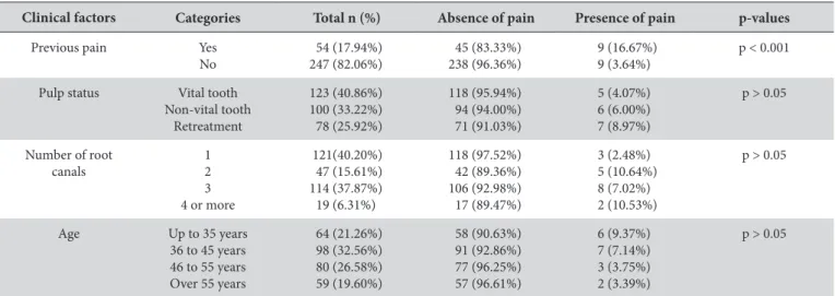
he descriptive frequency distribution of clinical factors analyzed in the sample is given in Table 1 showing that, of the factors assessed, only previous pain had a signiicant inluence on the presence of postoperative pain (p < 0.001). Among those patients with previous pain, 16.67% experienced postoperative pain versus only 3.64% of patients without previous pain.
he auxiliary chemical substances used had no statistically signiicant inluence on the outcome of postoperative pain, irrespective of pulp status of the teeth (p > 0.05). Of the 123 vital teeth, 4.07% (5/123) had postoperative pain, compared with 6% of non-vital teeth (6/100) and 8.87% (7/78) of endodontically retreated teeth. Analysis of postoperative pain by patient age and number of root canals revealed that these factors had no statistically signiicant inluence on postoperative pain. Likewise, the chemical substances used had no efect on pain outcome (p > 0.05).
Table 2 depicts the analyses of the inluence of the auxiliary chemical substances in teeth with and without preoperative pain. Neither of the auxiliary chemical substances reduced or exacerbated postoperative pain ater 24 hours.
DISCUSSION
Postoperative pain following endodontic treatment remains common, with a prevalence of 3% to 58%18, and can stem from chemical, mechanical or microbiological injuries in periradicular tissues. Psychological factors have also been suggested as a possible cause of postoperative pain9,10.
Unfortunately, the etiological factors are not yet clearly elucidated, and results available in the literature fail to clarify the true role of the suggested etiological factors of postoperative pain.
he results of the present prospective randomized clinical study revealed a rate of 93.7% for absence of postoperative pain, irrespective of the associated factors analyzed such as history of previous pain, auxiliary chemical substance, pulp status and number of root canals. hese results difer from the indings of
Table 1. Frequency distribution of descriptive clinical factors analyzed in the sample
Clinical factors Categories Total n (%) Absence of pain Presence of pain p-values
Previous pain Yes
No 54 (17.94%) 247 (82.06%) 45 (83.33%) 238 (96.36%) 9 (16.67%) 9 (3.64%)
p < 0.001
Pulp status Vital tooth
Non-vital tooth Retreatment 123 (40.86%) 100 (33.22%) 78 (25.92%) 118 (95.94%) 94 (94.00%) 71 (91.03%) 5 (4.07%) 6 (6.00%) 7 (8.97%)
p > 0.05
Number of root canals
1 2 3 4 or more
121(40.20%) 47 (15.61%) 114 (37.87%) 19 (6.31%) 118 (97.52%) 42 (89.36%) 106 (92.98%) 17 (89.47%) 3 (2.48%) 5 (10.64%) 8 (7.02%) 2 (10.53%) p > 0.05
Age Up to 35 years
36 to 45 years 46 to 55 years Over 55 years
64 (21.26%) 98 (32.56%) 80 (26.58%) 59 (19.60%) 58 (90.63%) 91 (92.86%) 77 (96.25%) 57 (96.61%) 6 (9.37%) 7 (7.14%) 3 (3.75%) 2 (3.39%)
p > 0.05
previous studies in the literature reporting rates of between 15.2% and 69%3,4,19-22.
he present study included vital, non-vital and endodontically retreated teeth. Pulp status and modality of treatment had no signiicant inluence on postoperative pain. his result corroborates the indings of investigations in the literature2,3,19,21,22 that found no diference between original endodontic treatments and retreatments as regards the occurrence of postoperative pain.
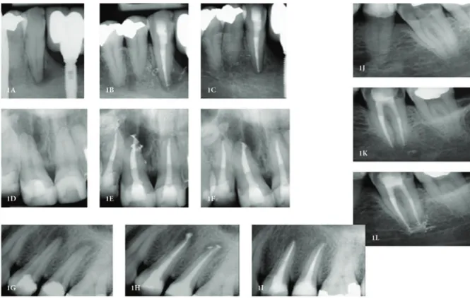
he results of this study demonstrated that apical overinstrumentation and overilling had scant inluence on postoperative pain given that all teeth assessed were overinstrumented and overilled (Figure 1). hese data are in line with previous investigations showing that achieving apical patency and overinstrumentation with enlargement of the apical foramen11,15 did not increase the incidence, severity or duration of postoperative pain8.
Although enlargement of the apical foramen predisposes to extrusion of irrigant, no statistically signiicant diference in the incidence of postoperative pain was evident ater use of 5.25%
sodium hypochlorite, considered a highly toxic substance in the literature, when extruded into the periapical region. his result may be explained by the fact that the chemicals served as auxiliary substances during instrumentation and not as irrigants. he risk of extrusion was reduced given that the root canals and pulp chamber were instrumented with the auxiliary chemical substances within the root canal. Active irrigation using physiological saline solution, which is compatible with periapical tissues, may have helped cleanse the apical and periapical areas since these were intentionally irrigated under pressure within the canal.
With regard to the factors analyzed, previous pain was found to have a signiicant inluence on postoperative pain (p < 0.001). Of the 54 teeth with previous pain, 9 (16.67%) had postoperative pain, whereas of the 247 teeth without previous pain, 9 (3.64%) had postoperative pain, a result in agreement with those of previous studies conirming the inluence of this factor on postoperative pain1-3, 22.
he incidence of severe spontaneous pain (lare-up) was 0.66%, lower than the general incidence of lare-up in the literature, which
Figure 1. Examples of cases treated and followed-up with patency, foraminal enlargement and periodontal illing. Figures 1A, D, G, J depict initial radiographs. Figures 1B, E, H, K depict radiographs taken immediately ater endodontic treatment. Figures 1C, F, I, L depict follow-up radiographs taken 6 months ater completion of endodontic treatment.
Table 2. Inluence of auxiliary chemical substances on teeth with (n = 54) and without (n = 247) previous pain
Auxiliary chemical
substance Total n (%) Absence of Pain Presence of Pain p-values
Teeth with previous pain
CHX 27 (50%) 21 (77.78%) 6 (22.22%) p > 0.05
NaOCl 27 (50%) 24 (88.89%) 3 (11.11%) p > 0.05
Teeth without previous pain
CHX 118 (47.77%) 112 (94.92%) 6 (5.08%) p > 0.05
NaOCl 129 (52.23%) 126 (97.67%) 3 (2.33%) p > 0.05
reports rates ranging from 1.5% to 12%2-5,19,23. his disparity may be explained by the design of the studies, some of which were retrospective and others prospective, or by the undeined variables in a small number of patients23. Additional explanatory factors include the heterogeneous populations, variation in treatment modality and alternative assessment methods described. According to Walton, Fouad2, lare-ups are positively correlated with the presence of previous symptoms, but not with age or number of visits.
Our results difer from those of Georgopoulou et al.24, who showed increased lare-up when teeth were overinstrumented, and from those of Nobuhara et al.25, who reported that endodontic instruments forced beyond the apical foramen can extrude a variety of irritants into periapical tissues and increase both the incidence and severity of pain. Progressive crown-apex decontamination was decisive in preventing this occurrence.
he teeth were treated in a single visit because, given the combination of efective mechanical instrumentation, use of antimicrobial irrigant and three-dimensional obturation of the root canal, the single-visit treatment can efectively reduce the intracanal microbiota and allow a favorable outcome6. In addition, a number of previous studies failed to ind any diference in incidence of pain between single- and multiple-visit treatments. Other studies have found that single-visit treatments are associated with a lower rate of postoperative pain1,2,5,6,22, although some authors have found the opposite results4. Sathorn et al.18 found no convincing evidence of difering incidences of lare-up in single- or multiple-visits treatment.
In the present study, the rate of postoperative pain of 6.3% may be explained by factors associated with failures in occlusal adjustment of the coronal restoration, or by the pressure exerted on the periodontium by the rubber dam clamp during treatment.
he severe pain found in 0.66% (2/301) may have been caused by iatrogenic technical procedures associated with chemical7 or traumatic mechanical8 injuries, as well as those caused by microorganisms and their products9,10. Patients’ individual pain thresholds and emotional factors may also have inluenced outcomes. Patients can experience a wide range of diferent emotional responses for very similar levels of stimuli intensity, depending on their perceptions of the event.
In view of the dearth of clinical studies in the literature investigating the inluence of auxiliary chemical substances on the postoperative pain related to endodontic treatment, there is a need for future prospective clinical trials correlating postoperative pain with diferent preparation and obturation techniques as well as illing materials.
CONCLUSION
Based on the results of this study, it can be concluded that the chemical substances used in the single-visit endodontic treatments or retreatments with enlargement of the apical foramen and apical extrusion of the endodontic sealer had no inluence on spontaneous postoperative pain.
REFERENCES
1. ElMubarak AH, Abu-bakr NH, Ibrahim YE. Postoperative pain in multiple-visit and single-visit root canal treatment. J Endod. 2010 Jan;36(1):36-9. http://dx.doi.org/10.1016/j.joen.2009.09.003. PMid:20003932
2. Walton R, Fouad A. Endodontic interappointment flare-ups: a prospective study of incidence and related factors. J Endod. 1992 Apr;18(4):172-7. http://dx.doi.org/10.1016/S0099-2399(06)81413-5. PMid:1402571
3. Glennon JP, Ng YL, Setchell DJ, Gulabivala K. Prevalence of and factors affecting postpreparation pain in patients undergoing two-visit root canal treatment. Int Endod J. 2004 Jan;37(1):29-37. http://dx.doi.org/10.1111/j.1365-2591.2004.00748.x. PMid:14718054
4. Ng YL, Glennon JP, Setchell DJ, Gulabivala K. Prevalence of and factors affecting post-obturation pain in patients undergoing root canal treatment. Int Endod J. 2004 June;37(6):381-91. http://dx.doi.org/10.1111/j.1365-2591.2004.00820.x. PMid:15186245
5. Eleazer PD, Eleazer KR. Flare-up rate in pulpally necrotic molars in one-visit versus two-visit endodontic treatment. J Endod. 1998 Sept;24(9):614-6. http://dx.doi.org/10.1016/S0099-2399(98)80122-2. PMid:9922752
6. Su Y, Wang C, Ye L. Healing rate and post-obturation pain of single- versus multiple-visit endodontic treatment for infected root canals: a systematic review. J Endod. 2011 Feb;37(2):125-32. http://dx.doi.org/10.1016/j.joen.2010.09.005. PMid:21238790
7. Bashetty K, Hegde J. Comparison of 2% chlorhexidine and 5.25% sodium hypochlorite irrigating solutions on postoperative pain: a randomized clinical trial. Indian J Dent Res. 2010 Oct;21(4):523-7. http://dx.doi.org/10.4103/0970-9290.74225. PMid:21187618
8. Arias A, Azabal M, Hidalgo JJ, Macorra JC. Relationship between postendodontic pain, tooth diagnostic factors, and apical patency. J Endod. 2009 Feb;35(2):189-92. http://dx.doi.org/10.1016/j.joen.2008.11.014. PMid:19166770
9. Seltzer S, Naidorf IJ. Flare-ups in endodontics: I. Etiological factors. J Endod. 1985 Nov;11(11):472-8. http://dx.doi.org/10.1016/S0099-2399(85)80220-X. PMid:3868692
10. Seltzer S, Naidorf IJ. Flare-ups in endodontics: II. Therapeutic measures. 1985. J Endod. 2004 July;30(7):482-8, discussion 475. http://dx.doi. org/10.1097/00004770-200407000-00006. PMid:15220642
11. Gurgel-Filho ED, Castelo-Branco YN, Maniglia-Ferreira C, Souza-Filho FJ, Coutinho-Filho T. Avaliação in vivo da dor pós-operatória em dentes vitais após o alargamento do forame apical. RFO. 2010 May;15(2):145-9.
12. Souza-Filho FJ, Teixeira FB, Hyzatugu R, Zaia AA. The evaluation of postoperative pain following apical foramen enlargement: a clinical study. J Endod. 1998; 24(4):291.
13. Monsef M. Effect of apical patency on the apical seal of obturated canals. J Endod. 1998; 24(4):284.
15. Fox J, Atkinson JS, Dinin AP, Greenfield E, Hechtman E, Reeman CA, et al. Incidence of pain following one-visit endodontic treatment. Oral Surg Oral Med Oral Pathol. 1970 July;30(1):123-30. http://dx.doi.org/10.1016/0030-4220(70)90021-6. PMid:5269799
16. Buchanan LS. Continuous wave of condensation technique. Endod Prac. 1998 Dec;1(4):7-10. PMid:10220310.
17. Figini L, Lodi G, Gorni F, Gagliani M. Single versus multiple visits for endodontic treatment of permanent teeth: a Cochrane systematic review. J Endod. 2008 Sept;34(9):1041-7. http://dx.doi.org/10.1016/j.joen.2008.06.009. PMid:18718362
18. Sathorn C, Parashos P, Messer H. The prevalence of postoperative pain and flare-up in single- and multiple-visit endodontic treatment: a systematic review. Int Endod J. 2008 Feb;41(2):91-9. PMid:17956561.
19. Siqueira JF Jr, Rôças IN, Favieri A, Machado AG, Gahyva SM, Oliveira JC, et al. Incidence of postoperative pain after intracanal procedures based on an antimicrobial strategy. J Endod. 2002 June;28(6):457-60. http://dx.doi.org/10.1097/00004770-200206000-00010. PMid:12067129
20. Harrison JW, Baumgartner CJ, Zielke DR. Analysis of interappointment pain associated with the combined use of endodontic irrigants and medicaments. J Endod. 1981 June;7(6):272-6. http://dx.doi.org/10.1016/S0099-2399(81)80006-4. PMid:6942083
21. Harrison JW, Baumgartner JC, Svec TA. Incidence of pain associated with clinical factors during and after root canal therapy. Part 1. Interappointment pain. J Endod. 1983 Sept;9(9):384-7. http://dx.doi.org/10.1016/S0099-2399(83)80190-3. PMid:6579198
22. Ince B, Ercan E, Dalli M, Dulgergil CT, Zorba YO, Colak H. Incidence of postoperative pain after single- and multi-visit endodontic treatment in teeth with vital and non-vital pulp. Eur J Dent. 2009 Oct;3(4):273-9. PMid:19826598.
23. Walton R. Interappointment flare-ups: incidence, related factors, prevention, and management. Endod Top. 2002 Nov;3(1):67-76. http:// dx.doi.org/10.1034/j.1601-1546.2002.30107.x.
24. Georgopoulou M, Anastassiadis P, Sykaras S. Pain after chemomechanical preparation. Int Endod J. 1986 Nov;19(6):309-14. http://dx.doi. org/10.1111/j.1365-2591.1986.tb00495.x. PMid:3466868
25. Nobuhara WK, Carnes DL, Gilles JA. Anti-inflammatory effects of dexamethasone on periapical tissues following endodontic overinstrumentation. J Endod. 1993 Oct;19(10):501-7. http://dx.doi.org/10.1016/S0099-2399(06)81491-3. PMid:8120485
CONFLICTS OF INTERESTS
he authors declare no conlicts of interest.
*CORRESPONDING AUTHOR
Marcelle Louise Sposito Bourreau, Faculdade de Odontologia São Leopoldo Mandic, Rua José Rocha Junqueira, 13, Ponte Preta, 13045-755 Campinas - SP, Brasil, e-mail: [email protected]