INTRODUCTION
Article/Artigo
1. Departamento de Ciências Farmacêuticas, Universidade de Blumenau. Blumenau-SC. 2. Departamento de Análises Clínicas, Centro de Ciências da Saúde, Universidade Federal de Santa Catarina, Florianópolis, SC.
Address to: Dr. Caio Mauricio Mendes de Cordova. Deptº de Ciências Farmacêuticas/FURB. Rua São
Paulo 2171, Campus III, Itoupava Seca, 89030-000 Blumenau, SC, Brasil. Phone: 55 47 3321-7318
e-mail: [email protected]
Received in 08/03/2011
Accepted in 19/09/2011
Drug resistance of
Mycobacterium tuberculosis
strains in Southern Brazil
Estudo da resistência de cepas de
Mycobacterium tuberculosis
aos antimicrobianos no Sul do Brasil
Laynara Katize Grutzmacher
1, Eduardo Monguilhot Dalmarco
2, Solange Lucia Blat
1and Caio Mauricio
Mendes de Cordova
1ABSTACT
Introduction: he aim of this work was to evaluate the prevalence of Mycobacterium tuberculosis
(MT) strains with mutations that could result in resistance to the main drugs used in treatment in a region with one of the highest numbers of tuberculosis (TB) cases in southern Brazil. Methods: Deoxyribonucleic acid (DNA) from 120 sputum samples from diferent patients suspicious of pulmonary tuberculosis who atended the Municipal Public Laboratory for
Mycobacterium sp. diagnosis was directly ampliied and analyzed by PCR-SSCP. he DNA was ampliied in known hotspot mutation regions of the genes rpoB, ahpC, embB, katG, inhA, and
pncA. Results: he percentage of samples positive by culture was 9.2% (11/120); 5% (6/120) were positive by bacilloscopy and MT-PCR, and DNA fragments of the aforementioned resistance genes could be ampliied from seven (7) of the eleven (11) samples with positive results, either by culture or PCR/bacilloscopy. All presented a SSCP patern similar to a native, nonresistant genotype, with the ATCC strain 25177 as control, except for one sample (0.01%), which presented a SSCP proile demonstrating mutation at the embB gene. Conclusions: hese results are consistent with the empirical observations by physicians treating TB patients in our region of a low occurrence of cases that are refractory to conventional treatment schemes, in contrast to other parts of the country. Continued surveillance, especially molecular, is essential to detect and monitor the outbreak of MT-resistant strains.
Keywords: Antibiotic. Mycobacterium. PCR-SSCP. Resistance. Tuberculosis.
RESUMO
Introdução: O objetivo deste trabalho foi avaliar a prevalência de cepas de Mycobacterium tuberculosis (MT) com mutações que podem resultar em resistência às principais drogas utilizadas no tratamento em uma das regiões com o maior número de casos de tuberculose (TB) no Sul do Brasil. Métodos: O ácido desoxiribonucleico (DNA) de 120 amostras de escarro de diferentes pacientes com suspeita de TB pulmonar que procuraram o serviço público de saúde do município sede da região para o diagnóstico de MT foi diretamente ampliicado e analisado por PCR-SSCP. Foram ampliicadas regiões conhecidas onde ocorrem a maioria das mutações nos genes rpoB, ahpC, embB, katG, inhA, and pncA. Resultados: Nove virgula dois por cento (11/120) das amostras apresentaram resultado positivo por cultura, 5% (6/120) foi positiva por bacilscopia e PCR para MT, e os fragmentos dos genes mencionados puderam ser ampliicados em sete (7) dos onze (11) casos com resultado positivo, seja por cultura ou PCR/baciloscopia. Todos estes casos apresentaram um padrão de SSCP similar ao genótipo nativo, não resistente, por comparação com a cepa controle ATCC 25177, com exceção de uma amostra (0,01%), que apresentou um padrão de SSCP mutante no gene embB. Conclusões: Estes resultados são consistentes com as observações empíricas por parte dos clínicos que tratam os pacientes com TB na região, de uma baixa ocorrência de casos refratários ao tratamento convencional, em contraste com outras partes do país. Porém, a vigilância contínua, especialmente molecular, é essencial para identiicar e monitorar o aparecimento de cepas de MT resistentes.
Palavras-chaves: Antibióticos. Mycobacterium. PCR-SSCP. Resistência. Tuberculose.
Because tuberculosis (TB) has deep social roots intimately linked to unequal income distribution, the problem with the disease reflects the level of development of a country. Causes related to poverty, the weak organization of health systems, and administration deiciencies limit technology implementation and, as a consequence, contribute to the dissemination of the etiological agent and the increase in population susceptibility. Since 1993, the World Health Organization (WHO) has considered TB a global emergency, thus supporting measures for its control1,2. However, the batle against the disease is far from over.
he region of the Itajaí Valley in the State of Santa Catarina, which has about 500,000 inhabitants, is among the regions with the highest number of TB cases in southern Brazil (source: htp://portalses. saude.sc.gov.br).
Reports of Mycobacterium tuberculosis (MT) strains resistant to the antibiotics used in treatment have been noted everywhere in the world3. Besides empirical observations of the absence of resistant
Mycobacterium strains in our region, reported by the physicians who treat the TB patients, it is obviously important to offer and perform active research to monitor an eventual rise in resistance using fast and efficient tools, such as molecular methods. As is well known, MT cultures may take up to 8 weeks to yield results, and in the case of positivity, antibiotic sensitivity testing by traditional microbiologic techniques takes about 4 weeks more. During this time, the patient may already be spreading MT-resistant strains to close contacts. Using methods capable of ofering results in a couple of days allows physicians to be readily informed and to take the appropriate therapeutic measures on time. Based on empirical information about the observation, obtained from the Municipality Laboratory staf, the number of patients with more than one year of TB treatment still presenting positive bacilloscopy results may be significant.
METHODS
RESULTS
Laboratory in the capital of each state by microbiologic methods. It is interesting to note that before 2009, the guidelines accepted by the public health system allowed requests for these tests only by patients with certain conditions, including HIV patients, symptomatic patients with negative bacilloscopy, and any patient who is not responsive to
current therapy, among other special circumstances4. An immune
competent patient eventually infected by a resistant MT strain had to wait for treatment failure and 8 more weeks on average for laboratorial evidence that could improve his outcome. Only after the implementation of new national guidelines, published in August 2009, has culture with antibiotic sensitivity testing for any respiratory symptomatic patient with suggestive radiographic findings been advised5. However, this is not the case observed in clinical practice, which continues to be based on bacilloscopy indings for primary diagnosis. In this context, through the Clinical Laboratory of the University Ambulatory Center of our institution, which assists patients in the whole region as a reference service, we aimed to ofer the possibility of detection of mutations that may lead to MT resistance to irst-line oral antibiotics used for TB treatment in Brazil, through a molecular method such as polymerase chain reaction followed by single-strand conformational polymorphism (PCR-SSCP). his methodology is a robust, fast, and reliable technique that may allow a medical decision in up to a couple of days. Furthermore, to date, no updated information about the prevalence of Mycobacterium-resistant strains is available in our region. As a starting point, in this study we have evaluated the eventual presence and prevalence of resistant TB strains in our public health system (SUS) with the use of PCR-SSCP. hese results may contribute to the evaluation of the current TB treatment scheme used in the system, as well as provide new information about the prevalence of resistant strains in our population. Such information is crucial for evaluating the management and surveillance policies on the tuberculosis control efort.
Patients and samples
We analyzed 120 sputum samples from different patients suspicious of pulmonary tuberculosis who have attended the Municipal Public Laboratory for Mycobacterium sp. diagnosis. Ater processing for bacilloscopy at the site, the samples were sent to the Clinical Laboratory of the University Ambulatory of our institution for culture and PCR.
Sample treatment
he samples were treated by a conventional modiied method
using 1% NaOH, SDS, and phosphoric acid6. he treated samples
were then used for culture and PCR. DNA was extracted and puriied as described elsewhere7.
Mycobacterium tuberculosis diagnosis
Bacilloscopy was performed directly on sputum sample slides stained by the Ziehl-Neelsen method. he culture was prepared by inoculation of samples in Löwenstein-Jensen medium and incubation at 37ºC for up to 8 weeks. For PCR, DNA was ampliied with a
primer pair speciic to M. tuberculosis (MT1: 5'-CCT.GCG.AGC.
GTA.GGC.GTC.GG-3' and MT2: 5'-CTC.GTC.CAG.CGC.CGC. TC.GG -3'), resulting in a 123bp fragment of the insertion element
IS61108. Positive samples obtained by any of the methodologies used for MT diagnosis were used in PCR-SSCP tests.
Polymerase chain reaction
For detection of MT strains harboring mutations that may lead to resistance to rifampicin, isoniazid, pyrazinamide, and ethambutol, we ampliied hotspot sites at the genes rpoB, katG, inhA, ahpC, pncA, and embB. he amplicons were later submited to SSCP-PCR
(single-strand conformation polymorphism-polymerase chain reaction)9.
he primers were synthesized according to the sequences described by others10. Ampliication reactions were performed in a volume of 25uL, consisting of 0.5uL of each 10uM primer (Invitrogen), 0.2uL
of 100uM dNTPs (Invitrogen), 1.0uL of 50mM MgCl2 (Invitrogen),
and 1.0U of Taq DNA polymerase in the provided reaction bufer
(Invitrogen). he annealing temperatures were 64ºC for the rpoB
and embB, 57ºC for the pncA, and 55ºC for the ahpC, katG, and
inhA genes. As naïve control, puriied DNA from the ATCC 25177
M. tuberculosis strain was ampliied.
Single-strand conformation polymorphism
he PCR amplicons of the aforementioned gene hotspots were resolved by 1% agarose gel electrophoresis with ethidium bromide staining under UV light. hose samples with positive ampliication were submited to native 5.5% polyacrylamide gel electrophoresis after denaturation of the double-strand DNA. Briefly, 18μL of formamide dye mix was added to 2μL of each PCR product, followed by boiling for 5min and immediate cooling on ice; 2μl of 10x sucrose gel loading bufer was added, ater which the mixture was applied to the gel and submited to 300V (45mA) for 5h. he gel was stained with AgNO3 and photographed11.
Ethical considerations
This project was approved by the Committee on Ethics in Research with Human Beings of our institution (Protocol no. 032/03) under the guidelines of the Helsinki Declaration of 1964 and further revisions.
Among the 120 patient samples tested for MT diagnosis, six (5%) were positive either by bacilloscopy or PCR with primers speciic to the M. tuberculosisIS6110 element. On the other hand, eleven (9.2%) were positive by culture.
he samples that were positive by either method were tested by PCR-SSCP for detection of mutations in the hotspot regions of the genes responsible for the phenotypes resistant to the main antibiotics used in treatment.
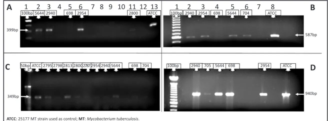
he 399bp fragment of the embB gene related to ethambutol
resistance could be amplified from five (5) positive samples (Figure 1A). Regarding resistance to isoniazide, the 457bp fragment of the inhA gene, the 703bp fragment of the katG gene, and the 587bp product of the ahpC gene could also be equally ampliied from ive (5) samples (Figure 1B).
Regarding resistance to rifampicin, the 349bp fragment of the
rpoB gene could be ampliied from ten (10) samples (Figure 1C). The 940bp hotspot fragment of the gene responsible for pyrazinamide resistance, pncA, could be ampliied from ive (5) positive samples (Figure 1D).
A
1 2 3 4 5 6 7 8 9 10 11 12 13 1 2 3 4 5 6 7 8100bp 5644 2940 698 2954 2800 ATCC 100bp 2940 2954 698 5644 704 ATCC
B
587bp 399bp
C
50bp ATCC 2795 2798 2813 2800 2787 2954 2940 5644 698 704 100bp 2940 705 5644 698 2954 ATCC940bp
D
349bp
FIGURE 1 -Polymerase chain reaction ampliication of hotspot mutation regions of the genes embB (A), ahpC (B), rpoB (C), and pncA
(D), from samples with a positive result for MT diagnosis, either by culture, IS6110 PCR, or bacilloscopy.
ethambutol resistant phenotype (Figure 2), all the samples from which it was possible to amplify a hotspot fragment of the genes responsible for M. tuberculosis resistance evaluated in this study presented a SSCP patern similar to a native, nonresistant genotype, using the ATCC strain 25177 as control.
FIGURE 2 – Single-strand conformation polymorphism of polymerase chain reaction amplicons from the genes embB (1), ahpC (2), rpoB (3), and pncA (4), from samples with a positive result for MT diagnosis, either by culture, IS6110 PCR, or bacilloscopy.
DISCUSSION
Estimates from the WHO indicate that nearly one third of the world’s population is infected by the TB bacilli. About 8.8 million new cases are recorded every year, with 1.6 million deaths. Twenty-two countries harbor about 80% of all cases in the world, and Brazil occupies the 16th position in this ranking, with 70% of the cases concentrated in 315 of its 5,570 cities12-14. Strategies to stop the advance of this epidemic, however, are threatened not only by its synergic efect with HIV/AIDS but mainly by the increasing number of multiresistant strains. With the risk of the wide spread of resistant bacilli, the lack of stringent control and monitoring of patients may exponentially increase the rise of resistance, a drawback in the eforts on tuberculosis control15,16.
A few reports point out a decrease in multiresistant TB cases in the last decade; however, most studies indicate the opposite. A study conducted in Brazil from 1996 to 1997 reported that 31% of analyzed strains were resistant to the main drugs used in treatment.
M. tuberculosis presents a signiicant frequency of naturally resistant mutants: 1:106 for isoniazide, 1:108 for rifampicin, but less than 1:1014 for both simultaneously. Misuse of the drugs certainly contributes to the selection of resistant strains13,17.
There are four known mechanisms through which bacterial resistance is acquired (conjugation, transformation, transduction, and mutation), but it is mainly by mutationthat M. tuberculosis
becomes resistant to drugs. Genetic and molecular analyses of resistant bacilli also suggest that resistance is acquired by mutations in the genes that codify the drug target. Unlike many other bacteria, MT does not perform horizontal gene transfer. he mechanism of multidrug resistance is still uncertain as no single gene involved has been identiied; rather, it has been observed that multiresistant phenotypes arise from mutation in diferent genes18.
Rifampicin binds to the beta subunit of RNA polymerase, which is codiied by the rpoB gene, inhibiting the transcription process.
Mutations at the rpoB produce conformational enzymatic changes
and, therefore, resistance. About 95% of the rifampicin-resistant isolated MT strains have mutations in a 69bp region of this gene13,19,20.
Several mutations, affecting one or more genes, have been found to be associated to isoniazide resistance; these include point mutations in genes such as the one that codiies the
catalase-peroxidase enzyme (katG); the enoyl-ACP reductase, involved in
the biosynthesis of mycolic acid (inhA); the alkyl hydroperoxide reductase, involved in cellular response to oxidative stress (ahpC), or, with lower frequency, even the β-ketoacyl-ACP synthase (kasA). Mutations in the katG and inhA genes are found in 75-85% of M. tuberculosis isolates resistant to this drug20,21.
Pyrazinamide is a highly speciic drug against M. tuberculosis, with no or minor efect against other Mycobacterium species but with a high level of intrinsic resistance observed. More than 70% of the M. tuberculosis strains resistant to pyrazinamide have mutations in the
gene pncA, which codiies a pyrazinamidase enzyme that converts
the drug to its active form. On the other hand, there is no perfect correlation between loss of pyrazinamidase activity and pyrazinamide resistance; mycobacteria may be resistant to the drug in spite of having enzyme activity20,22.
ATCC: 25177 MT strain used as control; MT: Mycobacterium tuberculosis.
he authors declare that there is no conlict of interest.
CONFLICT OF INTEREST
FINANCIAL SUPPORT
REFERENCES
he genetic bases of ethambutol resistance are associated with modiications in the embB gene in 70% of the resistant isolated strains. Mutations in multiple codons have been found, resulting in two, three, or four amino acid changes in the EmbB protein. he most frequent, however, were found in codon 306, a conserved methionine coding region20,23.
In our study, we found no PCR-SSCP pattern that might correspond to a phenotype resistant to the main drugs used in MT treatment, except for one sample with a polymorphism in the embB gene that could correspond to resistance to ethambutol. For diagnostic purposes, that genotypic proile would have to be conirmed, ideally by sequencing; however, that is beyond the scope of this study. Our aim was to evaluate the PCR-SSCP technique as a screening tool for detection of M. tuberculosis resistance and, as a consequence, to obtain a notion of the prevalence of resistant strains in our population; the existing data lack scientiic evidence, being only empirically suspected by the physicians in the public health system. he low prevalence of genotypic alterations is probably due to the eiciency and seriousness with which TB treatment is performed in our region. Tuberculosis drugs, as well as laboratory tests, are given at no cost to the patients, and medication is given through health agents, who personally deliver the drug to the house or workplace of the patient and then wait to see if the medicine is appropriately taken. Adhesion to treatment is very high. Only those patients who demonstrate commitment and capacity to take the medicine in the appropriate manner who earn the trust of the health agents are allowed to take home doses suicient for a few weeks and take the drugs by themselves. However, surveillance and monitoring by the health agents continue to the end of the treatment period.
he failure of our procedure to detect some mutations must be taken into account. he detection of isoniazide-resistant strains, for instance, is quite complex, due to the number of genes involved and of regions to be analyzed. It is well known that the prevalence of the described mutations that may confer isoniazide resistance can vary according to the geographic location of the isolates; studies with M. tuberculosis strains in Brazil have demonstrated a signiicant variation, from 50 to 80%, depending on the region where the bacilli have been isolated. Mutations in the katG gene are responsible for 58 to 68% of the resistant strains, while 32% of the mutations appear in the inhA
gene. Mutations in other sites, such as in the promoter region of the
ahpC gene, are less frequent but do exist10. One must also consider that these apparently high rates of resistance actually do not represent the global prevalence of mutants in our population. In Brazil, requests for culture and antibiotic susceptibility testing in the public health system are only allowed for patients with conventional treatment failure or immunosupressed ones, which obviously produces a bias in these studies.
Another point to be considered in our study is that some mutations may not be detected because of limitations of the method. It should be noted that the sensitivity of the method is inversely proportional to the size of the ampliied fragment. hus, the number of mutations detected decreases when fragments smaller than 150 to 200bp are analyzed. Also, depending on the polyacrylamide concentration, the temperature, pH, or even the presence of additives, such as glycerol, the electrophoretic mobility, and the resolution may be afected24.
It is also noteworthy that it was not possible to amplify the DNA of the studied genes from every sample with a positive result
by culture. One reasonable explanation for this is the sensitivity diference between PCR with primers directed at the mentioned genes and PCR developed for diagnostic purposes. Even the ampliication of the IS6110 region of M. tuberculosis did not achieve the same sensitivity of culture for TB diagnosis. It is therefore expected that PCR designed to amplify regions of the genes where the mutations usually responsible for MT resistance are found would not be able to amplify the DNA of every positive sample.
Furthermore, as has already been demonstrated, in our population, samples from patients suspicious of having TB with a positive result by culture may actually be infected by non-
M. tuberculosis species7. The genetic diversity among the
Mycobacterium species sequences may prevent the studied genes from being ampliied in PCR-SSCP.
It has been reported that the prevalence of resistant TB has significantly increased in the last years and, according to the Brazilian Society of Pneumology and hisiology, it is possible that 50 million people around the world may be infected with resistant
M. tuberculosis. herefore, there is an obvious need for the availability of rapid and accurate tests capable of detecting resistant MT in a certain population. Automated culture methods with liquid media and antibiotic susceptibility testing have been validated and are accepted, and may reduce the wait for results to 10-40 days for culture and more than 10 days for drug sensitivity5; however, the cost of these methods largely supplant the value of the tests received by laboratories in Brazil. Polymerase chain reaction-single-strand conformation polymorphism may contribute to illing in this gap, with the increasing availability of laboratories capable of performing molecular diagnosis across the country.
According to the WHO, a prevalence of resistant bacilli higher than 2% has the inefficacy of the therapeutic scheme and the ineiciency of local TB control programs as principal causes. he prevalence of genotypic alterations indicated is only 0.01%, which certainly does not represent an additional problem in local control of the disease. In some parts of Brazil, a large proportion of patients fail to follow the adequate treatment regime25. In any case, it is obviously necessary to maintain surveillance for the purpose of monitoring and detecting an eventual rise in MT resistance rates in our population. In spite of its limitations, PCR-SSCP constitutes an easy, reliable, and fast method in this context.
Fundação de Amparo à Pesquisa e Inovação do Estado de Santa Catarina - FAPESC grant no. 14594/2005-7.
1. Borella VR, Sato DN, Fonseca BAL. Problems in the standardization of the polymerase chain reaction for the diagnosis of pulmonary tuberculosis. Rev Saude Publica 1999; 33:281-286.
2. Ministério da Saúde. Tuberculose: guia de vigilância epidemiológica. Brasília: Ministério da Saúde; 2002.
4. Castelo Filho A, Kritski AL, Barreto AW, Lemos ACM, Ruffino Netto A, Guimarães CA, et al. II Consenso Brasileiro de Tuberculose. J Bras Pneumol 2004; 30:S1-56.
5. Conde MB, Melo FAF, Marques AMC, Cardoso NC, Pinheiro VGF, Dalcin PTR, et al. III Brazilian horacic Association Guidelines on Tuberculosis. J Bras Pneumol 2009; 35:1018-1048.
6. Campinas LLSL, Ferrazoli L, Telles MAS, Matsumoto NF, Biagolini AM, Ferraz SMP. Tuberculose, manual de orientação. São Paulo: Secretaria da Saúde. Divisão de Tuberculose; 2002.
7. Marchi AM, Jutel ID, Kawacubo EM, Dalmarco EM, Blat SL, Cordova CMM. Evaluation of methods for detection and identiication of Mycobacterium species in patients suspected of having pulmonary tuberculosis. Brazilian Journal of Microbiology 2008; 39:613-618.
8. Eisennach KD, Cave MD, Crawford JT. PCR detection of Mycobacterium tuberculosis. In: Pershing DH, Smith TF, Tenover FC, White TJ, editors. Diagnostic Molecular Microbiology, Principles and Applications. Rochester: Mayo Foundation; 1993. p. 191-196.
9. Kim BJ, Kim SY, Park BH, Lyu MA, Park IK, Bai GH, et al. Mutations in the rpoB gene of Mycobacterium tuberculosis that interfere with PCR-single-strand conformation polymorphism analysis for rifampin susceptibility testing. J Clin Microbiol 1997; 35:492-494.
10. Chan RCY, Hui M, Chan EWC, Au TK, Chin ML, Yip CK, et al. Genetic and phenotypic characterization of drug-resistant Mycobacterium tuberculosis isolates in Hong Kong. Journal of Antimicrobial Chemotherapy 2007; 59:866-873. 11. Sambrook J, Russel D. Molecular cloning: a laboratory manual, 3rd ed. New York:
Cold Spring Harbor Laboratory; 2001.
12. Barreira D, Grangeiro A. Evaluation of tuberculosis control strategies in Brazil. Rev Saude Publica 2007; 41:4-8.
13. Carvalho WS, Miranda SS, Pesquero JL, Gomes MA. Diagnóstico de resistência do Mycobacterium tuberculosis à rifampicina utilizando-se da reação em cadeia da polimerase. Rev Bras Ciênc Farm 2007; 43:31-38.
14. Maciel ELN, Vieira RCA, Milani EC, Brasil M, Fregona G, Dietze R. Community health workers and tuberculosis control: knowledge and perceptions. Cad Saude Publica 2008; 24:1377-1386.
15. Natal S. Emergência da resistência às drogas.Bol Pneumol Sanit 2002; 10:57-70. 16. Clark-Curtiss JE, Haydel SE. Molecular Genetics of Mycobacterium Tuberculosis
pathogenesis. Ann Rev Microbiol 2003; 57:517-549.
17. Barroso EC, Mota RMS, Santos RO, Sousa ALO, Barroso JB, Rodrigues JLN. Risk factors for acquired multidrug-resistant tuberculosis. J Bras Pneumol 2003; 29:89-97.
18. Dalcomo MP, Andrade MK, Picon PD. Multiresistant tuberculosis in Brazil: history and control. Cad Saude Publica 2007; 41:34-42.
19. Telenti A, Philipp WJ, Sreevatsan S. The emb operon, a gene cluster of
Mycobacterium tuberculosis involved in resistance to ethambutol. Nature Med 1997; 3:567-570.
20. Rosseti MLR, Valim ARM, Silva MSN, Rodrigues VS. Resistant tuberculosis: a molecular review. Cad Saude Publica 2002; 36:525-532.
21. Musser JM, Kapur V, Williams DL, Kreiswirth BN, van Soolingen D, van Embdem JDA. Characterization of the catalase-peroxidase gene (katG) and inhA locus in isoniazid-resistance and susceptible strains of Mycobacterium tuberculosis by automated DNA sequencing: restricted array of mutations associated with drug resistance. J Infect Dis 1996; 173:196-202.
22. Sreevatsan S, Pan X, Zhan Y, Kreiswirth BN, Musser JM. Mutations associated with pyrazinamide resistance in pncA of Mycobacterium tuberculosis complex organisms. Antimicrob Agents Chemoter 1997; 41:636-640.
23. Telenti A, Honore N, Bernasconi C, March J, Ortega A, Heym B, et al. Genotypic assessment of isoniazid and rifampin resistance in Mycobacterium tuberculosis: a blind study at reference laboratory level. J Clin Microbiol 1995; 35:719-723. 24. Salazar LA, Hirata MH, Hirata RDC. Increasing the Sensitivity of Single-
Strand Conformation Polymorphism Analysis of the LDLR Gene Mutations in Brazilian Patients with Familial Hypercholesterolemia. Clin Chem Lab Med 2002;40:441-445.