INTRODUCTION
Article/Artigo
1. Programa de Pós-Graduação em Ciências Veterinárias, Faculdade de Veterinária, Universidade Estadual do Ceará, Fortaleza, CE. 2. Núcleo Regional de Oiologia, Universidade Federal do Ceará, Fortaleza, CE. 3. Laboratório de Fisiofarmacologia Cardio-Renal, Instituto Superior de Ciências Biomédicas, Universidade Estadual do Ceará, Fortaleza, CE. 4. Centro de Controle de Zoonoses de Fortaleza, Fortaleza, CE. Address to: Dr. José Claudio Carneiro de Freitas. Programa de Pós-Graduação em Ciências Veterinárias/ FAVET/UECE. Av. Paranjana 1700, Campus do Itaperi, Serrinha, 60740-000 Fortaleza, CE, Brasil. Phone: 55 85 31019840
e-mail: [email protected]
Received in 21/01/2011
Accepted in 29/09/2011
Clinical and laboratory alterations in dogs naturally infected by
Leishmania chagasi
Alterações clínicas e laboratoriais em cães naturalmente infectados por
Leishmania chagasi
.
José Cláudio Carneiro de Freitas
1,
Diana Célia Sousa Nunes-Pinheiro
1, Belarmino Eugênio Lopes Neto
1,
Glauco Jonas Lemos Santos
1, Cyntia Rafaelle Amaral de Abreu
1, Roberta Rocha Braga
2, Rafael de Morais Campos
3and Ligene Fernandes de Oliveira
4ABSTACT
Introduction: Canine visceral leishmaniasis (CVL) is a zoonotic disease with diferent clinical manifestations. Parasitism oten occurs in bone marrow, but changes have been observed in peripheral blood and serum biochemical parameters. he aim of this study was to evaluate the hematological and biochemical parameters in dogs naturally infected by Leishmania chagasi.
Methods: Eighty-ive adult dogs of both sexes and various weights and ages from the Zoonosis Control Center of Fortaleza (CCZ) were used, selected by immunoluorescence assay (IFA) and considered positive with IFA titers greater than 1:40 and by visualizing amastigotes of
Leishmania chagasi in smears obtained by bone marrow aspiration. he dogs (n = 85) were grouped according to clinical signs: negative (CN = 7), subclinical (CS = 10), and clinical (CC = 68). Blood samples were collected for determination of hematological and biochemical serum values. he experimental protocol was approved by the CEUA/UECE. Results: he most frequent clinical signs were cachexia (77.9%), keratitis (61.8%), and lymphadenopathy (55.9%), and 86.8% of the animals showed more than one clinical sign characteristic of CVL. In CC were observed reductions in red blood cells (63%), hematocrit (72%), and hemoglobin (62%), as well as leukocytosis (33%), neutropenia (28%), thrombocytopenia (50%), uremia (45%), hyperproteinemia (53%, p<0.05), hypergammaglobulinemia (62%, p<0.01), and hypoalbuminemia (58%). Conclusions: Animals with the clinical form of the disease demonstrate hematological and biochemical changes consistent with anemia, uremia, hyperproteinemia, and hyperglobulinemia, which present themselves as strong clinical markers of visceral leishmaniasis associated with the signs previously reported.
Keywords: Dogs. Canine visceral leishmanisis. Biomarkers. Anemia. Uremia. Hyperglobulinemia.
RESUMO
Introdução: A leishmaniose visceral canina (LVC) é uma zoonose com diferentes manifestações clínicas. O parasitismo ocorre frequentemente na medula óssea e têm sido relatadas alterações hematológicas e bioquímicas. Objetivou-se avaliar os parâmetros clínicos, hematológicos e bioquímicos de cães naturalmente infectados por Leishmania chagasi. Métodos: Utilizaram-se 85 cães adultos, ambos os sexos, peso e idade variados, oriundos do Centro de Controle de Zoonoses de Fortaleza, selecionados pela reação de imunoluorescência indireta (RIFI), sendo considerados positivos os animais com títulos de RIFI ≥ 1:40 e pelo exame parasitológico das formas amastigotas de Leishmania chagasi em esfregaços de medula óssea. Os cães foram agrupados conforme os sinais clínicos associados à doença: negativos (CN=7); subclínicos (CS=10) e clínicos (CC=68). Amostras de sangue foram coletadas para determinação dos parâmetros hematológicos e bioquímicos séricos. O protocolo experimental foi aprovado pelo CEUA/UECE, protocolo n° 08622833-1. Resultados: Os sinais clínicos mais frequentes foram caquexia (77,9%), ceratoconjuntivite (61,8%) e linfadenopatia (55,9%), sendo que 86, 8% dos animais apresentaram mais de um sinal clínico característico de LVC. Em CC foram observadas reduções nas hemácias (63%), hematócrito (72%) e hemoglobina (62%), leucocitose (33%), neutropenia (28%), trombocitopenia (50%), uremia (45%), hiperproteinemia (53%, p<0,05), hiperglobulinemia (62%, p<0,01) e hipoalbuminemia (58%). Conclusões: Concluiu-se que os animais com a forma clínica da doença apresentam alterações condizentes com anemia, uremia, hiperproteinemia e hiperglobulinemia, as quais se apresentam como marcadores clínicos da leishmaniose visceral, associados aos sinais previamente relatados.
Palavras-chaves: Cães. Leishmaniose visceral canina. Biomarcadores. Anemia. Uremia. Hiperglobulinemia.
Visceral leishmaniasis is a zoonosis that afects humans when they come into contact with the
transmission cycle of the parasite1. It is one of the
most relevant emerging diseases worldwide, and Brazil is among the countries of Latin America that present the greatest number of human cases, about
90% of annual cases2.
Although humans can also act as reservoirs of the agent and play a role in the transmission cycle, the dog is considered one of the most important
links in the epidemiological chain of leishmaniasis3.
Canine visceral leishmaniasis (CVL) is transmited through the bite of insects known as sandlies, mainly
the species Lutzomyia longipalpis and L. cruzi, which
convey the infective promastigotes. he main agent
of visceral leishmaniasis in Brazil is Leishmania
(Leishmania) chagasi1,4.
he pathogenesis of CVL involves several factors, and a decisive factor in the disease progression is associated with the immune response that the
animal develops against the parasite5-7.In this case,
the antibodies, rather than having a protective function, become highly harmful, participating in inlammatory processes and being responsible for
most of the clinical signs associated with CVL6,8,9.
he infection may present itself in clinical form (clinical dogs), in which dogs show clinical signs and/or typical clinical and laboratory changes with
conirmation of Leishmania chagasi, or in subclinical
form (subclinical dogs), in which dogs show no clinical and laboratory changes, but the presence of Leishmania chagasi is confirmed by routine diagnostic tests10.
The hematological and serum biochemical parameters, although limited in the diagnosis of CVL, are very useful in evaluating the clinical status of the animal and the extent of lesions and might give
indications on the animal prognosis11,12. However,
there is litle information on these parameters and on biomarkers of leishmaniasis.
METHODS
RESULTS
and biomarkers of CVL, we carried out this study to evaluate the hematological and biochemical aspects of dogs naturally infected by Leishmania chagasi.
Animals
Adult dogs (n = 85), varying in age and weight and of no deined breeds, were used. he dogs were from the Zoonosis Control Center
of Fortaleza (CCZ), collected through the program SOS Cão.
Immunoluorescence assay for selection of animals
Animals suspected of being infected by Leishmania chagasi were
selected by the immunoluorescence assay (IFA) technique, with those having IFA titers greater than 1:40 considered seropositive.
he serological diagnosis of CVL was performed in the CCZ of Fortaleza using standardized kits supplied by Bio-Manguinhos. he principle of the test used consists of the reaction of sera eluted
with antigens from Leishmania chagasi set on microscope slides.
Subsequently, we used a fluorescent conjugate to elucidate the reaction, considering the sera that showed luorescence as reactive and the sera that showed no luorescence as nonreactive. hese were used as positive and negative reference controls, respectively.
Parasitological diagnosis
With the animal anesthetized, a puncture was made in the bone marrow to obtain smears, which were placed on microscope slides set in methanol and stained with fast dye using the principle of eosin. he smears were observed under an optical microscope under immersion oil (1,000x magniication), and samples that showed the
presence of amastigotes of Leishmania chagasi in bone marrow were
considered positive.
Experimental groups
All dogs were examined by observing the typical clinical signs of CVL, such as onychogryphosis, apathy, keratoconjunctivitis, hepatosplenomegaly, cachexia, lymphadenopathy, skin ulcers, fever, alopecia, mucosal ulceration, peeling, eczema, vomiting, and rectal bleeding and edema formation.
he dogs were divided into three groups according to
Solano-Gallego et al.10: negative dogs (ND = 7), which did not show
clinical and laboratory alterations (hematology and biochemistry) and were negative for leishmaniasis by serology and parasitology; subclinical dogs (SD = 10), which did not show clinical and
laboratory alterations but were positive for Leishmaniachagasi
infection; and clinical dogs (CC = 68), which showed clinical and laboratory alterations in routine testing and had infection conirmed by serological and parasitological diagnosis.
Collection of blood samples
Blood (10mL) was collected from dogs in the diferent groups by jugular venipuncture with a sterile syringe; 5ml of blood was placed into a tube containing anticoagulant EDTA (ethylenediamine tetraacetic acid) for hematological evaluation, and another 5mL into a tube containing separation gel, without anticoagulant, for serum biochemistry evaluation. Sera were obtained by centrifugation, aliquoted, and stored at -20°C until biochemical analysis.
Hematological assessment
he blood samples in EDTA were mixed and subjected to an automated blood analyzer (Cell Dyn 3600) for complete blood count. he hematological parameters evaluated were white blood
cells (in x103/dL), including total leukocytes (TL) and diferential
leukocytes, neutrophils (Neu), eosinophils (Eos), monocytes (Mon), basophils (Bas), and lymphocytes (Lym); red blood cells;
erythrocytes (RBC, in x106/dL); hemoglobin (Hb, in g/dL);
hematocrit (Ht, in %); and total platelets (Plt, in x103/mm3). he
results of the blood tests were compared to the reference values for
canine species according to Meyer et al.13.
Biochemical evaluation
In the serum samples from dogs, the levels of urea (U, in mg/dL), creatinine (Crea, in mg/dL), total protein (TP, in g/dL), albumin (Alb, in g/dL), and globulin (Glob, in g/dL); and the enzyme activity of glutamic oxaloacetic transaminase (GOT, in U/L) and glutamic pyruvic transaminase (GPT, in U/L) were determined. For total protein, the albumin/globulin (A/G) ratio was used. he serum dosage was determined by an automated system (Konelab
60i) using speciic commercial kits (Wiener Lab®), according to
the manufacturer's methodology.
he biochemical evaluation results obtained were compared to
the reference values for canine species according to Kaneko et al.14.
Statistical analysis
he results were expressed as means and standard deviations. For comparison between groups, an analysis of variance (ANOVA) for parametric data was performed. Tukey's test was used to determine diferences between groups (p < 0.05). he results on the A/G ratio were compared between groups using the Kruskal-Wallis test and Dunn's test (p < 0.05).
Ethical considerations
The experimental protocol was approved by the Ethics Commitee for Animal Use of the State University of Ceará (CEUA/ UECE), protocol SPU 08622833-1.
Clinical signs of dogs positive for Leishmania chagasi he results of the evaluation of typical clinical signs of CVL were
expressed in percentages (%) and are shown in Table 1. he more
frequent clinical signs were cachexia (77.9%), keratoconjunctivitis (61.8%), and lymphadenopathy (55.9%), and 86.8% of the animals showed more than one typical clinical sign of CVL.
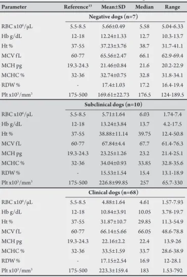
Hematological changes in dogs positive for Leishmania chagasi he results of the evaluation of red blood cells from animals
in groups ND, SD, and CD are presented in Table 2. here was
a reduction in the mean values of erythrocyte (4.88 x 10⁶/mL),
hematocrit (31.87%), and hemoglobin (10.84g/dL) in group CD compared to the reference values for dogs. here was no signiicant diference between groups. It was observed that among the animals belonging to the CD group, 63% had reduced erythrocyte counts
(below 5.5x10⁶/µL), 72% had decreased hematocrit levels (below
TABLE 1 - Clinical signs observed in dogs in group CD (clinical dogs) naturally infected by Leishmania chagasi, from the Zoonosis Control Center in Fortaleza, Ceará, Brazil.
Clinical sign Number Percentage
Onychogryphosis 23 33.8
Hepatosplenomegaly 31 45.6
Cachexia 53 77.9
Lymphadenopathy 38 55.9
Keratoconjunctivitis 42 61.8
Injuries by ectoparasites 35 51.5
Skin ulcers 24 35.3
Fever 22 32.4
Apathy 19 27.9
Alopecia 21 30.9
Mucosal ulceration 8 11.8
Peeling and eczema 22 32.4
Vomiting 6 8.8
Rectal bleeding 5 7.4
Edemaciation 23 33.8
More than one clinical sign 59 86.8
TABLE 2 - Hematology and platelet parameters in dogs with clinical and subclinical infections, naturally infected by Leishmania chagasi.
Parameter Reference13 Mean±SD Median Range
Negative dogs (n=7)
RBC x106/µL 5.5-8.5 5.66±0.49 5.58 5.04-6.33 Hb g/dL 12-18 12.24±1.33 12.7 10.3-13.7 Ht % 37-55 37.23±3.76 38.7 31.7-41.1 MCV f L 60-77 65.56±2.47 66.1 62.9-69.4 MCH pg 19.3-24.3 21.46±0.84 21.6 20.2-22.9 MCHC % 32-36 32.74±0.75 32.8 31.8-34.1
RDW % - 17.4±1.03 17.2 16.4-19.4
Plt x103/mm3 175-500 169.61±22.73 176.5 124-189.5
Subclinical dogs (n=10)
RBC x106/µL 5.5-8.5 5.71±1.64 6.03 1.74-7.4 Hb g/dL 12-18 13.24±3.84 13.7 4.2-17.5 Ht % 37-55 38.88±11.14 39.75 12.4-50.8 MCV f L 60-77 67.84±4.4 67.7 61.4-76.3 MCH pg 19.3-24.3 23.25±1.26 23.2 21.4-25.1 MCHC % 32-36 34.04±0.93 33.85 32.8-35.6
RDW % - 15.53±1.54 15.4 13.1-18.9
Plt x103/mm3 175-500 226.8±99.85 257 65.7-330
Clinical dogs (n=68)
RBC x106/µL 5.5-8.5 4.88±1.64 4.61 1.57-7.93 Hb g/dL 12-18 10.84±3.91 10.05 3.78-19.7 Ht % 37-55 31.87±10.7 29.85 11.3-54.9 MCV f L 60-77 66.14±5.66 66.05 48.6-78.8 MCH pg 19.3-24.3 22.16±2.2 22.4 13.9-26 MCHC % 32-36 33.5±1.59 33.7 28.6-38.9
RDW % - 17.15±2.54 16.9 12-28.1
Plt x103/mm3 175-500 223.3±159.4 183 1.53-792
SD: standard deviation; RBC: red blood cells; Hb: hemoglobin; Ht: hematocrit;
MCV: mean corpuscular volume; MCH: mean corpuscular hemoglobin;
MCHC: mean corpuscular hemoglobin concentration; RDW: anisocytosis index;
Plt: platelet.
TABLE 3 - Leukocyte parameters in dogs with clinical and subclinical infections, naturally infected by Leishmania chagasi.
Parameter Reference13 Mean±SD Median Range
Negative dogs (n=7)
TL x103/mm3 6-17 9.78±3.75 8.93 5.74-16.9 Neu x103/mm3 3-11.5 4.11±1.81 3.96 2.14-7.68 Lym x103/mm3 1-4.8 3.02±1.25 3.11 1.31-4.56 Mon x103/mm3 0.1-1.3 0.71±0.86 0.035 0.02-1.8 Eos x103/mm3 0.1-1.3 0.27±0.35 0.11 0-0.898 Bas x103/mm3 Rare 1.11±1.29 0.53 0-3.45
Subclinical dogs (n=10)
TL x103/mm3 6-17 11.67±3.79 11.25 6.3-17 Neu x103/mm3 3-11.5 9.81±8.11 7.82 1.26-43.7 Lym x103/mm3 1-4.8 5.53±4.48 3.04 1.47-13 Mon x103/mm3 0.1-1.3 0.56±0.52 0.39 0.003-1.4 Eos x103/mm3 0.1-1.3 0.16±0.19 0.08 0-0.52 Bas x103/mm3 Rare 0.26±0.7 0 0-2.24
Clinical dogs (n=68)
TL x103/mm3 6-17 15.24±9.77 12.9 2.5-52.9 Neu x103/mm3 3-11.5 5.42±3.98 5.07 1.03-13.6 Lym x103/mm3 1-4.8 5.17±5.62 3.2 0.65-31.6 Mon x103/mm3 0.1-1.3 0.65±0.87 0.27 0.005-4.27 Eos x103/mm3 0.1-1.3 0.24±0.41 0.04 0-1.8 Bas x103/mm3 Rare 0.23±1.22 0 0-9.86
SD: standard dev iation; TL: total leukoc y tes; Neu: neutrophils;
Lym: lymphocytes; Mon: monocytes; Eos: eosinophils; Bas: basophils.
he average platelet counts were within the normal limits among
the groups (Table 2). However, 50% of group CD showed a reduction
in the number of platelets.
he results of the evaluation of white blood cells from animals
in groups ND, SD, and CD are presented in Table 3. he CD and
SD groups showed, on average, a mild lymphocytosis in relation to the reference values for the species. he average counts of total leukocytes, neutrophils, monocytes, eosinophils, and basophils in both groups showed no changes compared to the reference values. he groups did not difer statistically (p < 0.05). However, among the animals belonging to the CD group, 33% had total leukocyte counts
exceeding 17x103/dL, and 28% had neutrophil counts greater than
11.5x10³/dL. Among the SD group animals, there were no changes
observed in the parameters of the white blood cells.
Biochemical changes in serum of dogs seropositive for
Leishmania chagasi
he average levels of GOT and GPT in animals from diferent
groups are shown in Table 4. In all the groups, the activity of
transaminases was within the normal range for dogs (GOT: 23 to 66 IU, GPT: 23 to 66 IU), and there were no signiicant diferences between groups (p <0.05).
he levels of urea and creatinine are presented in Table 4. It can
DISCUSSION
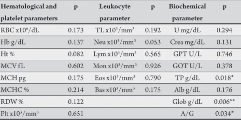
The average levels of total protein, globulin, albumin, and
albumin/globulin (A/G) are shown in Table 4. he total protein was
increased in the CD group (7.36g/dL) compared to the reference values (5.4 to 7.1g/dL), and the change is signiicant for the ND
and SD groups (p < 0.05) (Table 5). In the CD group, 53% of the
animals showed elevated levels of total protein, which is associated with increased levels of globulin fractions (4.81g/dL) compared to the reference values (2.7 to 4.4g/dL), while the albumin fraction (2.54g/dl) was low compared to the reference values for dogs (2.6 to 3.3g/dL). here were signiicant diferences in the levels of globulin in group CD compared to those in the ND and SD groups (p < 0.01)
(Table 5). Hyperglobulinemia was presented by 62% of group CD, while hypoalbuminemia was reported in 58% of the animals.
here were no changes in the A/G ratio between the groups when compared to the reference values (0.59 to 1.11). Although the average A/G ratios in the SD group showed signiicant changes compared to those in the ND and CD groups (p<0.05), the changes were not relevant since the values were within the normal limits.
TABLE 5 - P value of hematological, platelet, leukocyte, and biochemical assessments of clinical dogs naturally infected by Leishmania chagasi, in relation to subclinical and negative dogs.
Hematological and p Leukocyte p Biochemical p
platelet parameters parameter parameter
RBC x106/dL 0.173 TL x103/mm3 0.192 U mg/dL 0.294 Hb g/dL 0.137 Neu x103/mm3 0.053 Crea mg/dL 0.131 Ht % 0.082 Lym x103/mm3 0.565 GPT U/L 0.746 MCV f L 0.602 Mon x103/mm3 0.926 GOT U/L 0.378 MCH pg 0.175 Eos x103/mm3 0.790 TP g/dL 0.018* MCHC % 0.214 Bas x103/mm3 0.175 Alb g/dL 0.176
RDW % 0.122 Glob g/dL 0.006**
Plt x103/mm3 0.651 A/G 0.034*
RBC: red blood cells; Hb: hemoglobin; Ht: hematocrit; MCV: mean corpuscular volume; MCH: mean corpuscular hemoglobin; MCHC: mean corpuscular hemoglobin concentration; RDW: anisocytosis index; Plt: platelet; TL: total leukocytes; Neu: neutrophils; Lym: lymphocytes; Mon: monocytes;
Eos: eosinophils; Bas: basophils; U: urea; Crea: creatinine; GPT: glutamic pyruvic transaminase; GOT: glutamic oxaloacetic transaminase; TP: total proteins;
Alb: albumin; Glob: globulin; A/G: albumin-globulin ratio.
*Represents diferences at a signiicance level of 5%. ** Represents diferences at a signiicance level of 1%.
TABLE 4 - Biochemical parameters in dogs with clinical and subclinical infections, naturally infected by Leishmania chagasi.
Parameter Reference13 Mean±SD Median Range
Negative dogs (n=7)
U mg/dL 21-60 33.43±9.47 28 25-49
Crea mg/dL 0.5-1.5 0.69±0.15 0.7 0.5-0.9 GPT U/L 23-66 40.14±14.95 43 23-60 GOT U/L 23-66 52.43±25.09 48 25-94 TP g/dL 5.4-7.1 6.47±1.11 6.4 5.3-8.2 Alb g/dL 2.6-3.3 2.68±0.44 2.65 2.6-3.3 Glob g/dL 2.7-4.4 3.98±1.21 3.55 2.8-5.5 A/G 0.59-1.1 0.73±0.26 0.75 0.4-1.1
Subclinical dogs (n=10)
U mg/dL 21-60 25.6±4.4 24.5 21-33
Crea mg/dL 0.5-1.5 0.78±0.19 0.75 0.5-1.2
GPT U/L 23-66 28±11.55 24.5 21-60
GOT U/L 23-66 37.9±11.95 39.5 19-65 TP g/dL 5.4-7.1 6.54±0.54 6.75 5.4-7.1 Alb g/dL 2.6-3.3 2.99±0.28 3.05 2.6-3.3 Glob g/dL 2.7-4.4 3.6±0.5 3.65 2.8-4.4 A/G 0.59-1.1 0.86±0.13 0.85 0.6-1.1
Clinical dogs (n=68)
U mg/dL 21-60 65.12±60.78 44 21-356 Crea mg/dL 0.5-1.5 1.14±0.81 0.9 0.4-5.1 GPT U/L 23-66 59.38±144.74 29.5 2-1107 GOT U/L 23-66 59.67±50.71 41.5 9-287 TP g/dL 5.4-7.1 7.36±1.12 7.2 5.2-10.3 Alb g/dL 2.6-3.3 2.54±0.76 2.4 1.4-4.5 Glob g/dL 2.7-4.4 4.81±1.26 4.8 2.5-8 A/G 0.59-1.1 0.59±0.34 0.5 0.3-1.7
SD: standard deviation; U: urea; Crea: creatinine; GPT: glutamic pyruvic transaminase; GOT: glutamic oxaloacetic transaminase; TP: total proteins;
Alb: albumin; Glob: globulin; A/G: albumin-globulin ratio.
Visceral leishmaniasis is a chronic infectious disease that can be characterized by the development of a symptomatic or asymptomatic
infection accompanied by the appearance of various typical clinical signs1.
he high percentage of animals with typical clinical signs of
leishmaniasis (Table 1) demonstrates that a clinical form of the
disease may evolve with signs such as vomiting and cachexia and involve more than one clinical sign, as observed in this study (86.8% of the animals). hese data conirm the clinical indings that have
been reported in the literature15,16. It is noteworthy that Matos Jr
et al.15 found 88.8% of animals with leishmaniasis presenting more
than one clinical sign.
In this study we found alterations consistent with anemic conditions in animals belonging to group CD. Anemia in dogs
naturally infected with Leishmania chagasi is one of the most common
laboratory indings, as reported by Reis et al.12 in symptomatic
dogs, and by Ciaramella and Corona6 in about 60% of infected
animals, but the factors involved in its pathogenesis are complex and poorly known. he reason for anemia may be related to bleeding, hemolysis, inlammation, renal failure, chronic disease,
and marrow aplasia or hypoplasia17. However, no correlation has
been found between anemia and the appearance of clinical signs18.
The hematocrit and hemoglobin levels were below the reference values in the CD group; nevertheless, there were no
signiicant changes found between the groups. Costa-Val et al.18
reported signiicant changes in hematocrit and hemoglobin in dogs with leishmaniasis regardless of the presence of multiple, few, or no typical signs of CVL in the animals.
Although the average platelet counts found in this study were within the normal range independent of the evaluated group, 50% of the animals belonging to the CD group had thrombocytopenia. Some studies have reported thrombocytopenia as a typical sign of CVL19,20. Moreover, in a study by Costa-Val et al.18 with 42 dogs
positive for CVL, only 15% of the animals showed a decrease in the platelet counts.
he authors declare that there is no conlict of interest.
CONFLICT OF INTEREST
FINANCIAL SUPPORT
REFERENCES
and neutropenia (28%) were reported. Amusategui et al.21 reported
that the leukocyte counts of symptomatic, oligosymptomatic, and asymptomatic dogs did not difer statistically among themselves, and there was no correlation between leukocyte count and clinical signs found in the studied groups. However, this study veriied a trend towards increased levels of total leukocytes on the basis of clinical symptoms. With respect to the lymphocyte count, there was a slight increase in the averages in the SD and CD groups compared to the reference
values for dogs. Moreover, Paludo et al.22 reported that the main
alteration found in the white blood cell count of asymptomatic and symptomatic animals was a reduction in the average levels of lymphocytes.
The leishmaniases are a complex of diseases that involve immunological mechanisms, and as such, its worsening has been associated with increased antibody production. As a result of this production, formation of soluble and circulating immune complexes may occur; these complexes are deposited in organs and tissues,
making them targets and leading to tissue damage23.
Assessment of liver function was performed in this study by measuring the plasma activity of transaminases. In general, there was no great activity observed for both GPT and GOT in all tested groups. And analyzing the data from this study, we found that only 11% of the animals belonging to the CD group had increased levels
of GPT. CVL generally does not cause severe liver injuries6 because
most liver lesions are due to the spread of infected macrophages, thus
causing a chronic infection in this organ24. In this regard, these results
do not corroborate the indings from studies done by Ciaramella
et al.25, which revealed a considerable increase in the concentration
of GPT in animals with clinical symptoms.
In this study we observed an increase in the average levels of urea in group CD, which could mean a probable renal compromise, although the average creatinine level in all groups was within the normal limits, according to the reference values for the species. It was found that only 17% of the animals belonging to the CD group presented creatinine levels above 1.5mg/dL, thereby demonstrating that the disease was still in the acute phase. hese results are similar to
those found by Abreu-Silva et al.26 which demonstrated that uremia
is a major inding typical in dogs naturally infected by Leishmania
chagasi. his uremia may have contributed to the anemia in the CD group, because urea, which has toxic efects on red blood cells, may
decrease the half-life of erythrocytes18. Regarding renal function, it
is important to determine the degree of injury and the prognosis of
dogs with leishmaniasis by assessing the levels of creatinine and urea6.
he renal damage may also be atributed to deposits associated with
the speciic IgM and IgG antibodies27.
In this work, the CD group had high average levels of total protein and globulin, and low levels of albumin. his increase may be
associated with an increase in the levels of anti-Leishmania antibodies,
related to the symptoms of the disease. he proile of proteins in plasma is considered one of the most reliable markers for monitoring CVL. he levels of total protein in serum are substantially increased in dogs with CVL and can reach levels above 10g/dL, due mainly to
high levels of β- and γ-globulin6. Furthermore, it has been observed
that both hyperproteinemia and hypergammaglobulinemia are the
most common indings in dogs seropositive for Leishmania spp12, 26.
As CVL is a chronic disease that leads to an increase in the total protein concentration and its globulin fraction, a decrease in the
albumin concentration can be observed as well6.
he animal infected with Leishmania spp can develop a cellular
immune response mediated by h1 cells secreting IFN-γ and TNF-α, which are the predominant cytokines in asymptomatic dogs that show apparent resistance to visceral leishmaniasis. Moreover, there is evidence of a strong correlation between progression of the disease
and the IL-4 and IL-10 from h2 cells28. here are reports linking
the development of symptoms of CVL with the increased amount
of immunoglobulins24, indicating a direct correlation between high
titers of IgG1 anti-Leishmania and the appearance of clinical signs,
while IgG2 has been associated with asymptomatic dogs29.
It is noteworthy that 58% of group CD showed a reduction in the levels of serum albumin, which can be directly correlated with the edema formation observed in 33.8% of the animals. his has
been observed in dogs with the appearance of clinical signs12 and
can be explained by the migration of albumin into the extravascular regions, associated with luid accumulation, with consequent edema
formation30.
herefore, it can be concluded that animals with the clinical form of the disease show hematological and biochemical changes consistent with anemia, uremia, hyperproteinemia, and hypergammaglobulinemia, which present themselves as strong markers for canine leishmaniasis associated with the signs previously reported.
he irst author has a scholarship provided by the Fundação
Cearense de Apoio ao Desenvolvimento Científico e Tecnológico (FUNCAP).
1. Brasil MS. Manual de Vigilância e Controle da Leishmaniose Visceral. 1ª ed. Brasília: Ministério da Saúde; 2006.
2. Monteiro EM, Silva JCF, Costa RT, Costa DC, Barata A, Paula EV, et al. Leishmaniose visceral: estudo de lebotomíneos e infecção canina em Monte Claros, Minas Gerais. Rev Soc Bras Med Trop 2005; 38:147-152.
3. Ribeiro VM. Leishmaniose visceral canina: aspectos de tratamento e controle. Clín Vet 2007; 71:66-76.
4. Camargo-Neves VLF, Glasser CM, Cruz LL, Almeida RG. Manual de Vigilância e Controle da Leishmaniose Visceral Americana do Estado de São Paulo. 1ª ed. São Paulo: Secretaria de Estado da Saúde; 2006.
5. Ferrer L. Canine Leishmaniosis: Evalution of the immunocompromised patient. Wsava Congress Chooses 8 2002. [Acesso nov. 2010] Disponível em: htp;//www. vin.com/proceedings/Proceedings.plx? CID=WSAVA2002&PID=PR02653. 6. Ciaramella P, Corona M. Canine leishmaniasis: clinical and diagnostic aspects.
VetLearn 2003; 25:358-368.
7. Miranda S, Martorell S, Costa M, Ferrer L, Ramis A. Characterization of circulating lymphocyte subpopulation in canine leishmaniasis throughout treatment with antimonials and allopurinol. Vet Parasitol 2007; 144:251-260. 8. Ciaramella P, Oliva G, De Luna L, Gradoni R, Ambrosio L, Cortese A, et al.
A retrospective clinical study of canine leishmaniasis in 150 naturally infected by Leishmania infantum. Vet Rec 1997; 141:539-543.
9. Trotz-Williams L, Gradoni L. Disease risks for the travelling pet Leishmaniasis. In Practice 2003; 25:190-197.
11. Ikeda FA, Luvizoto MCR, Gonçalves ME, Feitosa MM, Ciarlini PC, Lima VMF. Peril hematológico de cães naturalmente infectados por Leishmania chagasi
no município de Araçatuba, São Paulo (Brasil): um estudo retrospectivo de 191 casos. Clín Vet 2003; 47:42-48.
12. Reis AB, Martins-Filho OA, Teixeira Carvalho A, Carvalho MG, Mayrink W, França-Silva JC et al. Parasite density and impaired biochemical/hematological status are associated with severe clinical aspects of canine visceral leishmaniasis. Res Vet Sci 2006; 81:68-75.
13. Meyer DJ, Coles EH, Rich LJ. Medicina de Laboratório Veterinário – interpretação e diagnóstico. 1ª ed. São Paulo: Roca; 1995.
14. Kaneko JJ, Harvey JW, Bruss ML. Clinical biochemistry of domestic animals. 6ª ed. San Diego: Academic Press; 2008.
15. Matos Jr DG, Pinheiro JM, Menezes RC, Costa DA. Aspectos clínicos e de laboratório de cães soropositivos para leishmaniose. Arq Bras Med Vet Zootec 2004; 56:119-122.
16. Baneth G, Koutinas AF, Solano-Gallego L, Bourdeau P, Ferrer L. Canine leishmaniosis - new concepts and insights on an expanding zoonosis: part one. Trends Parasitol 2008; 24:324-330.
17. Koutinas AF, Polizopoulou ZS, Saridomichelakis MN, Argyriadis D, Fytianou A, Plevraki K. Clinical considerations on canine visceral leishmaniasis in Greece: a retrospective study of 158 cases (1989 - 1996). J Am Anim Hosp Assoc 1999; 35:376-383.
18. Costa-Val AP, Cavalcanti RR, Gontijo NF, Michalick MSM, Alexander B, Williams P, et al. Canine visceral leishmaniasis: Relationships between clinical status, humoral immune response, haematology and Lutzomyia (Lutzomyia) longipalpis infectivity. Vet J 2007; 174:636-643.
19. Soares MJV, Moraes JRE, Palmeira-Borges V, Miyazato LG, Moraes FR. Alterações renais em cães com leishmaniose visceral. J Ven Anim Toxins Includ Trop Dis 2005; 11:579-593.
20. Cortese L, Terrazzano G, Piantedosi D, Sica M, Prisco M, Ruggiero G, et al. Prevalence of anti-platelet antibodies in dogs naturally co-infected by Leishmania infantum and Erlichia canis. Vet J 2011; 188:118-121.
21. Amusategui I, Sainz A, Rodriguez F, Tesouro MA. Distribution and relationships between clinical and biopathological parameters in canine leishmaniasis. Eur J Epidemiol 2003; 18:147-156.
22. Paludo GR, Aquino LC, Lopes BCC, Silva PHC, Borges TS, Dias CA, Castro MB. Laboratorial indings of canine visceral leishmaniosis in Brasília, Brazil. Vet Clin Pathol 2007; 36:382-398.
23. Tizard IR. Imunologia Veterinária - Uma Introdução. 8ª ed. Rio de Janeiro: Elsevier; 2009.
24. Silva FS. Patologia e patogênese da leishmaniose visceral canina. Rev Trop: Ci Agr Biol 2007; 1:20-31.
25. Ciaramella P, Pelagalli A, Cortese L, Pero ME, Corona M, Lombardi P, et al. Altered platelet aggregation and coagulation disorders related to clinical indings in 30 dogs naturally infected by Leishmania infantum. Vet J 2005; 169: 465-467. 26. Abreu-Silva AL, Lima TB, Macedo AA, Moraes-Júnior FJ, Dias EL, Batista ZS, et al. Soroprevalência, aspectos clínicos e bioquímicos da infecção por Leishmania
em cães naturalmente infectados e fauna de lebotomíneos em uma área endêmica na ilha de São Luís, Maranhão, Brasil. Rev Bras Parasitol Vet 2008; 17:197-203. 27. Soares MJV, Moraes JRE, Moraes FR. Renal involvement in canine leishmaniasis:
a morphological and immunohistochemical study. Arq Bras Med Vet Zootec 2009; 61:785-790.
28. Barbiéri CL. Immunology of canine leishmaniasis. Parasite Immunol 2006; 28: 329-337.
29. Iniesta L, Gállego M, Portús M. Immunoglobulin G and E responses in various stages of canine leishmaniosis. Vet Immunol Immunopathol 2005; 103:77-81. 30. Kumar V, Abbas AK, Fausto N. Robins e Cotran: Patologia: Bases Patológicas