INTRODUCTION
Article/Artigo
1. Programa de Pós-graduação em Doenças Infecciosas e Parasitárias, Faculdade de Medicina, Universidade Federal de Mato Grosso do Sul, Campo Grande, MS. 2. Departamento de Patologia, Centro de Ciências Biológicas e da Saúde, Universidade Federal de Mato Grosso do Sul, Campo Grande, MS. 3. Laboratório de Imunologia, Departamento de Patologia, Centro de Ciências Biológicas e da Saúde, Universidade Federal de Mato Grosso do Sul, Campo Grande, MS. 4. Centro de Prevenção ao Câncer, Campo Grande, MS. Address to: Dra. Inês Aparecida Tozeti. Depto de Patologia/CCBS/UFMS. Caixa Postal 549, 79070-900
Campo Grande, MS, Brasil. Phone: 55 67 3345-7388 e-mail: [email protected] Received in 08/06/2011 Accepted in 10/08/2011
S100, CD68, and MHC class II molecule expression in cervical high-
and low-grade HPV-induced lesions
Expressão de S100, CD68 e moléculas de MHC classe II em lesões cervicais de alto e baixo
grau induzidas por HPV
Fernanda Cassandri
1, Inês Aparecida Tozeti
1, Carlos Eurico dos Santos Fernandes
2, Flávia Gato de Almeida
3,
Gustavo Ribeiro Falcão
4, Ilzia Doraci Lins Scapulatempo
2,4, hiago heodoro Martins Prata
3,
Cacilda Tezelli
Junqueira Padovani
2, Daniella Borges Alves
1, Alda Teixeira Ferreira
2and Maria Auxiliadora Gomes Sandim Abdo
2ABSTACT
Introduction: Some human papillomavirus (HPV) types are involved in malignant processes in the cervical epithelium, with 99% of cases atributed to oncogenic HPV infection. his study aimed to detect S100, CD68, and major histocompatibility complex class II (MHC-II) molecules in cervical uterine epithelial samples in patients with high- and low-grade lesions induced by HPV. Methods: Fity-eight samples from patients who were conirmed positive or negative for high-risk oncogenic HPV DNA, had histopathological diagnosis of cervical intraepithelial neoplasia (CIN) of grades I, II, or III, or were negative for intraepithelial lesion or malignancy were subjected to immunohistochemistry reaction to S100 protein, CD68, and MHC-II (HLA-DR alpha chain). Results: he presence of MHC-II predominated in samples exhibiting histopathological alterations (p < 0.05). S100 detection was more numerous in carcinoma samples (CIN III) (75%). Presence of this protein correlated signiicantly (p < 0.05) with histopathological indings and viral load. Conclusions: A small expression of CD68 was observed, which may be explained by the observation in our study having been made on random microscopic ields and not on speciic areas. he indings, such as the presence of S100 protein and MHC-II expression in samples with histological alterations, could suggest that the immune system fails to control HPV replication at the early stages of infection. Further studies with larger prospective data are necessary to conirm this result.
Keywords: Human papillomavirus. Immune response. Immunohistochemistry.
RESUMO
Introdução: Alguns tipos de papilomavirus humano (HPV) estão envolvidos em processos malignos no epitélio cervical, com 99% dos casos atribuídos à infecção por HPV oncogênico. O objetivo deste estudo foi detectar a proteína S100, CD68 e moléculas de MHC-II (complexo principal de histocompatibilidade classe II) em amostras de epitélio cervical uterino, de pacientes com lesões de alto e baixo grau induzidas pelo HPV. Métodos: Cinquenta e oito amostras de pacientes positivos ou negativos, conirmados, para DNA de HPV de alto ou baixo risco oncogênico, e que tiveram diagnóstico histopatológico de neoplasia intraepithelial cervical (NIC) de graus I, II ou III ou foram negativas para lesão intraepithelial e malignidade (NILM), foram submetidas à reação de imunohistoquímica (IHQ) para proteína S100, CD68 e MHC-II (HLA-DR cadeia alfa). Resultados: A presença da molécula MHC-II predominou em amostras exibindo alterações histopatológicas (p < 0,05). A detecção de S100+ foi mais numerosa em amostras com carcinoma (NIC III) (75%). A presença dessa proteína correlacionou-se signiicantemente (p < 0,05) com achados histopatológicos e a carga viral.
Conclusões: Pequenaexpressão CD68+ foi observada, uma possível explicação seria que em nosso estudo as observações foram feitas em campo microscópicos aleatórios e não em áreas especíicas. Os achados como a presença de S100 e a expressão de MHC-II, em amostras com alterações histológicas, podem sugerir que o sistema imune falha em controlar a replicação do HPV nas fases iniciais da infecção. Maiores estudos, com mais dados prospectivos, são necessários para conirmar esses resultados.
Palavras-chaves: Papilomavírus humano. Resposta immune. Imunohistoquímica.
Cervical cancer is the second most frequent cause of death from cancer among women. An estimated half a million cases occur each year, almost 80% of these in developing countries1.
Human papillomavirus (HPV) infection is sexually transmitted, with effects ranging from benign verrucae to invasive cancer. Some HPV types are involved in malignant processes in the cervical epithelium, with 99% of cases atributed to oncogenic HPV infection2. In the anogenital tract,
HPV infects basal and parabasal squamous epithelial cells. In cases of latent infection, no cytopathic efects are observed and the epithelium is normal. Cases of productive infection may involve clinical (Condyloma acuminatum, invasive carcinoma) or subclinical manifestations (high- or low-grade cervical intraepithelial neoplasia)3.
Immunodeficiency is associated with low numbers of Langerhans cells and the presence of HPV-induced lesions4,5. he presence of
antigen-presenting cells (APC), like Langerhans cells and macrophages, and the activity of such cells by major histocompatibility complex class II (MHC-II) molecule expression at the early stages of infection are therefore important factors for disease resolution. We know that Langerhans cells are the most eicient APCs to initiate primary response by naïve T-lymphocyte activation6 and that macrophages are
the APCs that diferentiate T-helper lymphocytes in the efector phase of cellular immunity and humoral response7.
Evidence suggests that E5, a small, highly hydrophobic protein observed in cell cultures and located in internal compartments of the endoplasmic reticulum and Golgi complex, inhibits the transport of proteins, including class I and class II MHC molecules, to the cell surface8,9.
METHODS
RESULTS
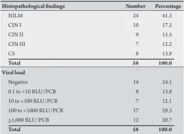
TABLE 1 - Histological classiication of uterine cervical epithelial lesion and viral load of HPV DNA of the samples.
Histopathological indings Number Percentage
NILM 24 41.3
CIN I 10 17.2
CIN II 9 15.5
CIN III 7 12.2
CS 8 13.8
Total 58 100.0
Viral load
Negative 14 24.1
0.1 to <10 RLU/PCB 8 13.8
10 to <100 RLU/PCB 7 12.1
100 to <1000 RLU/PCB 17 29.3
>1,000 RLU/PCB 12 20.7
Total 58 100.0
HPV: human papillomavirus; DNA: deoxyribonucleic acid; NILM: negative for intraepithelial lesions and malignancy; CIN: cervical intraepithelial neoplasia; CS: carcinoma samples; RLU/PCB: relative light units/positive control to group B.
and inhibition of immune response10, the exact mechanisms
responsible for activating effective immune response to HPV remain unknown. Systemic immune abnormalities have been demonstrated in cases of uterine cervical cancer11,12. Moreover,
the tumor microenvironment may be capable of controlling local immune response, thereby interfering with disease progression13,14.
In local immune response, the initial mechanisms of presentation of antigens and their components, like MHC-II expression, are essential for activating and maintaining efective immune responses capable of promoting virus elimination or limiting replication to low levels. he purpose of the present study was to describe the presence of S100 protein, CD68 marker, and MHC-II molecules (HLA-DR+) in epithelial lesions of the uterine cervix of patients with high- and low-grade HPV-related lesions. As these markers can be found in Langerhans cells, macrophages, and others cells, characterizing the antigen presentation events is important for eicient immune response.
Subjects
he study included samples of uterine cervical lesions collected from women aged 18 to 65 years, assisted for routine analysis or for clinical suspicion of HPV infection from 2000 to 2002 at the Centro de Prevenção ao Câncer, a cancer prevention center located in Campo Grande, the capital city in the State of Mato Grosso do Sul, Brazil.
Fity-eight samples of parain-embedded cervical stroma were used. Of these, 44 were positive for high-risk oncogenic HPV DNA, as detected using the hybrid capture II (HCII) test (Digene), and had a negative histopathological diagnosis for intraepithelial lesion or malignancy (NILM) or cervical intraepithelial neoplasia (CIN) grade I, II, or III. In this last group we normally have the samples classiied as in situ or invasive carcinoma, but in this work the samples considered carcinoma in situ or invasive were considered separately, as with other authors15, because the immune response to the neoplastic
process can be diferent from that to the pre-neoplastic process. In this study we included yet another 14 samples obtained from patients who were negative for HPV DNA, as revealed by HCII, and with NILM results. Viral load, quantiied by HCII, was classiied into four groups, according to Lorincz et al.16: from 0.1 to less than 10, from
10 to less than 100, from 100 to less than 1,000, and from 1,000 or more relative light units/positive control to group B (RLU/PCB). he data for histopathological diagnosis and viral load (Table 1)
were provided by a previous study17.
Immunohistochemistry
he immunohistochemistry (IHC) reaction was performed as proposed by Santos and colleagues18. Antigen retrieval was performed
by heating sections in 10mM citrate bufer at pH 6.0. To detect IHC markers, the following monoclonal antibodies were used: mouse anti-human S100 protein (Zymed, ref. 18-0046); mouse anti-human CD68, clone KP1 (Dako, ref. M0814); and mouse anti-human HLA-DR, alpha chain, clone TAL.1B5 (Dako, ref. M0746). he detection system was a Universal LSAB + kit/-HRP (Dako, ref. K0690), and diaminobenzidine (Dako, ref. K3468) was used as chromogen. All sections were examined under a 10× objective with conirmation at 40×, and ten randomly chosen microscopy ields/per region were observed. Only epithelium was considered for S100 protein, only
stroma for CD68, and both epithelium and stroma for MHC-II. he percentage of cells stained was categorized as follows: 0 for the absence of cells (negative), 1 for a small number of cells, and 2 for a large number. Two previously calibrated independent observers (κ = 0.091) carried out the observation; conlicting results were resolved by consensus.
Data analysis
he data were organized in spreadsheets for statistical analysis (chi-square test), carried out using SPSS 10.0 sotware. For statistical analysis, the histopathology results were grouped as CIN I, CIN II/III, and carcinoma samples (CS). he last CS was considered a separate group because immune responses to neoplastic processes can difer from those to pre-neoplastic conditions. he data were analyzed for possible correlations between histological or viral load groups and the presence of IHC markers.
Ethical considerations
The investigation was approved by the Research Ethics Commitee of the Universidade Federal de Mato Grosso do Sul (UFMS) (protocol 1355).
Relationship between the presence of IHC markers and histopathological indings
Table 2 shows data on MHC-II expression and the presence
of CD68 and S100 for each type of histopathological inding. Low amounts of MHC-II molecules were expressed in 79.2% of NILM samples. Greater expression of this molecule was found in CIN I, reaching maximum values in CS (57.1%). Histopathological indings correlated signiicantly (p< 0.05) with the presence of MHC-II molecules.
TABLE 2 - Classiication of uterine cervical epithelial lesion samples by presence of MHC-II molecules, CD68, and S100+ cells.
Immunohistochemistry MHC-II
0 1 2 Total
Histopathological indings n % n % n % n %
NILM 4 16.7 19 79.2 1 4.2 24 100.0 CIN I 0 0.0 6 60.0 4 40.0 10 100.0 CIN II/III 3 18.8 8 50.0 5 31.3 13 100.0* CS 0 0.0 3 42.9 4 57.1 7 100.0*
Total 7 12.3 36 62.2 14 24.7 58 100.0
CD68
0 1 2 Total
Histopathological indings n % n % n % n %
NILM 4 16.7 18 75.0 2 8.3 24 100.0 CIN I 1 10.0 8 80.0 1 10.0 10 100.0 CIN II/III 1 6.3 14 87.5 1 6.3 16 100.0 CS 0 0.0 4 50.0 4 50.0 8 100.0
Total 6 10.3 44 75.9 8 13.8 58 100.0
S100
0 1 2 Total
Histopathological indings n % n % n % n %
NILM 4 16.7 17 70.8 3 12.5 24 100.0 CIN I 0 0.0 5 50.0 5 50.0 10 100.0* CIN II/III 0 0.0 6 37.5 10 62.5 16 100.0 CS 0 0.0 2 25.0 6 75.0 8 100.0
Total 4 6.9 30 51.7 24 41.4 58 100.0
MHC-II: major histocompatibility complex; CD68: cluster of diferentiation 68; S100: S100 protein;
NILM: negative for intraepithelial lesions and malignancy; CIN: cervical intraepithelial neoplasia;
CS: carcinoma samples. Statistical analysis: c2 test. * p < 0.05.
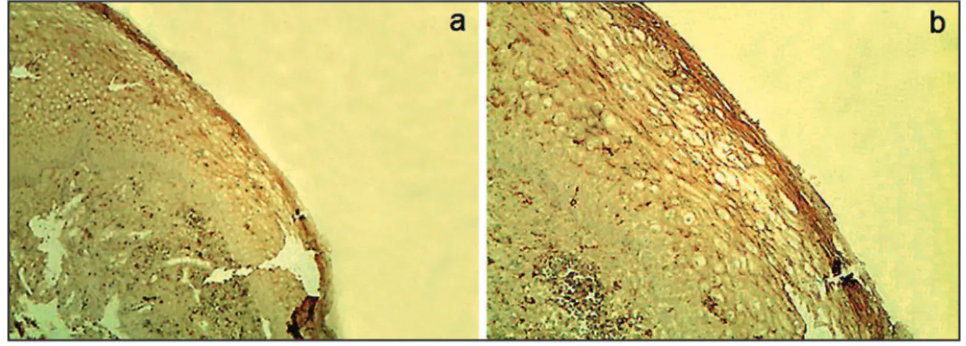
S100 protein, on the other hand, was present in small numbers in 70.8% of NILM samples, but in high quantities in CIN I and CIN II/III samples (50-60%), as well as in CS samples (75%). Presence of S100 correlated signiicantly with histopathological indings (p < 0.05). Figure 1 shows S100 and CD68, in addition to MHC-II molecules, detected using the IHC reaction. Figure 2 shows NILM samples with marker absence.
Relationship between the presence of IHC markers and viral load
No significant correlation was found between MHC-II expression, CD68 presence, and viral load. As shown in Table 3, most samples revealed low levels of MHC-II molecules and CD68 (60-80%), regardless of viral load, with a slight predominance of MHC-II expression (42.9%) and CD68 presence at high levels (28%) among samples having a viral load of 10 to less than 100 RLU/ PCB.
S100 protein was found in large numbers in 62.5% of the samples having 1 to less than 10 RLU/PCB and in 83.3% of samples with 1,000 or more RLU/PCB. Presence of S100 protein correlated signiicantly with viral load (p < 0.05).
FIGURE 1 -Macrophages (a: 100x; b: 400x), major histocompatibility complex-II molecules (c: 100x; d: 400x), and Langerhans cells (e: 100x; f: 400x) detected by immunohistochemistry reaction in chorium cells from cervical mucosa.
DISCUSSION
TABLE 3 - Classiication of samples by viral load by presence of MHC-II molecules, CD68, and S100+ cells. Immunohistochemistry
MHC-II
0 1 2 Total
Viral load n % n % n % n %
0 (negative) 3 21.4 11 78.6 0 0.0 14 100.0
1 (1 to <10 RLU/PCB) 1 12.5 4 50.0 3 37.5 8 100.0
2 (10 to <100 RLU/PCB) 0 0.0 4 57.1 3 42.9 7 100.0
3 (100 to <1,000 RLU/PCB) 2 11.8 10 58.8 5 29.4 17 100.0
4 (>1,000 RLU/PCB) 1 8.3 7 58.3 4 33.3 12 100.0
Total 7 12.1 36 62.1 15 25.9 58 100.0
CD68
0 1 2 Total
Viral load n % n % n % n %
0 (negative) 3 21.4 11 78.6 0 0.0 14 100.0
1 (1 to <10 RLU/PCB) 1 12.5 6 75.0 1 12.5 8 100.0
2 (10 to <100 RLU/PCB) 0 0.0 5 71.4 2 28.0 7 100.0
3 (100 to <1,000 RLU/PCB) 1 5.9 14 82.4 2 11.8 17 100.0
4 (>1,000 RLU/PCB) 1 8.3 8 66.7 3 25.0 12 100.0
Total 6 10.3 44 75.9 8 13.8 58 100.0
S100
0 1 2 Total
Viral load n % n % n % n %
0 (negative) 3 21.4 11 78.6 0 0.0 14 100.0
1 (1 to <10 RLU/PCB) 0 0.0 3 37.5 5 62.5 8 100.0
2 (10 to <100 RLU/PCB) 0 0.0 4 57.1 3 42.9 7 100.0
3 (100 to <1,000 RLU/PCB) 1 5.9 10 58.8 6 35.3 17 100.0
4 (>1,000 RLU/PCB) 0 0.0 2 16.7 10 83.3* 12 100.0
Total 4 6.9 30 51.7 24 41.4 58 100.0
MHC-II: major histocompatibility complex; CD68: cluster of diferentiation 68; S100: S100 protein; RLU/PCB: relative light units/positive control to group B. Statistical analysis: c2 test. *: p < 0.05.
FIGURE 2 - Negative immunohistochemistry reaction in chorium cells from cervical mucosa. (a: 100x; b: 400x).
Host defense mechanisms, even if limited, require TCD4 lymphocytes to recognize the antigen associated with MHC-II molecules presented by Langerhans cells or macrophages6,7.
Several markers, including OKT6, ATPase, and HLA-DR, have been identiied in Langerhans cells. In HPV infection, as well as in
CIN, reductions of as much as 60% have been found in the number of these cells, while those with the S100 marker have been reported as entirely absent. hese indings suggest that selective reduction of S100+ cells could be the cause of localized immunodeiciency6.
cells and low-grade lesions. In fact, this marker was present in large numbers in high- and low-grade lesions, but in low numbers in NILM samples. he later inding might stem from the absence of adequate stimulus for antigen presentation19,20.
Comparisons of histological indings and frequency of S100+ cells in HPV-negative cervix samples versus samples from women infected with oncogenic high-risk HPV21 have shown that the
presence of these cells in small numbers in HPV-positive patients may represent a mechanism of viral escape from immune response, thus reducing antigen presentation. A reduced presence of S100+ cells was also observed in the present study, revealing that these marked cells are prompted to migrate to afected areas only when histopathological alterations become more pronounced. he occurrence of Langerhans cells and other APC iniltration represent beter prognosis in cases of intraepithelial neoplasia and invasive and squamous carcinoma of the uterine cervix22, thyroid, lungs, and stomach23-25.
HLA-DR, a member of the MHC-II family of transmembrane receptors, is primarily expressed on B cells, onto which it presents antigenic peptides for recognition by CD4+ T-cell receptors. his interaction is central to antigen specificity in adaptive immune responses. here is a consensus that HLA-DR expression represents an intense local action of cytokine inductors26.
Malignant transformation of infected cells and neoplasia are effects of viral DNA integration, which at this infection stage might inhibit antigen presentation, since HPV interferes with MHC-I and MHC-II synthesis, hindering T-cell activation27. In
inflammatory conditions and viral infections, expression of higher levels of MHC-II molecules is responsible for more pronounced T-cell activation28.
Langerhans cells originally express MHC-II molecules, but HPV-16 and HPV-18 infections cause a decrease in these molecules in the cervical epithelium29. Loss of MHC-I molecules has been reported
in the progression of cervical lesions and can serve as a prognostic marker for vulvar disease, as can antigen expression by MHC-II30. Our results showed that higher levels of MHC-II expression
occurred at the stages in which viral replication was accompanied by histopathological changes, as was the case of CS.
Inflammator y infiltration by macrophages and CD4 T-lymphocytes has been observed in spontaneous regression of condyloma, and speciic CD4 T-lymphoproliferative response to HPV E2 antigen was associated with virus elimination. On the other hand, CD8 T-cells speciic for E6 and E7 antigens have been reported for patients with large lesions or cervical tumors. It was thus possible to observe that T-helper type 1 response is decreased when IL-2, INF-γ, and TNF-α production is low in patients with high levels of intraepithelial lesions31.
Murine cells expressing HPV-16 E6 protein have been found to induce lysis by macrophages, but not by natural killer cells32.
Low expression of CD68 molecules was observed in the present study, demonstrating reduced presence of this marker at all stages of infection, predominantly in CS.
In a study of 35 samples of cervical stroma following conization and with a four-year follow-up, no association was found between CD68+ cells and recurrence of CIN III, as detected by IHC reaction33.
Some authors suggest, however, that the inlammatory response is responsible for the worst prognostic in cervical disease, and showed that it had a direct relationship between the increasing grade of
the lesion and the number of macrophages in the epithelium34. In
Hammes and colleagues the authors showed that cases of low-grade squamous epithelial lesions with moderate to intense inlammatory regions had almost two times more macrophages than did those with negative to weak reactions34.
Other authors have found similar results with the signiicant increase of macrophages in carcinoma in situ and have suggested that this cell participates in malignant cell migration and invasion by production of angiogenic factors, once a direct relation was observed between macrophage and blood vessels15,35,36.
A small expression of CD68+ cells was observed in our study, demonstrating the low presence of this marker in all phases of infection; these cells are predominant only in carcinoma samples. One possible explanation for this discrepancy is that in our study the observation was made on ten random microscopic ields and not on speciic areas with CIN lesions and CS. herefore, our sample size was small, and further studies with larger prospective data are necessary to conirm this result.
By relating, on the one hand, the magnitude of viral load and, on the other, the presence of the structures linked to immune response activation, a predominance of S100 protein was found in samples from patients at the viral replication stage (≥1,000 RLU/PCB). he relationship between viral load and the presence of CD68 and MHC-II molecules revealed their distribution to be virtually uniform at all levels. Studies have demonstrated that viral load in HPV infection can serve as a marker for the progression of pre-cancerous lesions37,38. his is corroborated by the results of
the present investigation showing that, at non-replication stages of infection, the stimulus for immune response activation was not operating, allowing viral infection to persist, thus contributing to malignant transformation.
The evaluation of NILM samples, which were negative (n = 14) and positive (n = 10) for HPV DNA (data not shown), revealed few S100 or CD68+ cells and low levels of MHC-II molecules in both groups. In NILM samples that were positive for HPV DNA, S100 was present in large numbers only when the viral load was ≥1,000 RLU/PCB, suggesting that intense viral replication may be necessary for antigen presentation and immune response activation.
he presence of Langerhans cells at the initial stage of HPV infection could represent an indicator of infection control by the immune system. At more advanced stages, however, an increase in the number of these cells may be an indication that antigen presentation by these cells to T lymphocytes fails to occur at the initial stage. Periods of intense replication, when the virus causes characteristic cellular alterations and the infection is productive, are accompanied by the occurrence of immune response. he persistence of viral replication and cellular alterations become risk factors for malignancy. he indings of this article, such as the predominant presence of S100, CD68, and MHCII expression in samples with histological alterations, could suggest that the immune system fails to control HPV replication at the early stages of infection.
he authors declare that there is no conlict of interest.
CONFLICT OF INTEREST
FINANCIAL SUPPORT
REFERENCES
Fundação de Apoio ao Desenvolvimento do Ensino, Ciência e Tecnologia do Estado de Mato Grosso do Sul (Fundect-MS) and
Conselho Nacional de Pesquisa (CNPq) (Brazil).
1. International Agency for Research on Cancer (IARC). Monographs on the evolution of carcinogenic risks to humans: human papilomaviruses [Internet]. Vol 90. Lyon: IARC; 2009. [cited 2010 September 28] Available from: htp:// monographs.iarc.fr/ENG/Monographs/vol90/index.php.
2. Bosch FX, Manos MM, Muñoz N, Sherman M, Jansen AM, Peto J. Prevalence of Human papillomavirus in cervical cancer: a worldwide perspective. J Natl Cancer Inst 1995; 11:796-802.
3. McCance DJ. Human Papillomaviruses. Infect Dis Clin North A 1994; 8:751-767. 4. Uchimura NAS. Alterações das células de Langerhans e sua relação com lesão hispatológica do colo uterino por Papilomavírus Humano em pacientes com captura híbrida positiva [Tese]. [São Paulo]: Faculdade de Medicina Universidade Federal de São Paulo; 2002. 150p.
5. Girolamo P, Arcamone N, Pelagalli GV, Gargiulo G. Immunohistochemical localization of S100-like protein in non-mammalian kidney. Microscopy Res and Techn 2003; 60:652-657.
6. Tay SK, Jenkins D, Maddox P, Campion M, Singer A. Subpopulation of Langerhans cells in cervical neoplasia. Br J Obstet Gynaecol 1987; 94:10-15. 7. Rosenthal AS, Barcinski MA, Rosenwasser LJ. Function of macrophages in genetic
control of immune responsiveness. Fed Proc 1978; 37:79-85.
8. Dimaio D, Maton D. Mechanisms of cell transformation by papillomavirus E5 proteins. Oncogene 2001; 20:7866-7873.
9. Schapiro F, Sparkowski J, Adduci A, Suprynowicz F, Schlegel R, Grinstein S. Golgi alkalinization by the papillomavirus E5 oncoprotein. J Cell Biol 2000; 148:305-315.
10. Pinzon-Charry A, Ho CS, Caberty R, Maxwell T, Walker D, Gardiner A, et al. Population of HLA-DR+ immature cells accumulates in the blood dendritic cell compartment of patients with diferent types of cancer. Neoplasia 2005; 7:1112-1122. 11. Bais AG, Beckman I, Lindemans J, Ewing PC, Meijer CJ, Snijders PJ, et al. A shit to a peripheral h2-type cytokine patern during the carcinogenesis of cervical cancer becomes manifest in CIN III lesions. J Clin Patho 2005; 58:1096-1100. 12. Fernandes Jr PC, Gracia CB, Micheli DC, Cunha FQ, Murta EFC,
Tavares-Murta BM. Circulating neutrophils may play a role in the host response in cervical cancer. Int J Gynecol Cancer 2007; 17:1068-1074.
13. Balkwil LF, Mantovani A. Inlamation and cancer: back to Virchow? Lancet 2001; 357: 539-545.
14. Fine JS, Byrnes HD, Zavodny PJ, Hipkin RW. Evaluation of signal trasnduction pathways in chemoattractant-induced human monocyte chemotaxis. Inlamation 2001; 25:61-67.
15. Utrera-Barrilas D, Costa-Manrreza M, Castellanos E, Gutierrez-Rodríguez M, Arciniega-Ruiz de Esparza O, García-Cebada J, et al. he role of macrophage and mast cells in lymphangiogenesis and angiogeneis in cervical carcinogenesis. Exp Mol Path 2010; 89:190-196.
16. Lorincz A, Castle P, Sherman M, Scot D, Glass A, Wacholder S, et al. Viral load of Human Papillomavirus and risk of CIN 3 or cervical cancer. Lancet 2002; 360:228-229.
17. Tozeti IA, Scapulatempo IDL, Levi JE, Ferrreira AW. Determination of HPV DNA viral load by hybrid capture assay and its association with cytological indings. J Bras Patol Med Lab 2006; 42:449-453.
18. Santos ALF, Derchain SFM, Martins MR, Sarian LO, Martinez EZ, Syrjänen KJ. Procedimentos laboratoriais em imunohistoquímica e hibridização in situ.
In: Alves VAF, Bacchi C, Vassalo J, editors. Manual de imunohistoquímica. 1ª ed. São Paulo: Sociedade Brasileira de Patologia; 1999. p. 237-259.
19. Campoli M, Chang CC, Ferrone S. HLA class I antigen loss, tumor immune escape and immune selection. Vaccine 2002; 20:40-45.
20. Marincola FM, Wang E, Herlyn M, Seliger B, Ferrone S. Tumors as elusive targets of T-cell-based active immunotherapy. Trends Immunol 2003; 24:335-342. 21. Jimenez-Flores R , Mendez-Cruz R , Ojeda-Ortiz J, Muñoz-Molina R ,
Balderas-Carrillo O, Diaz-Soberanes ML, et al. High-risk Human Papillomavirus infection decreases the frequency the frequency of Dendritic Langerhans’ cells in the human female genital tract. Immunology 2006; 117:220-228. 22. Nakano T, Oka K, Arai T, Morita S, Tsunemoto H. Prognostic signiicance of
Langerhans cells iniltration in radiation therapy for squamous cell carcinoma of the uterine cervix. Arch Pathol Lab Med 1989; 113:507-511.
23. Tsujitani S, Furukawa T, Tamada R, Okamura T, Yasumoto K, Sugimachi K. Langerhans cells and prognosis in patients with gastric carcinoma. Cancer 1987; 59:501-505.
24. Schroder S, Schwarz W, Rehpenning W, Löning T, Böcker W. Dendritic/ Langerhans cells and prognosis in patients with papillary thyroid carcinomas. Am J Clin Pathol 1988; 89: 295-300.
25. Fox SB, Jones M, Dunnill MS, Gater KC, Mason DY. Langerhans cells in human lung tumors: and immunohistological study. Histopathology 1989; 14:269-275. 26. Adams TE, Bodmer JG, Bodmer WF. Production and characterization of monoclonal antibodies recognizing the alpha-chain subunits of human Ia alloantigens. Immunology 1983; 50:613-624.
27. O’Brien PM, Campo MS. Evasion of host immunity directed by papillomavirus-encoded proteins. Virus Res 2002; 88:103-117.
28. Servet C, Zitvogel L, Hosmalin A. Dendritic cells in innate immune responses against HIV. Curr Mol Med 2002; 2:739-756.
29. Goncalves MA, Soares EG, Fernandes AP, Fonseca BA, Betini JS, Simões RT, et al. Langerhans cell count and HLA class II proile in cervical intraepithelial neoplasia in the presence or absence of HIV infection. Eur J Obstet Gynecol Reprod Biol 2004; 114:221-227.
30. Zehbe I, Höhn H, Pilch H, Neukirch C, Freitag K, Maeurer MJ. Diferential MHC class II component expression in HPV-positive cervical cancer cells: Implication for immune surveillance. Int J Cancer 2005; 117:807-815.
31. Lee BN, Follen M, Shen DY, Malpica A, Adler-Storthz K, Shearer WT, et al. Depressed type 1 cytokine synthesis by superantigen-activated CD4+ T cells of women with human papillomavirus-related high-grade squamous intraepithelial lesions. Clin Diagn Lab Immunol 2004; 11:239-244.
32. Routes JM, Morris K, Ellison MC, Ryan S. Macrophages kill Human Papillomavirus type 16 E6-expressing tumor cells by tumor necrosis factor-Ü and nitric oxide-dependent mechanisms. J Virol 2005; 79:116-123.
33. Maluf PJ, Michelin MA, Etchebehere RM, Adad SJ, Murta EFC. T lymphocytes (CD3) may participate in the recurrence of cervical intraepithelial neoplasia grade III, Arch Gynecol Obstet 2008; 278:525-530.
34. Hammes LS, Tekmal RR, Naud P, Edelweiss MI, Kirma N, Valente PT, et al. Macrophages, inlammation and risk of cervical intraepithelial neoplasia (CIN) progression – clinocopathological correlation, Gynecol Oncol 2007; 105:157-165.
35. Mazibrada J, Rita M, Mundini M, Andrea M, Azzimonti B, Borgogna C, et al. Interaction between inlammation and angiogenesis during diferent stages of cervical carcinogenesis. Gynecol Oncol 2008; 108:112-120.
36. Kobayashi A, Weinberg V, Darragh T, Smith-McCune K. Evolving immuno-suppressive microenvironment during human cervical carcinogenesis. Mucosal Immunology 2008; 1:412-420.
37. Santos ALF, Derchain SFM, Martins MR, Sarian LO, Martinez EZ, Syrjänen KJ. Human Papillomavirus viral load in predicting high grade CIN in women with cervical smears showing only atypical squamous cells or low-grade squamous intraepithelial lesion. São Paulo Med J 2003; 121:238-243.